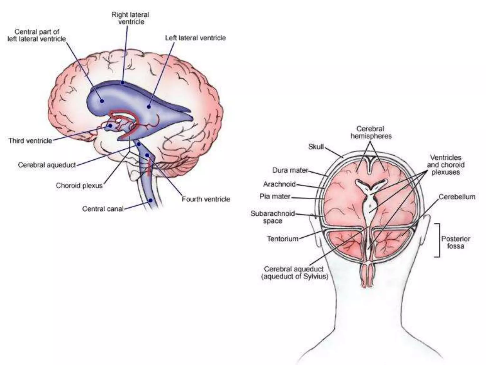
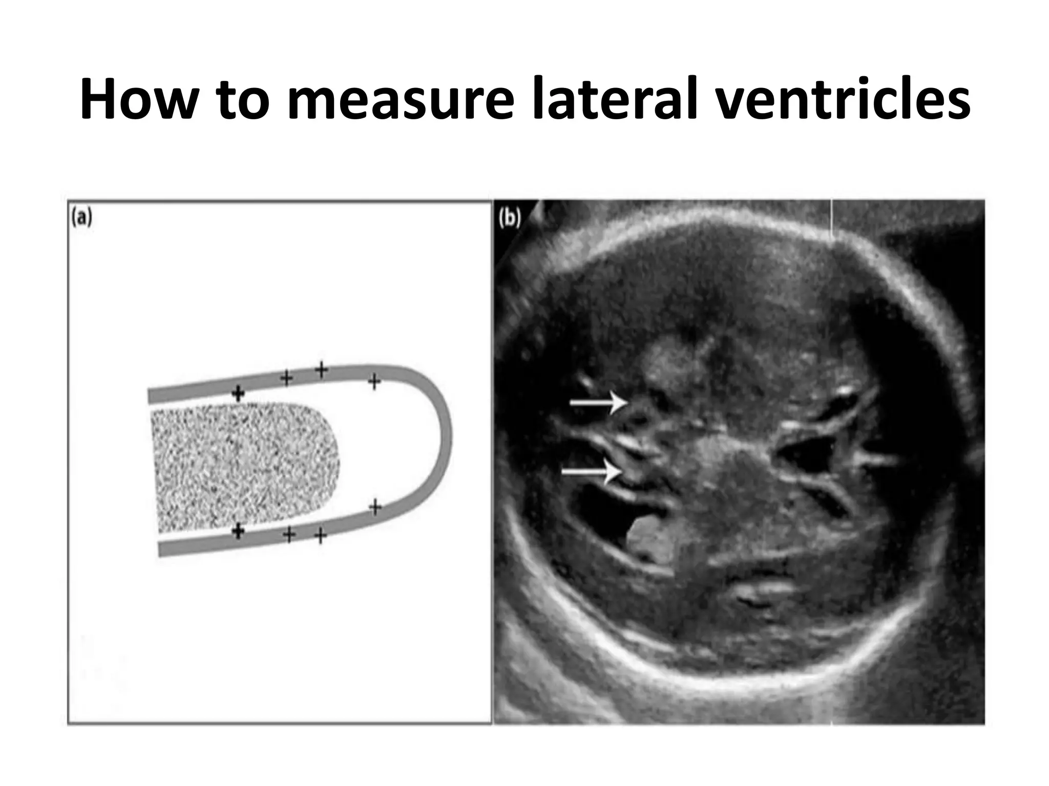
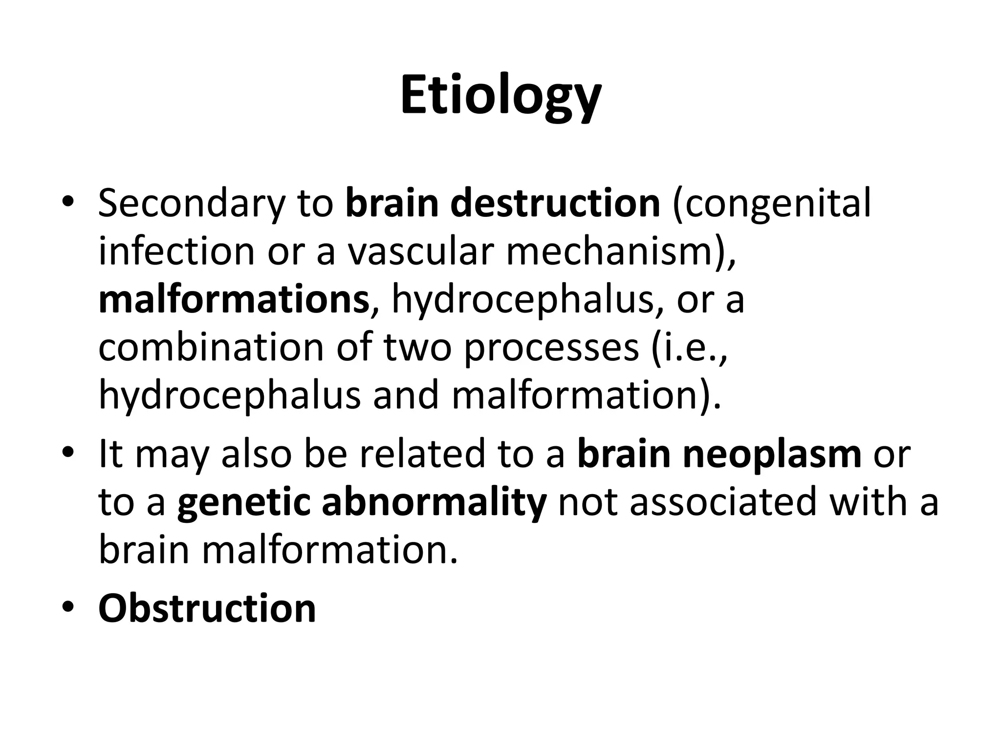
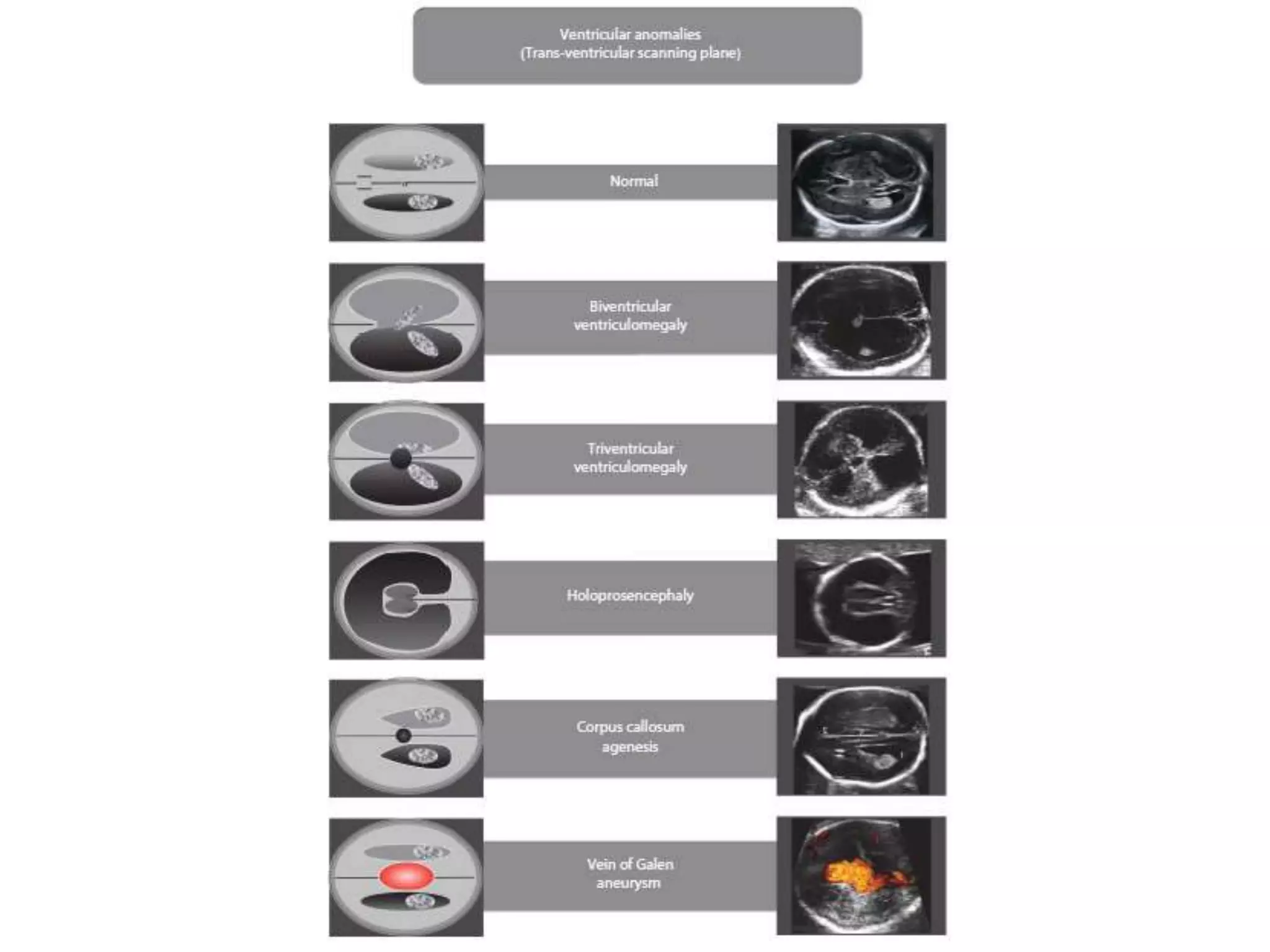
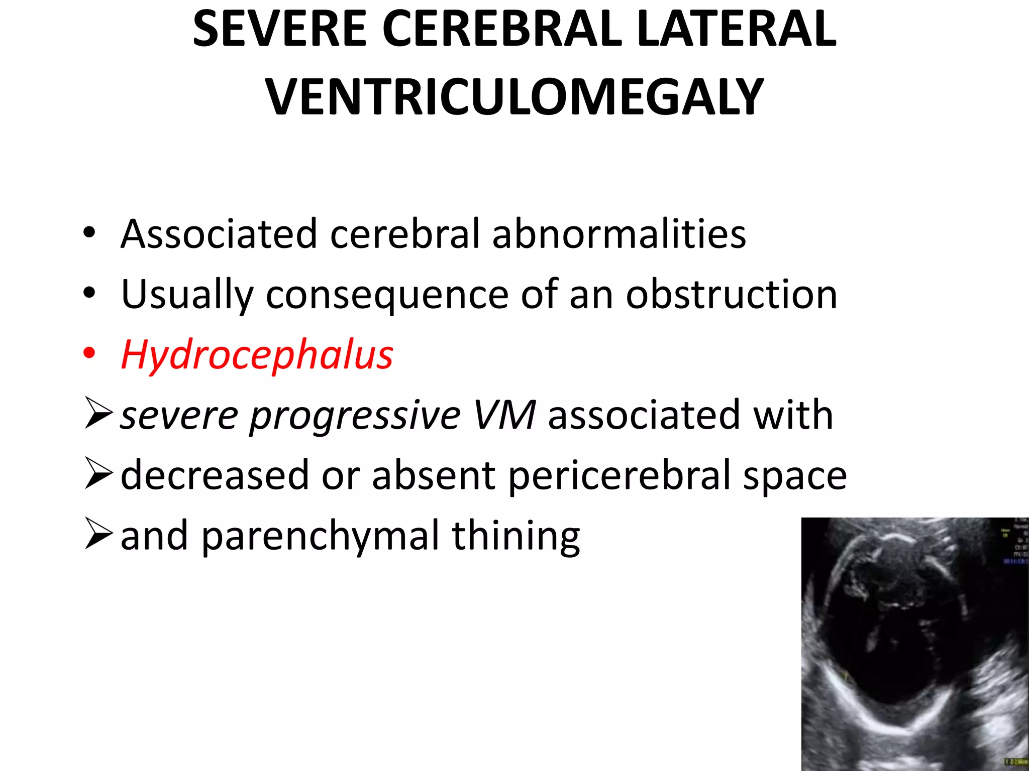
Ventriculomegaly
• Definition:
• VM may be the consequence of a variety of
conditions that can result in a dilatation of the
cerebral ventricular system.
• Incidence:
• Mild ventriculomegaly - 1%
• Severe VM- 1:1000 newborns
How to measure lateral ventricles
Etiology
• Secondary to brain destruction (congenital
infection or a vascular mechanism),
malformations, hydrocephalus, or a
combination of two processes (i.e.,
hydrocephalus and malformation).
• It may also be related to a brain neoplasm or
to a genetic abnormality not associated with a
brain malformation.
• Obstruction
Classification
• Unilateral, bilateral
• Mild, moderate, severe
• Etiology
• Obstructive, non obstructive
• Genetic, syndromes, acquired
Ventriculomegaly
• Most common abnormal fetal cerebral
diagnosis
• Enlargement of the lateral cerebral ventricles
• Ventriculomegaly is not a diagnosis, it is a sign
• Normal midtrimester exam does not exclude
VM, may develop later in gestation
• TAS/TVS neurosonography
• A detailed evaluation of the entire fetal
anatomy, including fetal echocardiography
• Maternal infection screen ( CMV and
toxoplasma)
Should MRI be considered???
• Useful in cases where USG picture is not clear
• Particular advantage of MRI – allows analysis
of gyration
• Best assessed in the third trimester
• Depends upon competence of the operators
Isolated mild fetal ventriculomegaly.
• Soft marker of trisomy 21 and likelihood ratio is
increased 3.8-fold over the general population,
invasive testing depends upon a priori risk, FTS or
combined screening risk
• Follow-up sonogram
• 16% of cases - progression of the ventricular
dilation
56 % remains stable
 34 % regresses
Ultrasound Obstet Gynecol. 2009;34(2):212–224
SEVERE CEREBRAL LATERAL
VENTRICULOMEGALY
• Associated cerebral abnormalities
• Usually consequence of an obstruction
• Hydrocephalus
severe progressive VM associated with
decreased or absent pericerebral space
and parenchymal thining
• 5% genetic cause
 X-linked hydrocephaly
caused by mutations in the gene L1CAM
recurrence risk 50% of males
Summary
• Confirm diagnosis
• Complete fetal evaluation
• Counselling
Investigations
• Detailed ultrasound examination, including
neurosonography.
• Invasive testing for karyotyping and array.
• TORCH test for fetal infections.
• Maternal blood testing for antiplatelet
antibodies in cases with evidence of brain
hemorrhage.
• Fetal brain MRI at ≥32 weeks for diagnosis of
abnormalities of neuronal migration, such as
lissencephaly.
Prognosis
• Isolated mild / moderate:
neurodevelopmental delay in 10% of cases,
this may not be higher than in the general
population.
• Isolated severe: 10 year survival 60%, severe
mental handicap 50%.
Recurrence
• Isolated <1%. Increases to 5% if there is a
history of affected fetus or sibling.
• Part of infection: no increased risk.
• Part of trisomies: 1%.
• X-linked hydrocephaly: 50% of males.
• Associated with alloimmune
thrombocytopenia and no treatment: 100%.
Fetal Ventriculomegaly

Fetal Ventriculomegaly

  • 1.
  • 3.
    • Definition: • VMmay be the consequence of a variety of conditions that can result in a dilatation of the cerebral ventricular system. • Incidence: • Mild ventriculomegaly - 1% • Severe VM- 1:1000 newborns
  • 4.
    How to measurelateral ventricles
  • 5.
    Etiology • Secondary tobrain destruction (congenital infection or a vascular mechanism), malformations, hydrocephalus, or a combination of two processes (i.e., hydrocephalus and malformation). • It may also be related to a brain neoplasm or to a genetic abnormality not associated with a brain malformation. • Obstruction
  • 6.
    Classification • Unilateral, bilateral •Mild, moderate, severe • Etiology • Obstructive, non obstructive • Genetic, syndromes, acquired
  • 7.
    Ventriculomegaly • Most commonabnormal fetal cerebral diagnosis • Enlargement of the lateral cerebral ventricles • Ventriculomegaly is not a diagnosis, it is a sign • Normal midtrimester exam does not exclude VM, may develop later in gestation
  • 9.
    • TAS/TVS neurosonography •A detailed evaluation of the entire fetal anatomy, including fetal echocardiography • Maternal infection screen ( CMV and toxoplasma)
  • 11.
    Should MRI beconsidered??? • Useful in cases where USG picture is not clear • Particular advantage of MRI – allows analysis of gyration • Best assessed in the third trimester • Depends upon competence of the operators
  • 13.
    Isolated mild fetalventriculomegaly. • Soft marker of trisomy 21 and likelihood ratio is increased 3.8-fold over the general population, invasive testing depends upon a priori risk, FTS or combined screening risk • Follow-up sonogram • 16% of cases - progression of the ventricular dilation 56 % remains stable  34 % regresses Ultrasound Obstet Gynecol. 2009;34(2):212–224
  • 14.
    SEVERE CEREBRAL LATERAL VENTRICULOMEGALY •Associated cerebral abnormalities • Usually consequence of an obstruction • Hydrocephalus severe progressive VM associated with decreased or absent pericerebral space and parenchymal thining
  • 15.
    • 5% geneticcause  X-linked hydrocephaly caused by mutations in the gene L1CAM recurrence risk 50% of males
  • 16.
    Summary • Confirm diagnosis •Complete fetal evaluation • Counselling
  • 17.
    Investigations • Detailed ultrasoundexamination, including neurosonography. • Invasive testing for karyotyping and array. • TORCH test for fetal infections. • Maternal blood testing for antiplatelet antibodies in cases with evidence of brain hemorrhage. • Fetal brain MRI at ≥32 weeks for diagnosis of abnormalities of neuronal migration, such as lissencephaly.
  • 18.
    Prognosis • Isolated mild/ moderate: neurodevelopmental delay in 10% of cases, this may not be higher than in the general population. • Isolated severe: 10 year survival 60%, severe mental handicap 50%.
  • 19.
    Recurrence • Isolated <1%.Increases to 5% if there is a history of affected fetus or sibling. • Part of infection: no increased risk. • Part of trisomies: 1%. • X-linked hydrocephaly: 50% of males. • Associated with alloimmune thrombocytopenia and no treatment: 100%.

Editor's Notes

  • #3 It flows from the lateral ventricles through the foramen of Monro into the third ventricle. It then flows from the third ventricle to the aqueduct of Sylvius through the fourth ventricle into the spinal subarachnoid space (foramen of Magendie) or to the basal cisterns (foramen of Luschka) over the cerebral hemispheres.
  • #5 calipers to be placed at the inner margin of the ventricular wall, at level of the parietooccipital fissure, an easily recognizable landmark beginning around 20 weeks’ gestation Normal width of atrium <10 mm
  • #7 The term mild (or borderline) VM is commonly used to indicate cases characterized by an atrial width of 10–15 mm. Some authors have reported a different rate of abnormal neurologic outcome in fetuses with atria >12 mm compared with those with atria measuring 10–12 mm
  • #8 signals to the possibility of associated anomalies of the brain or other organs. The final prognosis depends more on such anomalies than on the degree of ventricular dilation.
  • #12 As not much operators are familiar with fetal brain development according to gestation
  • #16 at Xq28 encoding for L1, a neural cell adhesion molecule X-linked hydrocephaly spectrum, or L1 spectrum, and include MASA (mental retardation, aphasias, shuffling gait, and adducted thumbs), complicated X-linked spastic paraplegia (SP 1), X-linked mental retardation–clasped thumb (MR-CT) syndrome, and some forms of X-linked agenesis of the corpus callosum. Couples with a previously affected child have a recurrence risk of 4%.