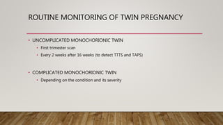
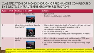
Ultrasound plays an important role in managing twin pregnancies by determining chorionicity, labeling and monitoring fetuses, screening for abnormalities, assessing growth and well-being, and detecting complications. Routine scans are recommended every 4 weeks for uncomplicated dichorionic twins and every 2 weeks for monochorionic twins. Additional monitoring may be needed based on conditions like selective fetal growth restriction. Management of complications is best done at tertiary centers.
1. The müllerian ducts normally develop into the fallopian tubes, uterus, cervix, and upper two-thirds of the vagina. Failures or abnormalities during development can result in müllerian duct anomalies.
2. Development occurs through three phases - organogenesis, fusion, and septal resorption. Failures in fusion can lead to bicornuate or didelphys uterus, while failed septal resorption causes septate uterus.
3. Müllerian duct anomalies have a variety of presentations including infertility, miscarriage, and obstructed reproductive systems. Diagnosis is made through ultrasound, hysterosalpingography, or laparoscopy.
Nuchal translucency
It is a sonographic pre natal screening scan to detect cardiovascular abnormality in a fetus.
NT can also detect altered extra cellular matrix composition and limited lymphatic drainage
Genetic sonogram and soft tissue markersMohit Satodia
This document discusses various soft tissue markers that can be detected on a genetic sonogram to screen for fetal aneuploidies like Down syndrome. It describes markers like nuchal fold thickness, absent nasal bone, echogenic intracardiac focus, choroid plexus cysts, short long bones, pyelectasis, and single umbilical artery. It provides details on the sensitivity and significance of each marker, as well as guidelines on when genetic amniocentesis is recommended based on the number and type of markers present. The document emphasizes that while these markers can help detect aneuploidies, they are often transient and nonspecific findings that also occur commonly in euploid fetuses.
MANAGEMENT OF VAULT PROLAPSE BY DR SHASHWAT JANIDR SHASHWAT JANI
This document discusses the management of vault prolapse, which is the descent of the vaginal cuff after a hysterectomy. It defines vault prolapse and lists risk factors. Conservative management includes pessaries but surgery is often needed. Surgical options include vaginal approaches like sacrospinous ligament fixation or abdominal approaches like sacral colpopexy. The document compares techniques and factors to consider in surgical planning like prolapse severity and patient factors. Prevention techniques like culdoplasty at time of hysterectomy are also discussed.
This document discusses the clinical uses of ultrasonography in twin pregnancies. It covers determining chorionicity and amnioticity, gestational age, screening for anomalies, preterm labor, assessing fetal growth, wellbeing, amniotic fluid, and complications unique to twins such as twin-to-twin transfusion syndrome. Ultrasonography is useful for monitoring growth, detecting anomalies and complications, and guiding management of high-risk twin pregnancies.
The document discusses the pelvic ureter and prevention of ureteric injuries during pelvic surgery. It provides details on the anatomy of the pelvic ureter and sites where it is vulnerable to injury. Ureteric injuries can occur during abdominal or vaginal hysterectomy or other pelvic operations, with an incidence ranging from 0.4-2.5% for benign conditions and up to 30% for malignancies. Preventive measures include adequate exposure of the ureter during surgery, identification of its location using surrounding structures as guides, and avoidance of blind clamping or coagulation near the ureter. Early diagnosis and management of any ureteric injuries is important to reduce morbidity.
The document discusses the approach to evaluating ovarian masses through imaging. It describes how ovarian masses can be categorized and that epithelial tumors are the most common type of malignant ovarian tumor. The evaluation involves considering patient factors like age and mass characteristics on ultrasound like size, wall thickness, and presence of septations or solid areas. Scoring systems can help characterize masses as benign or malignant, though some remain indeterminate. MRI may help in these cases by identifying tissue types and infiltrative features suggestive of malignancy. The goal is to determine if the mass is ovarian in origin and the degree of suspicion for malignancy to guide clinical management.
The document discusses the use of MRI in assessing female pelvic organs and genitourinary conditions. MRI provides detailed images of the uterus, ovaries, and surrounding tissues. It can accurately diagnose adenomyosis, uterine anomalies, and characterize fibroids and ovarian cysts. MRI is also useful for staging cervical, endometrial, and ovarian cancers by identifying the extent of tumor invasion and spread to nearby organs or lymph nodes. Due to its safety during pregnancy, MRI can also evaluate obstetric complications and differentiate between benign and malignant tumors that may complicate pregnancy.
This document discusses injuries to the ureter during gynecological surgeries. It provides details on the surgical anatomy of the ureter, risk factors for injury, common sites of injury, grading of injuries, prevention strategies and intraoperative diagnosis. The key points are:
1. The ureter's course makes it susceptible to injury during pelvic surgeries, especially at the pelvic brim.
2. Injuries occur in 0.3-1% of cases and are most common during hysterectomy.
3. Risk factors include large fibroids, adhesions, and prior pelvic surgery.
4. Prevention relies on proper surgical technique including identification and
This document discusses the role of color Doppler ultrasound in antepartum fetal surveillance. It begins by outlining the purposes of fetal surveillance, which include reducing fetal death and optimizing delivery timing. It then discusses various maternal and fetal conditions that require increased surveillance due to risks of chronic hypoxia. The document covers different methods of antepartum surveillance and provides detailed explanations of Doppler ultrasound principles, techniques like uterine and umbilical artery Doppler, and how abnormal Doppler readings can predict complications like fetal growth restriction.
In this presentation we will discuss role of Doppler US in Infertility, fertilization and assisted fertilization.
we will discuss the favorable and unfavorable RI and PI.
We will discuss role of doppler us in various gynecological malignancies.
This document discusses the interpretation of various types of Doppler ultrasound during pregnancy. It describes:
1. Umbilical artery Doppler which can detect placental hypoxia and increased resistance, predicting abnormal outcomes. Abnormal readings include increased resistance index and absent/reversed end diastolic flow.
2. Middle cerebral artery Doppler which can detect fetal anemia by increased blood flow to the brain. It is also used to time delivery of growth restricted infants.
3. Ductus venosus Doppler which has moderate predictive value for growth restriction in preterm infants.
4. Uterine artery Doppler has limited use in predicting fetal growth restriction but can identify maternal causes by abnormal readings.
This document discusses color Doppler ultrasound techniques for fetal surveillance. It describes the anatomy of fetal and placental circulation and examines the uterine, umbilical and middle cerebral arteries. Waveform analysis using indices like S/D ratio, resistance index and pulsatility index is discussed. Normal and abnormal Doppler findings are presented along with their clinical significance and management. Precise techniques for imaging and interpreting various fetal blood vessels are provided.
(usually in gestations of 7–8 w) to
provide pressure on the GS during US-guided
injection of MTX or Kcl into the GS.
AboubakrElnashar
3. Uterine artery embolization (UAE)
Indications:
1. Failed medical tt
2. Hemodynamically unstable
3. Ruptured CSP
4. Myometrial thickness <2 mm
5. Gestation >8 w
Complications:
1. Hge: 5-10%
2. Uterine atony: 5%
3. Infection: 1-2%
4. Nec
Fetal growth restriction (FGR), formerly called intrauterine growth restriction (IUGR), refers to a condition in which an unborn baby is smaller than it should be because it is not growing at a normal rate inside the womb.
Mild FGR usually doesn't cause long-term problems. In fact, most babies who have it catch up in height and weight by age 2. But severe FGR can seriously harm a baby before and after birth. The extent of the problems depends on the cause and how severe the growth restriction is. It also depends on what point in the pregnancy it starts.
MRI uses magnetism, radio waves, and computers to create images of areas inside the body. It involves four basic steps: (1) placing the patient in a magnetic field, (2) transmitting radio frequency pulses, (3) receiving signals from the patient, and (4) transforming the signals into images using computer processing. MRI provides superior soft tissue resolution compared to ultrasound and allows for multiplanar imaging. It is useful for evaluating various fetal and maternal conditions like brain abnormalities, tumors, placental issues, and complications in multiple pregnancies. While a valuable tool, MRI also has some limitations including high cost, inability to be used in early pregnancy or if metallic implants are present, and longer scan times than ultrasound.
This document provides information on Dr. Rupal N Shah, an experienced gynecologist and IVF consultant in Surat, India. It discusses her qualifications and experience in advanced infertility treatment, endoscopic surgeries, and her roles teaching and publishing on health topics. The rest of the document focuses on summarizing Dr. Shah's presentation on the uses of ultrasonography in infertility, including evaluating ovarian reserve, follicular development, polycystic ovary syndrome, ovarian cysts, uterine anomalies, and endometrial thickness.
Doppler ultrasound can be used in obstetrics and gynecology in several ways. It allows assessment of blood flow in various fetal and maternal vessels. In pregnancy, Doppler is commonly used to evaluate blood flow in the umbilical artery, middle cerebral artery, uterine arteries, ductus venosus and other vessels. Abnormal flow patterns in these vessels can indicate fetal growth restriction, hypoxia, or the risk of conditions like preeclampsia. Doppler provides important information about fetal well-being and helps manage high-risk pregnancies.
The document describes the Pelvic Organ Prolapse Quantification (POP-Q) system for evaluating and documenting pelvic organ prolapse. The POP-Q system uses specific anatomical points of reference to measure the degree of prolapse in centimeters in relationship to the hymen. It is the standard system used internationally for quantifying and comparing prolapse. The POP-Q allows for objective assessment of prolapse, comparison of surgical outcomes, and consistency in medical documentation and research.
This document discusses morbidly adherant placenta, also known as placenta accreta spectrum (PAS), which is becoming more common due to rising cesarean section rates. PAS occurs when the placenta invades deeply into the uterine wall and does not separate normally during delivery, potentially causing life-threatening hemorrhage. Early diagnosis through ultrasound screening and counseling of patients at high risk, such as those with prior uterine scarring, allows for improved maternal outcomes through preparedness and planned hysterectomy if needed. The key is anticipating PAS, making an accurate prenatal diagnosis, and being prepared to perform an emergency hysterectomy to control bleeding and save the mother's life if manual placental removal fails.
Uterus Transplantation Utx (obstetric and gynecology) D.A.B.M
Is the surgical procedure whereby a healthy uterus is transplanted into an organism of which the uterus is absent or diseased.
As part of normal mammalian sexual reproduction, a diseased or absent uterus does not allow normal embryonic implantation, effectively rendering the female infertile.
This phenomenon is known as Absolute Uterine Factor Infertility (AUFI).
Uterine transplant is a potential treatment for this form of infertility.
Uterus is a dynamic, complex organ. It is hugely blood-flow dependent.
More than 116,000 Number of men, women and children on the national transplant waiting list as of August 2017.
33,611 transplants were performed in 2016.
20 people die each day waiting for a transplant.
every 10 minutes another person is added to the waiting list.
Doppler ultrasound uses sound waves to evaluate blood flow and was first used in 1977 to study umbilical artery flow. It measures the Doppler shift in echoes from moving blood cells to determine flow velocity. Fetal vessels commonly assessed include the umbilical artery, which provides information on placental perfusion, and the middle cerebral artery, which indicates fetal oxygenation status. Doppler ultrasound can detect signs of fetal hypoxia like increased end-diastolic flow in the MCA, and meta-analyses found it reduces perinatal deaths and inductions when used for monitoring high-risk pregnancies.
Mullerian duct anomalies occur due to abnormal development of the paired mullerian ducts in females during embryological development. The three main phases of mullerian duct development are organogenesis, fusion, and septal resorption. When one or more of these phases are disrupted, it can lead to mullerian duct anomalies such as a bicornuate or septate uterus. Mullerian duct anomalies are diagnosed using imaging modalities like ultrasound, MRI, and hysterosalpingography which allow visualization of the uterine cavity and identification of the specific anomaly present. The most common anomalies include septate uterus, bicornuate uterus, and arcuate uterus.
Interventional ultrasound in obstetrics dr rabiRabi Satpathy
usg in pregnancy, interventional ultrasound, pregnancy ultrasound, obstetric ultrasound, congenital disease, intra uterine treatment of the fetus, fetal therapy,
This document discusses umbilical and uterine artery Doppler ultrasound. It notes that umbilical artery Doppler is useful for predicting abnormal fetal outcomes, with a resistance index above 0.72 outside normal limits after 26 weeks. Absent or reversed end diastolic flow in the umbilical artery indicates fetal distress and need for monitoring or delivery. Uterine artery Doppler has limited use in predicting fetal growth restriction but can suggest maternal versus fetal causes. An abnormal uterine Doppler with decreased diastolic flow or persistence of a diastolic notch after 24 weeks can help predict preeclampsia. Fortnightly umbilical artery Doppler scans are recommended when growth is not maintained or abdominal circumference is below the third percentile.
FIGO guidelines on Placenta Accreta Spectrum Disorders: Conservative manage...Aboubakr Elnashar
This document discusses guidelines for conservative management of placenta accreta spectrum disorders. It describes four primary conservative surgical methods: 1) extirpative technique, 2) leaving the placenta in situ, 3) one-step conservative surgery, and 4) triple-P procedure. It provides details on how to perform each technique and notes they can be used alone or combined with additional procedures. The document also reviews evidence for techniques like tamponade sutures and recommends close monitoring when leaving the placenta in situ.
Fetal surveillance in twin pregnancies is important due to increased risks of complications. Ultrasound is used to determine chorionicity and amnionicity, monitor growth, check for anomalies, and screen for conditions unique to twins like twin-twin transfusion syndrome. Dichorionic twins are monitored every 4 weeks after 16 weeks while monochorionic twins are monitored every 2 weeks due to higher risks. Complications require specialized management and timely intervention can help reduce poor outcomes for twins.
Twin pregnancies have an increased risk of complications compared to single pregnancies. They constitute approximately 1% of all pregnancies and over 97% of multiple pregnancies are twins. The risk of preterm birth and associated neonatal mortality and morbidity is much higher with twins. Factors such as chorionicity, zygosity, cervical length, fetal fibronectin levels and uterine activity can help predict the risk of preterm birth and guide management, but preventative interventions such as bedrest, activity restriction, and cervical cerclage have not been shown to significantly reduce rates of preterm birth.
This document discusses twin pregnancies. It begins by providing background information on twinning rates and risks. It then covers topics such as zygosity, chorionicity, complications of multiple pregnancies like preterm birth and pregnancy-induced hypertension. Specific complications for monozygotic twins like twin-to-twin transfusion syndrome are explained in detail. The roles of various tests and interventions for predicting and preventing preterm birth in twins are reviewed. Overall morbidity and mortality risks for twin pregnancies are higher than singleton pregnancies due to prematurity and growth restriction.
Invited lecture by Dr Sujoy dasgupta in the Annual Conference of the "Academy of Clinical Embryologists" (ACE) held in October 2021 in "Hybrid mode" (Kolkata and Webinar)
A multifetal pregnancy is a pregnancy in which there are two or more fetuses in the uterus at the same time. This can include twin pregnancies, triplet pregnancies, and higher-order multiple pregnancies.
The most common type of multifetal pregnancy is twin pregnancy, which can be either fraternal (dizygotic) twins, which are formed from two separate eggs fertilized by two separate sperm, or identical (monozygotic) twins, which are formed when a single fertilized egg splits and develops into two separate embryos.
Risk factors for multifetal pregnancy include:
Advanced maternal age
Assisted reproductive technologies (ART) such as in vitro fertilization (IVF)
A family history of twin pregnancies
Use of ovulation-inducing drugs
The management of multifetal pregnancies can be challenging and requires close monitoring and specialized care. It can include ultrasound monitoring to assess the growth and well-being of each fetus, and to detect any potential complications such as twin-to-twin transfusion syndrome (TTTS) or selective intrauterine growth restriction (sIUGR).
Due to the increased risk of complications, multifetal pregnancies are at a higher risk of preterm labor, cesarean delivery, and perinatal morbidity and mortality.
It's important to note that multifetal pregnancies should be managed by a team of specialists such as obstetricians, perinatologists, and pediatricians with experience in the care of multifetal pregnancies.
This document discusses twin pregnancies and provides information on:
1. The types of twins based on chorionicity and zygosity.
2. The maternal and fetal complications that can occur in twin pregnancies including preeclampsia and preterm birth.
3. The diagnosis and management of twin pregnancies including ultrasound examination and increased surveillance of monochorionic twins.
This document summarizes antepartum fetal surveillance and aneuploidy screening. It discusses:
1) The importance and benefits of antepartum fetal surveillance in predicting risks like perinatal death and detecting conditions like fetal growth restriction.
2) The various methods used in aneuploidy screening including ultrasound measurements, maternal serum markers, cell-free DNA testing, and invasive diagnostic tests.
3) The guidelines and best practices for aneuploidy screening including integrating first and second trimester screens and contingent screening models based on risk levels.
Multiple pregnancies can involve twins, triplets or more. Monitoring of monochorionic twins is important due to risks of twin-twin transfusion syndrome (TTTS), twin anemia-polycythemia sequence (TAPS), twin reversed arterial perfusion (TRAP) sequence, and selective growth restriction (sGR). TTTS occurs in 10-15% of monochorionic twins due to uneven blood flow through connecting blood vessels and can be diagnosed and treated with laser ablation. SGR can occur without fluid imbalance and is detected by significant fetal growth differences and abnormal Doppler readings of the umbilical artery. Close ultrasound monitoring every 2 weeks from 16 weeks is recommended for monochorionic twins.
The incidence of multiple gestation continues to increase, and now accounting for more than 3% of all live births.
Twin pregnancies and higher-order multiple births comprise an increasing proportion of the total pregnancies in the developed world due to the expanded use of fertility treatments and older maternal age at childbirth.
Multiple gestation is associated with:
Increase in neonatal morbidity and mortality rates.
Increase in maternal complications at least two folds.
The number of triplet, quadruplet, and higher-order multiple births peaked in 1998 and has dropped slightly recently, most likely because of limits in the number of embryos transferred and because of the availability and acceptance of multifetal pregnancy reduction (MFPR) procedures.
Prematurity, monochorionicity, and growth restriction pose the main risks to fetuses and neonates in multiple gestations.
The mean duration of pregnancy is 35.3 weeks for twin gestations, 31.9 weeks for triplets, and 29.5 weeks for quadruplets.
Stillbirth rates increase from 6.8 /1000 for singletons to 16.1 for twins and to 21.5 for triplets, and infant mortality rates increase from 5 to 23.4 and to 51.2 /1000 births, respectively.
Infants of multiple gestations comprise almost one quarter of very-low-birth-weight infants.
The incidence of severe handicap among neonatal survivors of multiple gestation is also increased: 34.0 and 57.5 /1000 twin and triplet survivors, respectively, compared with 19.7 /1000 singleton survivors.
Maternal morbidity is significantly increased in mothers with multiple gestations and is apparently related to the number of fetuses.
Multiple gestations are associated with significantly higher risks for:
Hypertension
Placental abruption
Preterm labor (78%)
Preeclampsia (26%);
HELLP syndrome (9%) (hemolysis, elevated liver enzymes, low platelets)
Anemia (24%)
Preterm premature rupture of membranes (pPROM) (24%)
Gestational diabetes (14%)
Acute fatty liver (4%)
Chorioendometritis (16%)
Postpartum hemorrhage (9%)
Twins can be dizygotic (DZ), resulting from the fertilization of two separate ova during a single ovulatory cycle.
DZ twins have dichorionic-diamniotic (DCDA) placentas, although these may fuse during pregnancy.
Monozygotic (MZ), resulting from a single fertilized ovum that subsequently divides into two separate individuals.
In MZ twins, the timing of egg division determines placentation (تكون المشيمة):
Diamniotic, dichorionic (DCDA) placentation occurs with division prior to the morula stage (within 3 days post fertilization).
Diamniotic, monochorionic (MCDA) placentation occurs with division between 4-8 days postfertilization.
Monoamniotic, monochorionic (MCMA) placentation occurs with division between 8-12 days postfertilization.
Division at or after day 13 results in conjoined twins.
Multiple gestation pregnancies are increasing in incidence due to assisted reproductive technologies and delayed childbearing. These pregnancies are at higher risk for maternal and fetal complications including hypertension, preterm birth, and growth discordance between twins. Prenatal surveillance includes regular ultrasounds to monitor fetal growth, well-being, chorionicity, and complications like twin-twin transfusion syndrome. Mode of delivery depends on fetal presentation, with vertex-vertex twins usually aiming for vaginal delivery while other presentations may require cesarean. Higher-order multiples generally require cesarean delivery for safety.
Prenatal diagnosis detects fetal abnormalities before birth through non-invasive and invasive techniques. Non-invasive techniques include ultrasound, fetal echocardiography, and magnetic resonance imaging to visualize the fetus. Maternal serum screening analyzes markers in the mother's blood to assess risk of fetal conditions like Down syndrome. Invasive techniques obtain fetal tissue through procedures like amniocentesis for cytogenetic or molecular testing to diagnose genetic disorders. Prenatal diagnosis aims to provide risk information to families for informed decision making during pregnancy.
ANTENATAL SURVEILLANCE AND MANAGEMENT OF MULTIPLE PREGNANCY – RECENT ADVANCES...Lifecare Centre
- Multiple pregnancies require enhanced antenatal monitoring including serial ultrasounds to check for complications which are more common than singletons such as prematurity and fetal growth issues.
- Determining chorionicity and amnionicity is important for management since monochorionic twins carry higher risks like twin-to-twin transfusion syndrome.
- Monitoring involves regular ultrasounds to check fetal size, growth, and Doppler studies of the umbilical artery depending on if there is size discordance or a small fetus. High risk monochorionic twins require every 2 week scans.
Prenatal evaluation and postnatal early outcomes of fetalJegon Varakala
This study analyzed 40 cases of prenatally diagnosed fetal ventriculomegaly (VM). It found that 60% were unilateral and 40% bilateral. Associated structural abnormalities occurred in 47.5% of cases. Most (92.5%) were classified as mild VM, while 7.5% were severe. The ventricle sizes of isolated mild VM cases resolved in most pregnancies, while sizes increased in all severe cases. The mortality rate was higher in cases with associated abnormalities or severe VM. Careful postnatal evaluation is important, as some isolated mild VM cases showed abnormalities after birth.
1. Multiple pregnancy refers to the presence of more than one fetus in the uterus. The majority of twin pregnancies are dizygotic, resulting from two separate fertilized eggs. Monozygotic twins result from the splitting of a single fertilized egg.
2. Complications of multiple pregnancies include risks to both the mother (such as preeclampsia and gestational diabetes) and fetuses (including preterm birth and fetal growth restriction). The risks are increased with monochorionic twins due to shared placental blood flow.
3. Major complications of monochorionic twins include twin-to-twin transfusion syndrome (TTTS), twin anemia-polycythemia sequence (T
The document discusses newer modalities for managing multiple gestations from infertility treatment, including limiting the number of embryos transferred and fetal reduction techniques like multifetal pregnancy reduction which terminates some fetuses to improve outcomes for the remaining pregnancies, and selective termination which terminates abnormal fetuses while preserving normal ones. Modern techniques for fetal reduction include radiofrequency ablation, bipolar diathermy cord coagulation, intrafetal laser ablation, and suture ligation.
This document discusses intrauterine growth restriction (IUGR), including definitions, causes, detection methods, and management. Key points include:
- IUGR, or small for gestational age (SGA), affects 10-15% of fetuses and is caused by placental insufficiency restricting nutrients/oxygen to the fetus.
- Ultrasound is used to monitor fetal growth parameters like abdominal circumference and estimated fetal weight against customized charts. Doppler ultrasound of umbilical and uterine arteries can also indicate placental insufficiency.
- If IUGR is detected, careful surveillance is required using biophysical profile, amniotic fluid volume, and Doppler ultrasound to determine optimal delivery timing weighing fetal vs. maternal
Prenatal diagnosis involves identifying fetal abnormalities through tests such as ultrasound scans and blood tests. Fetal therapy aims to improve the fetal environment through procedures like blood transfusions. First trimester screening for Down syndrome includes assessing the mother's age, measuring the fetal nuchal translucency, and testing maternal serum markers. Combining these factors provides a 79-87% detection rate. Additional markers like absent nasal bone and abnormal ductus venosus or tricuspid valve Doppler flows may further refine risk assessment. Genetic counseling is an important part of the screening and diagnostic process to help expectant families understand risks and make informed decisions.
Screening for fetal abnormality in first and second.pptxpoonamkirtipal
This document discusses screening methods for fetal abnormalities in the first and second trimesters of pregnancy. In the first trimester, screening involves assessing maternal age, measuring nuchal translucency, and testing maternal serum for beta-hCG and PAPP-A levels between 11-13 weeks. In the second trimester, screening analyzes maternal serum levels of beta-hCG, AFP, uE3, and inhibin-A between 15-22 weeks. Combining first and second trimester screening can detect up to 90% of cases of trisomy 21 with a 2% false positive rate. Screening aims to identify high risk pregnancies so diagnostic testing can be offered to confirm or rule out abnormalities.
This document provides an overview of the management of triplet pregnancies. Key points include:
- Triplet pregnancies are higher risk than twins or singletons due to higher rates of preterm birth and associated complications.
- Management involves frequent ultrasounds and office visits to monitor growth and complications like preeclampsia.
- Chorionicity determines specific monitoring protocols due to risks like twin-twin transfusion syndrome.
- Most triplet pregnancies are delivered via c-section before 37 weeks due to risks of prematurity.
- Maternal and fetal complications include growth restriction, cord accidents, and preterm birth. Close monitoring aims to reduce risks.
This case presentation discusses the diagnosis and management of congenital duodenal atresia in a 35-year-old pregnant patient. Prenatal ultrasound revealed signs of duodenal atresia including polyhydramnios. After being admitted in preterm labor at 34 weeks, the patient delivered a baby boy with duodenal atresia who underwent surgery on postnatal day 4. Duodenal atresia results from failure of embryonic development and is often associated with other anomalies. Prenatal diagnosis allows delivery at a center prepared to care for the newborn.
This case report describes the diagnosis and management of a fetus with a left-sided diaphragmatic hernia detected on ultrasound at 23 weeks gestation. Diaphragmatic hernias occur when abdominal organs protrude into the chest cavity through a defect in the diaphragm. The fetus was monitored closely with serial ultrasounds and delivered via cesarean section at 36 weeks for pre-eclampsia. The newborn was referred to the neonatal intensive care unit for surgery and management of pulmonary issues associated with the condition.
This document discusses ultrasound evaluation of the fetal face and neck. It outlines normal sonographic anatomy and various craniofacial anomalies that can be detected prenatally, including facial clefts, orbital defects, micrognathia, macroglossia, tumors, and ear abnormalities. It also mentions craniosynostosis and neck anomalies like nuchal cystic hygroma that can be identified. The conclusion emphasizes that identifying facial or neck anomalies indicates the need for a full fetal exam to check for associated conditions, and that prognosis depends on severity and presence of syndromes or neurological defects. Advanced ultrasound is improving detection and preparation for neonatal care.
The document discusses various fetal thoracic abnormalities that can be observed on ultrasound, including:
1. The 5 stages of lung development: embryonic, pseudoglandular, canalicular, saccular, and alveolar.
2. Specific lung abnormalities like lung hypoplasia, CCAM (congenital cystic adenomatoid malformation), bronchopulmonary sequestration, CHAOS (congenital high airway obstruction syndrome), and others.
3. Details on diaphragmatic development and abnormalities like CDH (congenital diaphragmatic hernia).
4. Other thoracic issues like congenital hydrothorax. Diagnostic features,
This document discusses ultrasound evaluation of the fetal musculoskeletal system. It begins by describing development of the fetal skeleton from various cell types and then discusses limb bud development. It describes the process of skeletogenesis and factors involved in patterning. It provides details on qualitative assessment of long bones and differential diagnosis of various skeletal dysplasias based on ultrasound findings. Key skeletal dysplasias discussed in detail include achondroplasia, thanatophoric dysplasia, achondrogenesis, osteogenesis imperfecta, and hypophosphatasia.
This document provides guidance on performing an ultrasound examination of the fetal heart. It begins by noting the importance of evaluating the fetal heart given the prevalence of congenital heart defects. The basic scan involves obtaining a four-chamber view of the heart to visualize the atria and ventricles. For a more detailed exam, the outflow tracts should also be examined. Proper scanning technique and fetal positioning are described for obtaining optimal views. Key features of normal ultrasound anatomy are outlined. Common abnormalities that may be detected on scans are also reviewed. The document provides a comprehensive overview of evaluating the fetal heart and lungs during the second trimester anatomy scan.
The document summarizes the development of the urinary tract in the fetus. It discusses the 3 sets of excretory organs during development - the pronephros, mesonephros, and metanephros. The pronephros develops in the 3rd week but degenerates by the 5th week. The mesonephros develops and eventually regresses during the 4th month, serving as the excretory organ during development of the metanephros. The metanephros is the definitive kidney that develops in the 5th week from the metanephric messoderm. The document also discusses normal development and abnormalities that can occur.
This document contains a list of patient records from a birth depression ward in August 2018. It includes information about each patient such as their age, admitting physician, admission and discharge details, pregnancy details, delivery outcome, newborn status and disposition. Several patients experienced birth asphyxia and related complications and had poor outcomes including expiration. Others were discharged after being treated for conditions like pneumonia.
This document presents a case of neonatal thrombocytopenia caused by maternal immune thrombocytopenia (ITP). The mother had a history of ITP diagnosed in 2015. The newborn was delivered at 37 weeks gestation via normal spontaneous delivery with good APGAR scores. Initial workup showed thrombocytopenia but no bleeding. The neonate was treated supportively with antibiotics and observed, with platelet counts improving before discharge on day 3. Maternal ITP can cause neonatal thrombocytopenia in 10-15% of cases via transplacental transfer of antiplatelet antibodies. The pathophysiology, differential diagnoses, and management of neonatal thrombocytopenia are discussed.
This document discusses the case of a 30-year-old pregnant woman with renal tubular acidosis and rickets. Renal tubular acidosis disrupts the body's ability to regulate acid-base balance and can cause bone abnormalities like rickets. The woman also had gestational diabetes. She received treatment including sodium bicarbonate and calcium supplements. She delivered a healthy baby via cesarean section. The document outlines the effects of renal tubular acidosis and vitamin D deficiency on both mother and fetus, as well as gestational diabetes' risks. It emphasizes the importance of multidisciplinary care and counseling for managing complex conditions in pregnancy.
Prenatal care involves regular checkups during pregnancy to monitor the health of the mother and baby. Checkups include a physical exam, ultrasound, and lab tests. They allow doctors to monitor weight gain, check fetal growth and heart rate, test for conditions, and identify potential complications. Regular prenatal care is important for having a healthy pregnancy and delivery. Reasons a pregnant woman may not get care include lack of awareness of the importance of checkups and barriers to accessing medical care.
Selective IUGR affects 10-15% of monochorionic twin pregnancies and presents a severe risk, including intrauterine demise and neurological adverse outcomes for the twins. It is defined as the estimated fetal weight of one twin falling below the 10th percentile with a weight discordance of over 25% between the twins. Selective IUGR is classified into three types based on umbilical artery Doppler findings, with Type I having positive diastolic flow in the small twin, Type II having persistently absent or reversed flow, and Type III having intermittent absent/reversed flow. Each type correlates to different risks and outcomes for the twins.
This document discusses the signs and symptoms of pregnancy and methods for confirming a pregnancy. It covers common early signs like amenorrhea, changes in the lower reproductive tract, uterus, breasts and skin, and later fetal movement. It also mentions pregnancy tests and sonographic recognition as ways to determine if a woman is pregnant in addition to examining physical signs and symptoms.
Skeletal dysplasia is a genetically heterogeneous group of over 350 disorders that affect the development of bone and cartilage, leading to abnormalities in bone size, shape, and mineralization. Prenatally diagnosed skeletal dysplasias have a high risk of stillbirth, neonatal death, and perinatal death. The four most common types prenatally diagnosed are thanatophoric dysplasia, achondrogenesis, achondroplasia, and osteogenesis imperfecta. Prenatal diagnosis involves 2D imaging to identify skeletal abnormalities like shortened long bones, followed by amniocentesis, fetal echocardiogram, and referral to genetic specialists for counseling and management planning.
This document discusses antepartum fetal surveillance, which involves assessing fetal well-being before the onset of labor to prevent injury and death. Various techniques are described for monitoring the fetus, including fetal movement counting, non-stress tests to check for fetal heart rate accelerations, and biophysical profiles. Abnormal test results can indicate issues like hypoxemia or acidemia. Maternal conditions like diabetes or hypertension are common indications for increased surveillance. The physiology behind factors regulating the fetal heart rate is also explained.
Placental respiratory gas exchange and fetal oxygenationmaricar chua
This document summarizes oxygen transport from the atmosphere to fetal tissues. It describes how oxygen diffuses across membranes in the lungs, placenta, and fetal tissues. While fetal oxygen levels are lower than maternal levels, high fetal cardiac output and blood flow to organs maintains oxygen delivery. The placenta facilitates gas exchange through a villous tree structure that increases surface area from stem to terminal villi. Fetal growth restriction is associated with a smaller, less developed placenta and lower umbilical oxygen levels. In response to acute hypoxia, the fetus redistributes blood flow to favor the brain and heart. Though oxygen therapy increases maternal oxygen levels, there is a smaller corresponding increase in fetal oxygen levels due to the placent
The document discusses amniotic fluid dynamics and regulation. Amniotic fluid provides space for fetal movement, allows fetal swallowing and breathing, and protects the fetus from trauma. Its volume increases until around 34 weeks gestation when it peaks at around 800mL, then decreases. Fluid is produced mainly through fetal urination in the second and third trimesters. Disorders of amniotic fluid volume include polyhydramnios and oligohydramnios, which are diagnosed using techniques like measuring the deepest vertical pocket or calculating the amniotic fluid index.
This document discusses hypertension in pregnancy, including classifications, diagnostic criteria, risk factors, pathophysiology, prevention, and management. It classifies hypertensive disorders in pregnancy into four categories: gestational hypertension, preeclampsia, chronic hypertension, and chronic hypertension with superimposed preeclampsia. It provides diagnostic criteria for each condition and indicators of severity. Risk factors include young maternal age, obesity, multifetal gestation, and genetic predispositions. The pathophysiology involves abnormal placental invasion and maternal inflammatory response. Management depends on severity of features and gestational age, ranging from expectant monitoring to expedited delivery.
Ultrasound can cause biological effects through thermal and non-thermal mechanisms. Thermal effects are caused by ultrasound energy being absorbed and converted to heat, while non-thermal effects include cavitation and direct mechanical effects. While some studies have reported associations between ultrasound exposure and fetal effects, most clinical studies have found no biological effects, even at higher intensities. Regulatory bodies have established safety indices and output limits, and recommend using the minimum exposure needed for diagnosis. Overall, diagnostic ultrasound is considered safe when used prudently according to established guidelines.
This document summarizes fetal growth disorders. It discusses the three phases of fetal growth and factors that can affect growth. It also describes fetal growth restriction and macrosomia (fetal overgrowth). For growth restriction, it covers diagnosis, risk factors, prevention, and management approaches. For macrosomia, it discusses risk factors and potential maternal and neonatal complications, as well as recommendations regarding diagnosis and management to prevent shoulder dystocia.
Here is the updated list of Top Best Ayurvedic medicine for Gas and Indigestion and those are Gas-O-Go Syp for Dyspepsia | Lavizyme Syrup for Acidity | Yumzyme Hepatoprotective Capsules etc
Promoting Wellbeing - Applied Social Psychology - Psychology SuperNotesPsychoTech Services
A proprietary approach developed by bringing together the best of learning theories from Psychology, design principles from the world of visualization, and pedagogical methods from over a decade of training experience, that enables you to: Learn better, faster!
Does Over-Masturbation Contribute to Chronic Prostatitis.pptxwalterHu5
In some case, your chronic prostatitis may be related to over-masturbation. Generally, natural medicine Diuretic and Anti-inflammatory Pill can help mee get a cure.
Osteoporosis - Definition , Evaluation and Management .pdfJim Jacob Roy
Osteoporosis is an increasing cause of morbidity among the elderly.
In this document , a brief outline of osteoporosis is given , including the risk factors of osteoporosis fractures , the indications for testing bone mineral density and the management of osteoporosis
Local Advanced Lung Cancer: Artificial Intelligence, Synergetics, Complex Sys...Oleg Kshivets
Overall life span (LS) was 1671.7±1721.6 days and cumulative 5YS reached 62.4%, 10 years – 50.4%, 20 years – 44.6%. 94 LCP lived more than 5 years without cancer (LS=2958.6±1723.6 days), 22 – more than 10 years (LS=5571±1841.8 days). 67 LCP died because of LC (LS=471.9±344 days). AT significantly improved 5YS (68% vs. 53.7%) (P=0.028 by log-rank test). Cox modeling displayed that 5YS of LCP significantly depended on: N0-N12, T3-4, blood cell circuit, cell ratio factors (ratio between cancer cells-CC and blood cells subpopulations), LC cell dynamics, recalcification time, heparin tolerance, prothrombin index, protein, AT, procedure type (P=0.000-0.031). Neural networks, genetic algorithm selection and bootstrap simulation revealed relationships between 5YS and N0-12 (rank=1), thrombocytes/CC (rank=2), segmented neutrophils/CC (3), eosinophils/CC (4), erythrocytes/CC (5), healthy cells/CC (6), lymphocytes/CC (7), stick neutrophils/CC (8), leucocytes/CC (9), monocytes/CC (10). Correct prediction of 5YS was 100% by neural networks computing (error=0.000; area under ROC curve=1.0).
8 Surprising Reasons To Meditate 40 Minutes A Day That Can Change Your Life.pptxHolistified Wellness
We’re talking about Vedic Meditation, a form of meditation that has been around for at least 5,000 years. Back then, the people who lived in the Indus Valley, now known as India and Pakistan, practised meditation as a fundamental part of daily life. This knowledge that has given us yoga and Ayurveda, was known as Veda, hence the name Vedic. And though there are some written records, the practice has been passed down verbally from generation to generation.
Muktapishti is a traditional Ayurvedic preparation made from Shoditha Mukta (Purified Pearl), is believed to help regulate thyroid function and reduce symptoms of hyperthyroidism due to its cooling and balancing properties. Clinical evidence on its efficacy remains limited, necessitating further research to validate its therapeutic benefits.
share - Lions, tigers, AI and health misinformation, oh my!.pptxTina Purnat
• Pitfalls and pivots needed to use AI effectively in public health
• Evidence-based strategies to address health misinformation effectively
• Building trust with communities online and offline
• Equipping health professionals to address questions, concerns and health misinformation
• Assessing risk and mitigating harm from adverse health narratives in communities, health workforce and health system
Basavarajeeyam is a Sreshta Sangraha grantha (Compiled book ), written by Neelkanta kotturu Basavaraja Virachita. It contains 25 Prakaranas, First 24 Chapters related to Rogas& 25th to Rasadravyas.
2. DATING OF TWIN PREGNANCY
• CRL OF 45 – 84 mm
• In pregnancies conceived spontaneously, the larger of the 2 CRL should be used
to estimate gestational age
• After 14 weeks, the larger head circumference should be used
• In-vitro fertilization should be dated using the oocyte retrieval date of the embryonic
age from fertilization
3. DETERMINING CHORIONICITY / AMNIONICITY
• Chrorionicity should be determined 11 to 13 weeks 6 days of gestation using the
membrane thickness at the site of insertion of the amniotic membrane into the
placenta
4.
5. DETERMINING CHORIONICITY / AMNIONICITY
• If not possible to determine chorionicity by transbdominal or transvaginal ultrasound in the
routine setting, a second opinion should be sought from a tertiary referral center
• After 14 weeks, chorionicity is best determined using the same ultrasound signs in particular by
counting the membrane layers and noting the discordant fetal sex.
• At the time chorionicity is determined, amnionicity should also be determined and documented
6. LABELING OF TWIN FETUSES
• The labeling of twin fetuses should follow a reliable and consistent strategy and
should be documented clearly inorder to ensure consistent labeling during the
follow up scans.
• Describe each twin using as many features as possible so as to enable to identify them
accurately.
• Perinatal switch phenomenon – twins labeled as Twin A and Twin B during antenatal
scans may not necessarily delivered in that order.
7. ROUTINE MONITORING OF TWIN PREGNANCY
• UNCOMPLICATED DICHORIONIC TWIN
• First trimester scan
• Detailed second trimester scan and scans every 4 weeks
• COMPLICATED DICHORIONIC TWIN
• Depending on the condition and its severity
8. ULTRASOUND MONITORING
PATHWAY
UNCOMPLICATED
DICHORIONIC TWIN
PREGNANCY
Khalil A, Rodgers M, Baschat A, Bhide A, Gratacos E, Hecher K, Kilby MD, Lewi L,
Nicolaides KH, Oepkes D, Raine-Fenning N, Reed K, Salomon LJ, Sotiriadis A,
Thilaganathan B, Ville Y. ISUOG Practice Guidelines: role of ultrasound in twin
pregnancy. Ultrasound Obstet Gynecol 2016; 47: 247–263.
9. ROUTINE MONITORING OF TWIN PREGNANCY
• UNCOMPLICATED MONOCHORIONIC TWIN
• First trimester scan
• Every 2 weeks after 16 weeks (to detect TTTS and TAPS)
• COMPLICATED MONOCHORIONIC TWIN
• Depending on the condition and its severity
10. ULTRASOUND
MONITORING PATHWAY
UNCOMPLICATED
MONOCHORIONIC TWIN
Khalil A, Rodgers M, Baschat A, Bhide A, Gratacos E, Hecher K, Kilby MD, Lewi L, Nicolaides KH,
Oepkes D, Raine-Fenning N, Reed K, Salomon LJ, Sotiriadis A, Thilaganathan B, Ville Y. ISUOG
Practice Guidelines: role of ultrasound in twin pregnancy. Ultrasound Obstet Gynecol 2016; 47:
11. IN EACH ULTRASOUND ASSESSMENT:
• Fetal biometry
• Amniotic fluid volume
• Umbilical artery doppler- from 20 weeks for both twins
• Discordance in estimated fetal weight from 20 weeks
12. MONOCHORIONIC TWINS
• Fetal biometry
• Amniotic fluid volume
• Umbilical artery doppler- from 20 weeks for both twins
• Discordance in estimated fetal weight from 20 weeks
• Cervical length assessment at 20-24 weeks may lead to better prediction of preterm birth
(less than 25 mm in asymptomatic twin)
• Middle cerebral artery PSV – peak systolic velocity - from 20 weeks ( screen for
TAPS)
• Monochorionic diamniotic twins – amniotic fluid volume (deepest vertical
pocket) to screen for TTTS
13. SCREENING FOR CHROMOSOMAL ABNORMALITIES
• First trimester screen for aneuploidy –
• nuchal translucency thickness
• Free beta HCG
• Pregnancy-associated plasma protein-A
• In case of vanishing twin – if there is still a measurable fetal pole, NT alone, in combination with
maternal age, should be used for risk estimation
• Detection rate of non invasive prenatal test- may be lower in twins than in singletons, but data are
still limited. DR for trisomy 21 in singleton – 99% FPR – 0.1%; DR for Trisomy 21 in twins 94.4%
and FPR - 0%
• Invasive testing-
• Pregnancy loss rate: CVS - 3.85%; Amniocentesis – 3.1%
14. IMPLICATION OF DISCORDANCE IN NT OR CRL
IN THE FIRST TRIMESTER
• The management of twin pregnancy with CRL discordance ≥ 10% or of NT
discordance ≥ 20%.
• Detailed ultrasound assessment and testing for karyotype abnormalities.
• 25% risk of fetal abnormalities for CRL discordance >10%
• CRL discordance at 7 + 0 to 9 + 6 weeks’ gestation is a predictor of the risk of
single fetal demise in the first trimester (DR of 74% for a FPR of 5%)
• An abnormal DV will pick up only 38% of all pregnancies that subsequently
develop TTTS, and, of those predicted to be at high risk, only 30% will ultimately
develop TTTS
15. SCREENING FOR STRUCTURAL ABNORMALITIES
• Assessed for the presence of any major
anomalies at the first trimester scan
Salomon LJ, Alfirevic Z, Bilardo CM, Chalouhi GE, Ghi T, Kagan KO, Lau TK, Papageorghiou AT, Raine-Fenning NJ, Stirnemann J, Suresh S, Tabor A, Timor-
Tritsch IE, Toi A, Yeo G. ISUOG Practice Guidelines: performance of first-trimester fetal ultrasound scan. Ultrasound Obstet Gynecol 2013; 41: 102–113.’.
16. SCREENING FOR STRUCTURAL ABNORMALITIES
• Routine second trimester anomaly scan (18-20 weeks)
• Cardiac screening assessment should be performed in monochorionic twins
• Laterality, Situs, 4 chamber, ventricular outflow tract and aortic arch views.
• IMPORTANT TO MAKE THE WOMAN AWARE OF THE LIMITATIONS OF ULTRASOUND
SCREEENING, WHICH VARY ACCORDING TO THE TYPE OF ANOMALY
17. SCREENING FOR RISK OF PRETERM BIRTH
• Cervical length measurement is the preferred method
• 25 mm is the cut-off most commonly used in the second trimester
• Cervical length of <25 mm at 18-24 weeks – moderate predictor of preterm birth before
34 weeks, but not before 37 weeks
• Cervical length of <20 mm at 20-24 weeks – most accurate predictor of preterm birth
before 32 and before 34 weeks
• No effective strategy to prevent preterm birth in these women.
• Bed rest, progesterone therapy, Arabin cervical pessary or oral tocolytics do not reduce the risk
of preterm delivery
• Progesterone therapy might reduce the risk of neonatal morbidity and mortality.
18. SCREENING, DIAGNOSIS AND MANAGEMENT OF
FETAL GROWTH RESTRICTION
• Selective FGR - one fetus has EFW <10th percentile and the intertwin EFW discordance is
>25%
• A discordance cut off of 20% - increased risk of adverse outcome
SCREENING:
• Combination of head, abdomen and femur measurements performs best in calculating
EFW
• Intertwin discordance > 25% referral should be made to a tertiary fetal medicine center.
• Once diagnosed – detailed anomaly scan and screening for viral infections (CMV, rubella and
toxoplasmosis)
• Amniocentesis – required to exclude chromosomal abnormalities
19. CLASSIFICATION OF MONOCHORIONIC PREGNANCIES COMPLICATED
BY SELECTIVE INTRAUTERINE GROWTH RESTRICTION
Type of sFGR Findings in the UMA Outcome
I Positive end diastolic flow • 90% survival
• In-utero mortality rates up to 40%
II Absent or reversed end diastolic
flow (AREDF)
• High risk of intrauterine death of growth restricted twin and
or very preterm delivery with associated risk of
neurodevelopmental delay
• IUD of either twin in up to 29%
• 15% risk of neurological sequelae if born prior to 30 weeks
III Cyclical / intermittent pattern of
AREDF
• 10 to 20% risk of unpredictable sudden death of FGR fetus
even with ultrasound features have been stable
• Up to 20% rate of neurological morbidity in surviving larger
twin
Gratacos E., Lewi L, Munoz B, Acosta-Rojas R, Hernandez-Andrade E, Martinez JM, Carreras E Deprest JA, Classification for selective intrauterine
growth restriction in monochorionic pregnancies according to umbilical artery Doppler Flow in the smaller twin, Ultrasound Obstet Gynecol 2007;
20. DICHORIONIC TWIN PREGNANCY COMPLICATED
BY SFGR
• Follow-up visits could be less frequent, as delivery is usually not recommended
before 32–34 weeks’ gestation.
• fetal Doppler should be assessed approximately every 2 weeks, depending on the
severity.
• Management of these cases is complex and should be coordinated by a tertiary-
level fetal medicine center
21. MONOCHORIONIC TWIN PREGNANCY COMPLICATED BY
SFGR
• fetal growth should be assessed at least every 2 weeks, and fetal Doppler
(umbilical artery and MCA) at least weekly.
• If the umbilical artery Doppler is abnormal, assessment of the DV blood flow
should be undertaken.
• The aim in managing these pregnancies is to prolong the pregnancy at least until
viability is achieved, while at the same time avoiding single IUD with its associated
serious consequences for the surviving cotwin.
22. MONOCHORIONIC TWIN PREGNANCY COMPLICATED
BY SFGR
• The timing of delivery based on assessment of fetal wellbeing, interval growth,
biophysical profile, DV waveform and/or computerized cardiotocography (CTG)
• Incidence of severe cerebral injury in monochorionic twins complicated by sFGR is
approximately 10% and is associated with abnormal umbilical artery Doppler,
single IUD and low gestational age at birth.
• Risks of neonatal morbidity (38% vs 19%), particularly respiratory distress
syndrome (32% vs 6%) and cerebral lesions, are higher in the larger than in the
smaller co-twin
23. MANAGING THE SURVIVING TWIN AFTER DEMISE
OF ITS CO-TWIN
The following complications are found in monochorionic and dichorionic
pregnancies.
Complication Monochorionic Dichorionic
Death of the co-twin 15% 3%
Preterm delivery 68% 54%
Abnormal postnatal cranial imaging of the surviving
co-twin
34% 16%
Neurodevelopmental impairment of the surviving co-
twin
26% 2%
Khalil A, Rodgers M, Baschat A, Bhide A, Gratacos E, Hecher K, Kilby MD, Lewi L, Nicolaides KH, Oepkes D, Raine-Fenning N, Reed K,
Salomon LJ, Sotiriadis A, Thilaganathan B, Ville Y. ISUOG Practice Guidelines: role of ultrasound in twin pregnancy. Ultrasound Obstet
Gynecol 2016; 47: 247–263.
24. MANAGING THE SURVIVING TWIN AFTER DEMISE
OF ITS CO -TWIN
• Fetal doppler studies – Middle cerebral artery – Peak systolic velocity to search for
signs of fetal anemia in the surviving co-twin.
• Parents advised of the possibility long term neurologic damage.
• If the fetus is remote from term
• fetal biometry is done every 2-4 weeks
• cardiotocography, doppler of the UMA and MCA done every 2-4 weeks,
• single course of maternal glucocorticoids is given
• elective delivery is done at 34-36 weeks
• Normal MCA-PSV in the first few days after the demise of the co-twin is reassuring
25. ROLE OF DOPPLER ULTRASOUND IN COMPLICATIONS
UNIQUE TO MONOCHORIONIC TWIN PREGNANCY
• Twin to twin perfusion syndrome
• Twin Anemia Polycythemia sequence
• Twin reversed arterial perfusion sequence
• Monochorionic monoamniotic twins
26. TWIN TO TWIN PERFUSION SYNDROME /
OLIGOHYDRAMNIOS-POLYHYDRAMNIOS SEQUENCE
• Affects 10-15%of monochorionic pregnancy
• Unequal hemodynamic and amniotic fluid balance caused by vascular anastomoses between the
two fetal circulations in a single big placenta.
• Results to diminution of amniotic fluid in the donor twin (SVP of <2 cm.) and a small or
absent urinary bladder.
• Recipient twin has SVP of more than 8 cm and a large bladder
• Fetal growth discordance is common but not an integral aspect
• Pathognomonic sign is the appearance of the donor as the stuck twin contained within the
collapsed inter-twin membrane
27. Khalil A, Rodgers M, Baschat A, Bhide A, Gratacos E, Hecher K, Kilby MD, Lewi L, Nicolaides KH, Oepkes D,
Raine-Fenning N, Reed K, Salomon LJ, Sotiriadis A, Thilaganathan B, Ville Y. ISUOG Practice Guidelines: role of
ultrasound in twin pregnancy. Ultrasound Obstet Gynecol 2016; 47: 247–263.
Quintero staging does not
always predict accurately
outcome or chronological
evolution of TTTS,
it remains the classification
system of choice
TWIN TO TWIN PERFUSION SYNDROME /
OLIGOHYDRAMNIOS-POLYHYDRAMNIOS SEQUENCE
28. TWIN TO TWIN PERFUSION SYNDROME /
OLIGOHYDRAMNIOS-POLYHYDRAMNIOS SEQUENCE
• In monochorionic twin pregnancy, screening for TTTS should start at 16 weeks, with
scans repeated every 2 weeks thereafter
• At every scan, note and record
• evidence of membrane folding and
• measure the DVP of amniotic fluid for each fetus.
If significant inequality in DVP exists or there is membrane infolding, then more frequent
ultrasound surveillance may be warranted
• less common in MCMA, compared with MCDA, twin pregnancy;
• diagnostic features include polyhydramnios in the common amniotic sac and
discordant bladder sizes.
29. TWIN TO TWIN PERFUSION SYNDROME /
OLIGOHYDRAMNIOS-POLYHYDRAMNIOS SEQUENCE
• Monochorionic twin pregnancies with uncomplicated amniotic fluid discordance
can be followed up on a weekly basis to exclude progression to TTTS
30. TREATMENT OF TTTS
• Laser ablation is the treatment of choice for TTTS at Quintero stages II and above
• Conservative management with close surveillance or laser ablation can be
considered for Quintero stage I
• When laser treatment is not available, serial amnioreduction is an acceptable
alternative after 26 weeks’ gestation
31. TWIN ANEMIA-POLYCYTHEMIA SEQUENCE (TAPS)
• The incidence occurring spontaneously in MCDA twins is up to 5%.
• may complicate up to 13% of cases of TTTS following laser ablation.
• TAPS is believed to be due to the presence of miniscule arteriovenous
anastomoses (< 1 mm) which allow slow transfusion of blood from the
donor to the recipient, leading to highly discordant hemoglobin
concentrations at birth
• The prenatal diagnosis of TAPS is based on the finding of discordant MCA
Doppler abnormalities
• There is little evidence about the outcome and optimal management of
TAPS
32. Khalil A, Rodgers M, Baschat A, Bhide A, Gratacos E, Hecher K, Kilby MD, Lewi L, Nicolaides KH, Oepkes D, Raine-Fenning N, Reed K, Salomon
LJ, Sotiriadis A, Thilaganathan B, Ville Y. ISUOG Practice Guidelines: role of ultrasound in twin pregnancy. Ultrasound Obstet Gynecol 2016; 47:
247–263.
33. • The polycythemic twin might have a ‘starry sky’
appearance of the liver pattern due to
diminished echogenicity of the liver parenchyma
and increased brightness of the portal venule
walls.
• Ultrasound image showing a starry sky liver in a
TAPS recipient with clearly identified portal
venules (stars) and diminished parenchymal
echogenicity (sky) that accentuates the portal
venule walls.
TWIN ANEMIA-POLYCYTHEMIA SEQUENCE (TAPS)
34. TWIN ANEMIA-POLYCYTHEMIA SEQUENCE (TAPS)
• Ultrasound image of a TAPS placenta showing
a difference in placental thickness and
echodensity. On the left side of the image the
hydropic and echogenic placental share of the
anemic donor and on the right side the normal
aspect of the placenta of the recipient is
depicted.
35. FLOWCHART WITH ANTENATAL MANAGEMENT
OPTIONS FOR TAPS
Tollenaar, L., Slaghekke, F., Middeldorp, J., Klumper, F., Haak, M., Oepkes, D., & Lopriore, E. (2016). Twin Anemia
Polycythemia Sequence: Current Views on Pathogenesis, Diagnostic Criteria, Perinatal Management, and Outcome. Tw
Research and Human Genetics, 19(3), 222-233. doi:10.1017/thg.2016.18
36. ALGORITHM FOR DIFFERENTIAL DIAGNOSIS IN MC TWINS
WITH APPARENT DISCREPANCY IN AF OR FETAL SIZE
Gratacós, E., Martinez, J.M., & Ortiz, J.U. (2012). A systematic approach to the differential diagnosis
and management of the complications of monochorionic twin pregnancies. Fetal diagnosis and
37. TWIN REVERSED ARTERIAL PERFUSION
(TRAP SEQUENCE)
• TRAP sequence is a rare complication of monochorionic twin pregnancy (1% of monochorionic
twin pregnancies and 1 in 35 000 pregnancies overall).
• It is characterized by the presence of a TRAP or acardiac mass perfused by an apparently normal
(pump) twin.
• The perfusion occurs in a retrograde fashion through arterioarterial anastomoses, usually
through a common cord insertion site
• This characteristic vascular arrangement predisposes to a hyperdynamic circulation and
progressive high-output cardiac failure in the pump twin.
• The risk of demise of the pump fetus in TRAP sequence managed conservatively is up to 30% by
18 weeks’ gestation
39. TWIN REVERSED ARTERIAL PERFUSION
(TRAP SEQUENCE)
• Pump twin maintains a normal pattern of fetal circulation.
• A portion of its cardiac output travels through placental arterial-arterial anastomoses to the umbilical
artery and eventually the systemic circulation of the recipient co-twin, thus creating a reversed
circulation in this twin.
• Acardiac twin lacks a functional heart whose pumping would normally provide forward flow and high
systemic pressure.
• Presence of arterial –arterial anastomoses allows blood to be pumped from the normal twin to the
acardiac twin without passing through a capillary bed. Venous- venous and arterio-venous
anastomoses also occur.
• Abnormal circulatory pattern provides perfusion of deoxygenated blood from the pump twin to the
lower half of the recipient twin via its iliac arteries but poor perfusion of the upper torso and head.
Unequal vascular perfusion from the pump twin may contribute to the evolution of a variety of
structural abnormalities in the recipient twin
40. TWIN REVERSED ARTERIAL PERFUSION
(TRAP SEQUENCE)
• Antenatal diagnosis may be made by the absence of the heart on ultrasound and
revealing vascular anastomosis with doppler despite present of trunk and extremity
movement in the twin that has multiple anomalies.
• Prenatal diagnosis of acardiac fetus may be made with ultrasound at first trimester
• Doppler show pathognomonic features flow of arterial –arterial anastomosis
• Definitive diagnosis : color doppler demonstrating reversal of blood flow within the
abnormal fetus
• Blood flow pattern reveals a paradoxical direction of arterial flow towards rather than
away from the acardiac twin and retrograde flow in the acardiac twins abdominal
aorta.
41. TWIN REVERSED ARTERIAL PERFUSION
(TRAP SEQUENCE)
• After diagnosis: fetal hemodynamic function should be assessed by fetal
echocardiography
• Hydrops in the pump twin is a poor prognostic feature
• Estimation of weight ratio of the acardiac to the pump twin should be established
42. INDICATORS OF POOR PROGNOSIS FOR TWIN
REVERSED ARTERIAL PERFUSION (TRAP SEQUENCE)
• Ratio of weight of the acardiac twin/weight of the pump twin greater than 0.70.
• weight (grams) = (-1.66 x longest length[cm]) + (1.21 x longest length[cm])
• Polyhydramnios (maximum vertical pocket ≥8 cm)
• Cardiac failure in the pump twin may be marked by abnormal Doppler studies including persistent
absent or reversed diastolic blood flow in the umbilical artery, pulsatile blood flow in the umbilical
vein, and/or reversed blood flow in the ductus venosus.
Ratio of Acardiac twin to the pump twin Risk of preterm delivery (%) Risk of polyhydramnios (%)
>0.70 90 40
<0.70 75 30
Moore, Thomas R. et al. Perinatal outcome of forty-nine pregnancies complicated by acardiac
twinning American Journal of Obstetrics & Gynecology , Volume 163 , Issue 3 , 907 - 912
43. INDICATORS OF POOR PROGNOSIS FOR TWIN
REVERSED ARTERIAL PERFUSION (TRAP SEQUENCE)
• Increase in relative size of the acardiac twin. The longest linear measurement
(length) of the acardiac twin is measured as an approximation of its growth.
• abdominal circumference (including the skin) of the acardiac twin at the level of
the stomach can be measured and compared to that of the pump twin.
• An acardiac pump twin ratio ≥1.0 is considered significant (ie, abdominal
circumference of the acardiac twin equal to or greater than that of the pump twin).
• Hydrops in the pump twin
• Monoamniotic pregnancy (cord entanglement risk)
44. TWIN REVERSED ARTERIAL PERFUSION
(TRAP SEQUENCE)
• weekly ultrasound surveillance of the pump twin to look for signs of fetal hydrops and
abnormal
• Doppler studies of the umbilical artery, umbilical vein and ductus venosus.
• Ultrasound surveillance is increased to two times a week if there is evidence of pre-
hydrops (ie, fluid in only one cavity-ascites, pleural effusion).
• Given the increased risk of preterm birth, antenatal corticosteroid administration
generally should be provided to all patients between 23 and 34 weeks of gestation.
• The timing within this range is based on evidence of preterm labor or impending
compromise of the pump twin which might necessitate delivery.
45. TWIN REVERSED ARTERIAL PERFUSION
(TRAP SEQUENCE)
• deliver TRAP pregnancies at 34 to 36 weeks of gestation.
• The timing of delivery within this range depends on the patient’s clinical scenario
and whether there are indications of compromise in the pump twin, which would
prompt earlier delivery.
• Cesarean delivery is indicated if there is a malpresentation, nonreassuring fetal
heart rate pattern or low biophysical profile score for the pump twin,
monoamniotic twins, or other contraindications to vaginal birth (eg, placenta
previa).
46. MONOCHORIONIC MONOAMNIONIC TWINS
• Monoamniotic twins are the least common type of twins, occurring just 1:10,000
pregnancies and complicating only 1%-5% of monozygotic twin gestations.
• It is the result of a single embryo dividing between 8-12 days post-fertilization.
• The perinatal mortality rate is very high, ranging between 10% - 40% (Murata, Van
Mieghem) . Most fetal losses are due to spontaneous miscarriage and congenital
anomalies
• TTTS and cerebral injury occurs in as much as 6% of cases
• Increased risk for intrauterine death compared to other types of twinning.
47. MONOCHORIONIC MONOAMNIONIC TWINS
• Because the frequency is so low, the management of monoamniotic
twins is controversial due to the lack of a strong evidence base for
antepartum management.
48. MONOCHORIONIC MONOAMNIONIC TWINS
ULTRASOUND CRITERIA:
• Same sex twins
• No evidence of dividing membrane, seen on at least two consecutive ultrasound
examinations
• Single placenta
• Normal amniotic fluid volume with 2 free-floating fetuses
• Unrestricted fetal movement (no evidence of stuck twin or conjoined twins)
• Cord entanglement, as seen by color Doppler and/or 3D ultrasound
• One yolk sac (first trimester)
• Visualization of 2 cord insertions into the placenta in close proximity is suggestive of
monoamnionicity.
50. MANAGEMENT OPTIONS AT VIABILITY:
• There is no consensus regarding optimal management of monoamniotic
twin pregnancies.
• These pregnancies warrant a high level of surveillance due to the high rates of
fetal loss, which is often due to cord entanglement.
• Patients should be informed of the risks and benefits of inpatient vs. outpatient
management and should be active participants in developing the surveillance
plan.
51. • Intensive inpatient management to begin at 26-28 weeks gestational age
1. Three times daily monitoring for two hours each time (total 6 hours CEFM daily)
2. Weekly biophysical profile with umbilical artery Doppler studies
3. Growth scan every 3 weeks
• Intensive outpatient management to begin at 26-28 weeks gestational age
1. May only be offered to patients with reliable transportation, who live within a
relatively close proximity to the office and who have the ability to attend visits regularly
2. Prolonged non-stress testing for one hour 4-7 times weekly
3. Twice weekly biophysical profile
4. Admission if any testing is non-reassuring, or if patient desires
52. • Evaluate early (24 to 28 weeks gestation) hospitalization of monoamniotic twins, with close fetal
surveillance, and delivery at 32 to 34 weeks gestation, would significantly improve the perinatal
morbidity and mortality compared with an outpatient management strategy.
• A multicenter, retrospective record review was undertaken. Records of all monoamniotic twins were
collected for a 10-year period. Monoamniotic twins were identified using intensive care nursery,
ultrasound, and pathology records. Data were collected on inpatient versus outpatient management
strategies and the perinatal/maternal outcomes and complications. Twenty-three sets of
monoamniotic twins were included in the study.
• Eleven sets were managed using an inpatient strategy and 12 sets were managed using an outpatient
strategy.
• There were no fetal deaths in the inpatient group and there were three fetal deaths in the outpatient
group.
• Inpatient management of monoamniotic twins should be considered.
INPATIENT VERSUS OUTPATIENT MANAGEMENT OF MONOAMNIOTIC TWINS
AND OUTCOMES
M DeFalco, Lisa & Sciscione, Anthony & Megerian, Garo & Tolosa, Jorge & Macones, George & O'Shea, Anne & A Pollock,
Marjorie. (2006). Inpatient versus Outpatient Management of Monoamniotic Twins and Outcomes. American journal of
perinatology. 23. 205-11. 10.1055/s-2006-934091.
53. Dias T, Thilaganathan B, Bhide A. Monoamniotic twin pregnancy. The Obstetrician &
Gynaecologist 2012;14:71–78.
54.
55. • Recommend routine (prophylactic) antenatal corticosteroids at 26-28 weeks
gestational age, or earlier if signs of fetal compromise are present.
• A second full course can be repeated prior to delivery at 32-34 weeks gestational age
or sooner if 4 weeks have elapsed since the first course and delivery is imminent.
Cesarean section at 32-34 weeks (sooner with complications);
• For single intrauterine death <23 weeks, outpatient observation.
• For single intrauterine death >24 weeks, admit for evaluation and CEFM. There
should be a low threshold for delivery of surviving twin
56. MONOCHORIONIC MONOAMNIONIC TWINS
Timing of delivery is a balance between the risk of preterm birth and the risk
of intrauterine death at a given gestation.
The basis for timed elective delivery is to prevent cord related deaths .
Majority uses cesarean birth as the preferred delivery mode for monoamniotic
twins
The disadvantage of using the smaller CRL is the potential of the operator believing that the larger twin is large for gestational age and therefore being falsely reassured that the smaller twin is still growing appropriately.
Chorionicity should be determnined between 11 and 13 weeks 6days using the membrane thickness at the site of insetion of the amniotic membrane into the placenta, identifying the T-sign or lambda sign and the number of placental masses visualized.
Perinatal switch phenomenon – twins labeled as twin a and twin b during antenatal scans may not necessarily delivered in that order. It is important to alert parents and healthcare professionals attending the delivery, particularly in pregnancies in which the twins are discordant for structural abnormalities that are not obvious by external examination.
In such cases, an ultrasound scan should be performed just prior to delivery and also before instigating any specific neonatal intervention.
In case of vanishing twin, if there is still a measurable fetal pole, B-HCG and PAPP-A measurements are biased and NT alone should be used for risk estimation.
The risk of Trisomy 21 in monochorionic twin pregnancy is calculated per pregnancy based on the average risk of both fetuses (the twins share the same karyotype), whereas in dichorionic twin pregnancy the risk is calculated per fetus ( as around 90% are dizygotic so have different karyotypes).
DR for Down syndrome may be lower in twin compared with singleton pregnancy. However, a recent meta-analysis reported similar performance (89% for singletons, 86% for dichorionic twins and 87% for monochorionic twins, at a false positive rate of 5%).
The risk of fetal abnormalities was found to be 25% in pregnancies with CRL discordance ≥ 10%, compared with 4% in pregnancies with CRL discordance < 10%.
The risk of fetal anomaly is greater in twin compared with singleton pregnancy.
As per rate, dizygotic twins is probably the same as that in singletons whereas it is 2-3x higher in monozygotic twins.
In around 1:25 dichorionic ; 1 in 15 MCDA and 1 in 6 monoamniotic twin, there is a major congenital anomaly that typically affects only one twin.
Therefore: screening for anomalies should be considered in monochorionic twin pregnancy, bearing in mind that brain and cardiac abnormalities might become more obvious in the third trimester.
Specific twin growth charts should be used for documenting, and monitoring growth in twin pregnancies. However, the use of specific twin for growth charts is controversial due to concern that the reduced growth in the third trimester observed in most twin pregnancies might be caused by some degree of placental insufficiency.
Selective fetal growth restriction in monochorionic twin pregnancy occurs mainly due to unequal sharing of the placental mass and vasculature.
As the risk of IUD in these pregnancies is increased, delivery might be indicated even before abnormalities in the DV Doppler or the computerized CTG become evident.
When one monochorionic twin dies in utero, the surviving twin may then lose part of its circulating volume to the dead twin, leading to potentially severe hypotension in the survivor. This can lead to hypoperfusion of the brain and other organs, which can cause brain damage or death.
The most serious consequence of a death of a fetus in twin pregnancy is the subsequent demise of the other fetus.
Severe hypotension may occur in the surviving twin because part of its blood volume is diverted to the dead twin. Thus the brain and other critical organs suffer from significant hypotension resulting in brain damage or death.
Parents advised of the possibility long term neurologic damage which may have started already such that even if an immediate cesarean delivery is done, the surviving twin could already have been injured.
If the fetus is remote from term fetal biometry is done every 2-4 weeks and cardiotocography, doppler of the UMA and MCA done every 2-4 weeks, single course of maternal glucocorticoids is given and elective delivery is done at 34-36 weeks
Normal MCA-PSV in the first few days after the demise of the co-twin is reassuring since fetal anemia of the surviving twin usually does not ensure.
Ultrasound or MRI of the brain of the surviving twin should be done after 4-6 weeks to assess brain damage
At 2 years of age, neurodevelopment of the child must be assessed.
Starting at 16 weeks, and every 2 weeks thereafter, monochorionic pregnancies should be scanned to watch out for the early findings of TTTS.
Presence of amniotic fluid volume discordance in an uncomplicated monochorionic twin pregnancy warrants a weekly scanning to watch our for progression to TTTS.
As long as the discordance in amniotic fluid falls within the normal range and UMA doppler findings ae normal, the outcome is good with overall survival of 93%, likewise a low risk for progression to severe TTTS.
In order to screen for TAPS, the MCA-PSV should be measured from 20 weeks onwards in both fetuses, and during the follow-up of cases treated for TTTS.
Prevention of TAPS by modification of the fetoscopic laser ablation technique remains the best way to prevent morbidity
After diagnosis of TRAP sequence, ratio of weight of the acardiac twin to pump twin should be considered to predict fetal prognosis.
The weight of acardiac twin can be calculated from its longest length by using following formula: weight (grams) = (-1.66 x longest length) + 1.21x (longest length)2 [7].
Otherwise, ratio of abdominal circumference of acardiac twin to normal twin can be measured to predict fetal outcome and its ratio of ≥1.0 is considered significant [7].
According to Moore et al., when the ratio of the weight of the acardiac twin to that of the pump twin exceeded 0.70, the risks of preterm delivery and hydramnios were 90 and 40%, respectively [7].
Fetal surveillance from 16 weeks onwards with doppler studies of middle cerebral artery help in early diagnosis of anemia in pump twin. Further, reversed diastolic blood flow in the umbilical artery and umbilical vein’s pulsatile blood flow, or abnormal ductus venosus blood flow in the pump twin are indicators of impending intrauterine fetal demise [8].
Other poor prognostic factors for normal twin are risk of cord entanglement and hydramnios in monoamniotic twin pregnancy.
Monoamniotic twin gestations are uncommon and most institutions will have limited experience in their direct management.
All published data are observational and open to individual practice and publication bias.
In the absence of the intertwin membrane, it is best confirmed by transvaginal scan.
Presence of cord entanglement which is universal in MCMA twin pregnancy using color and pulsed wave doppler ultrasound.
Using pulsed wave doppler, 2 distinct arterial waveform patterns with different heart rates are seen within the same sampling gate
Pulsed wave Doppler demonstration of the ‘galloping’ heart rates of monoamniotic twins before 12 completed weeks of gestation, confirming cord entanglement. One fetus had a heart rate of 148 and the other had a rate of 131 beats/min
Electronic fetal heart rate monitoring may be undertaken after fetal viability is achieved.
The rationale for the use of cardiotocography is that it may detect features of cord compression (bradycardia or repeated variable decelerations) before fetal demise ensues.
There is no evidence that this intervention is effective in preventing cord entanglement accidents and there is no consensus as to the optimal frequency of such monitoring (Table 4)
. Hospital admission from viability until delivery for monoamniotic twin pregnancy has also been reported. Heyborne et al.5 reported a 100% survival rate of inpatient versus 86% for outpatient-based management in a retrospective non-randomised cohort study. The authors used electronic fetal heart rate monitoring at variable frequency in both groups and did not explain how this form of monitoring would have prevented TTTS, which was responsible for the majority of outpatient losses in their study. In another retrospective analysis,7 a fetal survival of over 90% in nonanomalous twins was reported in both inpatient and outpatient groups. In the latter study,7 the outpatient group either had no monitoring or twice-weekly cardiotocography in a nonrandom allocation. Additionally, it is difficult to postulate how anything other than continuous cardiotocography throughoutthe antenatal period can diagnose imminent fetal demise early enough for effective intervention. Interestingly, improved perinatal survival has been reported in several series in which monoamniotic twins were managed on an outpatient basis.6, 1
Van Miegham, et al1 report the largest published series of monoamniotic twins to date. With four-times-a-week monitoring, the authors found a lower intrauterine fetal death rate than previously reported with outpatient care, although in our view it remains unacceptably high, with more than 1 in 18 fetuses lost.
The authors’ primary conclusions are that the optimal time for delivery is around 32 4/7 weeks of gestation and that inpatient and outpatient care yield similar outcomes.
Monoamniotic twins remain a clinical challenge, and strong conclusions should not be drawn from a single study.
We believe that women with monoamniotic twin
pregnancies should be referred to highly specialized tertiary care units and that these units should implement local management protocols for surveillance and then publish their unselected outcomes