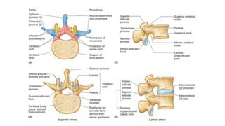
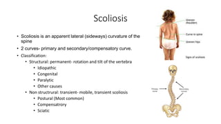
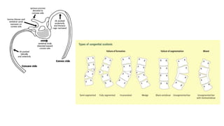
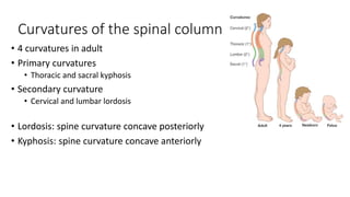
Anatomical basis of scoliosis,
lordosis, disc prolapse ,
spondylolistheis and spina
bidfida
Dr Siddhartha Sinha
Assistant Professor, Department of Orthopaedics
HIMSR & HAHC
Scoliosis
• Scoliosis is an apparent lateral (sideways) curvature of the
spine
• 2 curves- primary and secondary/compensatory curve.
• Classification:
• Structural: permanent- rotation and tilt of the vertebra
• Idiopathic
• Congenital
• Paralytic
• Other causes
• Non structrural: transient- mobile, transient scoliosis
• Postural (Most common)
• Compensatrory
• Sciatic
• Postural scoliosis (correctable
deformity, improves with patient sits)
• Short leg
• Pelvic tilt
• Hip contracture
• Structural Scoliosis (non correctable
deformity)
• Spinous processes swing round
towards the concavity of the curve
• Transverse processes on the
convexity rotate posteriorly.
• thoracic region the ribs on the convex
side stand out prominently, producing
the rib hump (characteristic)
Curvatures of the spinal column
• 4 curvatures in adult
• Primary curvatures
• Thoracic and sacral kyphosis
• Secondary curvature
• Cervical and lumbar lordosis
• Lordosis: spine curvature concave posteriorly
• Kyphosis: spine curvature concave anteriorly
Lordosis
• Cervical and lumbar lordoses
• Appear during the late fetal period but do not become
obvious until infancy due to extension from the flexed fetal
position
• Secondary curvatures -primarily by differences in thickness
between the anterior and the posterior parts of the IV discs.
• The cervical lordosis
• when infant begins to raise (extend) the head while prone and to hold
the head erect while sitting.
• The lumbar lordosis becomes
• when toddlers begin to assume the upright posture, standing and
walking.
• This curvature, generally more pronounced in females,
ends at the lumbosacral angle formed at the junction of L5
vertebra with the sacrum
Exaggerated lumbar lordosis (hollow back/
sway back)
• Anterior tilting of pelvis
• Increased extension of lumbar vertebrae
• Weak musculature of anterolateral abdominal
muscles
• Pregnancy, obseity
Kyphosis
• Thoracic and sacral kyphoses are primary
curvatures
• Develop due to the (flexed) fetal position.
• The primary curvatures are in the same
direction as the main curvatures of the fetal
vertebral column.
• The primary curvatures are retained
throughout life as a consequence of
differences in height between the anterior and
posterior parts of the vertebrae.
• Vertebral developmental defects
Excessive throcacic kyphosis (hunchback/
humpback)
• Vertebral column curves posteriorly
• Erosion of anterior part of vertebrae
• Osteoporosis – horizontal trabeculae
affected- failure in compression-
compression fractures
• Multiple compression fractures
• loss of height
• Increased AP dm of chest wall- decreased
dynamic pulmonary capacity
Disc prolapse
• 3 components
• Cartilage end-plates
• Nucleus pulposus (NP)
• Annulus fibrosus (AP)
• Ageing decreases nutrition and number of chondrocytes in NP
decrease- decreased proteoglycans & increase in proportion of
collagen to water in NP leading to stiffness of NP and less distribution
of load- facets bear more load (back pain)
• Eventually annulus fails causing disc herniation and radiculopathy
• After disc extrusion – disc
compress one or more spinal
roots in extrathecal course
• Often exiting nerve root
Spodylolistheisis
• Forward displacement of one
vertebra over another
• Normal:
• Forward displacement of
vertebral body prevented by
articular process with the
vertebra below it
• Disc, intervertebral ligaments to
a small extent
Spina bifida
• Neural arches fail to develop
normally and and fuse posterior
to the vertebral canal resulting in
an "open" vertebral canal
1. Spina bifida occulta
• Relatively common in population
(upto 24%)
• L5-S1 most commonly affected
2. Spina bifida cystica
• One or more vertebral arch fail to
develop completely
• Herniation
• Meninges (meningocoele)
• Meninges and Spinal cord
(meningomyelocoele)
3. Neural tube defects- failure of
closure of neural tube at 4th week of
embryonic development
Anatomical basis of common back problems

Anatomical basis of common back problems

  • 1.
    Anatomical basis ofscoliosis, lordosis, disc prolapse , spondylolistheis and spina bidfida Dr Siddhartha Sinha Assistant Professor, Department of Orthopaedics HIMSR & HAHC
  • 4.
    Scoliosis • Scoliosis isan apparent lateral (sideways) curvature of the spine • 2 curves- primary and secondary/compensatory curve. • Classification: • Structural: permanent- rotation and tilt of the vertebra • Idiopathic • Congenital • Paralytic • Other causes • Non structrural: transient- mobile, transient scoliosis • Postural (Most common) • Compensatrory • Sciatic
  • 5.
    • Postural scoliosis(correctable deformity, improves with patient sits) • Short leg • Pelvic tilt • Hip contracture • Structural Scoliosis (non correctable deformity) • Spinous processes swing round towards the concavity of the curve • Transverse processes on the convexity rotate posteriorly. • thoracic region the ribs on the convex side stand out prominently, producing the rib hump (characteristic)
  • 7.
    Curvatures of thespinal column • 4 curvatures in adult • Primary curvatures • Thoracic and sacral kyphosis • Secondary curvature • Cervical and lumbar lordosis • Lordosis: spine curvature concave posteriorly • Kyphosis: spine curvature concave anteriorly
  • 8.
    Lordosis • Cervical andlumbar lordoses • Appear during the late fetal period but do not become obvious until infancy due to extension from the flexed fetal position • Secondary curvatures -primarily by differences in thickness between the anterior and the posterior parts of the IV discs. • The cervical lordosis • when infant begins to raise (extend) the head while prone and to hold the head erect while sitting. • The lumbar lordosis becomes • when toddlers begin to assume the upright posture, standing and walking. • This curvature, generally more pronounced in females, ends at the lumbosacral angle formed at the junction of L5 vertebra with the sacrum
  • 9.
    Exaggerated lumbar lordosis(hollow back/ sway back) • Anterior tilting of pelvis • Increased extension of lumbar vertebrae • Weak musculature of anterolateral abdominal muscles • Pregnancy, obseity
  • 10.
    Kyphosis • Thoracic andsacral kyphoses are primary curvatures • Develop due to the (flexed) fetal position. • The primary curvatures are in the same direction as the main curvatures of the fetal vertebral column. • The primary curvatures are retained throughout life as a consequence of differences in height between the anterior and posterior parts of the vertebrae. • Vertebral developmental defects
  • 11.
    Excessive throcacic kyphosis(hunchback/ humpback) • Vertebral column curves posteriorly • Erosion of anterior part of vertebrae • Osteoporosis – horizontal trabeculae affected- failure in compression- compression fractures • Multiple compression fractures • loss of height • Increased AP dm of chest wall- decreased dynamic pulmonary capacity
  • 13.
    Disc prolapse • 3components • Cartilage end-plates • Nucleus pulposus (NP) • Annulus fibrosus (AP) • Ageing decreases nutrition and number of chondrocytes in NP decrease- decreased proteoglycans & increase in proportion of collagen to water in NP leading to stiffness of NP and less distribution of load- facets bear more load (back pain) • Eventually annulus fails causing disc herniation and radiculopathy
  • 16.
    • After discextrusion – disc compress one or more spinal roots in extrathecal course • Often exiting nerve root
  • 18.
    Spodylolistheisis • Forward displacementof one vertebra over another • Normal: • Forward displacement of vertebral body prevented by articular process with the vertebra below it • Disc, intervertebral ligaments to a small extent
  • 22.
    Spina bifida • Neuralarches fail to develop normally and and fuse posterior to the vertebral canal resulting in an "open" vertebral canal 1. Spina bifida occulta • Relatively common in population (upto 24%) • L5-S1 most commonly affected 2. Spina bifida cystica • One or more vertebral arch fail to develop completely • Herniation • Meninges (meningocoele) • Meninges and Spinal cord (meningomyelocoele) 3. Neural tube defects- failure of closure of neural tube at 4th week of embryonic development