This document provides an overview of infrared spectroscopy. It discusses the electromagnetic spectrum and how infrared spectroscopy uses infrared light to analyze chemical bonds in molecules based on their vibrational and rotational frequencies. Different sampling techniques are described for analyzing powders, liquids, and gases. Key points covered include the principles of infrared absorption, molecular vibrations, Hooke's law application to frequency determination, and methods for preparing samples like KBr pellets and diffuse reflectance.
This document discusses the theory, instrumentation, and applications of dispersive and Fourier transform infrared (FTIR) spectroscopy. It begins with an introduction to IR spectroscopy and the IR region. It then covers dispersive IR instrumentation, which uses prism or grating monochromators to separate wavelengths, and has limitations like slow scan speeds and limited resolution. The document introduces FTIR instrumentation, which uses an interferometer to simultaneously measure all wavelengths and overcomes the limitations of dispersive IR. It concludes that FTIR provides faster, more accurate and sensitive analysis compared to dispersive IR.
This document discusses the principles, instrumentation, and applications of a dispersive infrared spectrophotometer. It describes how this type of IR spectrometer works by using radiation sources like globars or Nernst glowers, monochromators to separate wavelengths, and detectors like photo detectors or thermal detectors to analyze the absorbed infrared wavelengths. Key applications of dispersive IR spectrophotometers include identifying organic and inorganic compounds by their functional groups and determining molecular structure and orientation. However, it also notes some disadvantages like slower scan speeds and less sensitivity compared to Fourier transform IR spectrometers.
X-ray crystallography uses X-rays to determine the atomic and molecular structure of crystals. When X-rays hit a crystal, they cause the crystalline atoms to diffract the X-rays into specific directions. By measuring the angles and intensities of these diffracted X-rays, the crystallographer can produce a three-dimensional picture of electron density within the crystal. From this electron density, the positions of atoms and chemical bonds in the crystal can be determined. There are several methods for X-ray crystallography including Bragg X-ray spectrometry, rotating crystal method, and powder crystal method. X-ray crystallography has many applications including determining crystal and molecular structures, and character
Spectrofluorimetry is a technique that measures fluorescence emitted from molecules. It involves exciting molecules with UV or visible light which causes electrons to transition to an excited state. The molecule then relaxes and emits light of a longer wavelength. Factors like concentration, quantum yield, path length, pH, temperature and presence of quenchers affect the intensity of fluorescence. Spectrofluorimeters are used to collect excitation and emission spectra of molecules to identify them.
spectrofluorometer is the instrument for recording fluorescence emission and absorption spectra When a beam of light is incident on certain substances they emit visible light or radiations. This is known as fluorescence. Fluorescence starts immediately after the absorption of light and stops as soon as the incident light is cut off. The substances showing this phenomenon are known as flourescent substances.
X- ray crystallography, Shriyansh Srivastava, M.Pharm (Department of Pharmaco...Shriyansh Srivastav
X-ray crystallography uses X-rays to determine the atomic and molecular structure of crystals. Wilhelm Röntgen discovered X-rays in 1895. X-rays are produced when high velocity electrons collide with a metal target. X-ray crystallography works by firing a beam of X-rays at crystalline solids and observing the diffraction pattern of scattered X-rays. Bragg's law describes the conditions under which constructive interference of X-rays occurs and can be used to determine crystal structures. Common methods include rotating crystal, powder diffraction, and using detectors like photographic film, Geiger-Müller counters, or scintillation counters. X-ray crystallography has applications in determining protein structures and identifying
Nuclear magnetic resonance (NMR) spectroscopyVK VIKRAM VARMA
SPECTROSCOPY
NMR SPECTROSCOPY
HISTORY
THEORY
PRINCIPLE
INSTRUMENTATION
SOLVENTS USED IN NMR(PROTON NMR)
CHEMICAL SHIFT
FACTORS AFFECTING CHEMICAL SHIFT
RELAXATION PROCESS
SPIN-SPIN COUPLING
푛+1 RULE
NMR SIGNALS IN VARIOUS COMPOUNDS
COUPLING CONSTANT
NUCLEAR MAGNETIC DOUBLE RESONANCE/ SPIN DECOUPLING
FT-NMR
ADVANTAGES & DISADVANTAGES
APPLICATIONS
REFERENCE
Types of crystals & Application of x raykajal pradhan
some basic information:-
A crystal lattice is a 3-D arrangement of unit cells.
Unit cell is the smallest unit of a crystal, By stacking identical unit cells, the entire lattice can be constructed
A crystal’s unit cell dimensions are defined by six numbers, the lengths of the 3 axes, a, b, and c, and the three interaxial angles, α, β and γ.
If a unit cell has the same type of atom at the corners of the unit cell but not also in the middle of the faces nor in the centre of the cell, it is called primitive and given by symbol P
7 types of crystal system details
14 bravis lattice
APPLICATION X-RAY CRYSTALLOGRAPHY
1. Structure of crystals
2. Polymer characterisation
3. State of anneal in metals
4. Particle size determination
a) Spot counting method
b) Broadening of diffraction lines
c) Low-angle scattering
5.Applications of diffraction methods to complexes
a) Determination of cis- trans isomerism
b) Determination of linkage isomerism
6.Miscellaneous applications
FT-NMR uses Fourier transforms to convert time domain signals from nuclear magnetic resonance into frequency domain spectra. The sample is placed in a strong magnet and exposed to pulses of radio frequency radiation, producing a free induction decay signal that is recorded over time. This time domain signal is then digitized and analyzed using a Fourier transform program on a computer to produce the frequency domain NMR spectrum. FT-NMR provides higher sensitivity than continuous wave NMR, allowing analysis of smaller sample sizes.
This document provides an introduction to infrared spectroscopy. It discusses the principle, theory, modes of molecular vibrations, instrumentation, factors influencing vibrational frequencies, and applications of infrared spectroscopy. Specifically, it explains that infrared spectroscopy analyzes the absorption of infrared radiation by molecules and the characteristic vibrational frequencies are dependent on the masses of atoms and the strength of bonds in a molecule. It also describes the different types of molecular vibrations that can be observed, including stretching and bending vibrations.
The document discusses atomic absorption spectroscopy. It begins with an introduction describing how atomic absorption spectroscopy measures the concentration of an element by measuring the amount of light absorbed at a characteristic wavelength when it passes through atoms of that element. It then describes the principle, instrumentation, applications, and sources of interference in atomic absorption spectroscopy. The key sources of interference discussed are non-spectral interferences such as matrix, chemical, and ionization interferences and spectral interferences such as background absorption.
PRINCIPLES of FT-NMR & 13C NMR
Fourier Transform
FOURIER TRANSFORM NMR SPECTROSCOPY
THEORY OF FT-NMR
13C NMR SPECTROSCOPY
Principle
Why C13-NMR is required though we have H1-NMR?
CHARACTERISTIC FEATURES OF 13 C NMR
Chemical Shifts
NUCLEAR OVERHAUSER ENHANCEMENT
Short-Comings of 13C-NMR Spectra
1. The document discusses various sample handling techniques for infrared spectroscopy, including techniques for solids, liquids, and gases.
2. For solids, it describes techniques like pelleting, mulling, casting thin films, and reflectance methods. For liquids, it discusses analyzing neat liquids, solutions, and reflectance techniques. For gases, it mentions using an absorbance cell or photoacoustic spectroscopy.
3. The document provides details on specific reflectance techniques like photoacoustic spectroscopy, specular and diffuse reflectance, and attenuated total reflectance. It also discusses how to prepare and analyze samples using these various infrared spectroscopy techniques.
Nuclear magnetic resonance (NMR) spectroscopy measures the absorption of radiofrequency radiation by atomic nuclei placed in a strong magnetic field. When placed in an external magnetic field, NMR active nuclei such as 1H and 13C can absorb radiation at frequencies characteristic of their isotopes. The resonant frequency and signal intensity are proportional to the magnetic field strength. NMR spectra plot chemical shift in δ units versus peak intensity. Applications of NMR include determining molecular structure, identification of organic compounds, and pharmaceutical analysis.
Mass spectroscopy
1.Introduction
2. Principle
3. Theory
4. Instrumentation
5. Different types of Ionization
6. FAB
7. MALDI
8. APCI
9. ESI
10.Quardapole
This document discusses flame emission spectroscopy. It begins with an introduction stating that flame emission spectroscopy uses a flame to provide energy and excite atoms introduced into the flame. It then covers the history, principle, instrumentation, applications and potential interferences of flame emission spectroscopy. The principle involves desolvation, vaporization, atomization, excitation and emission of light at characteristic wavelengths. Common instrumentation components include burners, atomizers, monochromators, detectors and readouts. Applications include analysis of chemicals, soils, plants, waters and more. Potential issues include matrix, chemical, ionization and spectral interferences.
This document discusses various ionization techniques used in mass spectrometry. It begins with an introduction to mass spectrometry and its basic principles. It then describes several ionization sources including gas phase sources like electron impact ionization and chemical ionization, and desorption sources like electrospray ionization, matrix-assisted laser desorption/ionization, and fast atom bombardment. The document proceeds to provide more detailed explanations of specific ionization techniques like electrospray ionization, atmospheric pressure chemical ionization, atmospheric pressure photoionization, matrix-assisted laser desorption ionization, and fast atom bombardment. It concludes with references used in the document.
The document provides an overview of mass spectrometry. It discusses the history, principles, instrumentation, ionization techniques, mass analyzers, and applications of mass spectrometry. Mass spectrometry involves converting sample molecules to ions, separating the ions based on their mass-to-charge ratio, and detecting the ions. Key components include an ion source, mass analyzer, and ion detector. Common ionization methods include electron ionization and chemical ionization. Common mass analyzers are magnetic sector, quadrupole, time-of-flight, and ion trap. Mass spectrometry has various applications in fields like proteomics, metabolomics, and environmental analysis.
IR interpretation and sample handling Afzaye Rasul
The document discusses sample handling and interpretation of infrared spectroscopy. It describes several methods for preparing solid, liquid, and gas samples for IR analysis. These include pressed KBr pellets for solids, liquid samples in thin films between windows, and gases in cells. The document then outlines how to interpret IR spectra by identifying key functional groups like carbonyl, hydroxyl, aromatic, and C=C bands. It provides examples of infrared absorptions for several classes of organic compounds including alkanes, alkenes, alcohols, ketones, and amides.
This document provides an overview of infrared spectroscopy. It discusses the principle that infrared spectroscopy involves absorption of infrared radiation which causes vibrational transitions in molecules. The instrumentation involves an infrared source, sample holder, and detector. Applications include identifying functional groups in organic molecules, determining drug formulations, and analyzing biological samples like urine.
Attenuated total reflectance (ATR) spectroscopy allows samples to be examined directly in the solid or liquid state without preparation by passing infrared radiation through an infrared-transmitting crystal with a high refractive index. The infrared beam undergoes total internal reflection within the crystal and evanescent waves penetrate into the sample in contact with the crystal, producing its infrared spectrum. ATR is useful for analyzing liquids, solids, powders, and other samples with little preparation and can be applied in fields like pharmaceuticals, chemicals, forensics, and biomedical research.
This document provides an overview of ultraviolet (UV) and visible spectroscopy. It begins by defining spectroscopy and discussing the difference between a spectrometer and a spectrophotometer. It then covers the electromagnetic spectrum and describes UV and visible spectroscopy. The document discusses Beer's Law and the instrumentation used. It explains electronic transitions that can occur, including σ-σ*, n-σ*, and π-π* transitions. Finally, it discusses applications of UV-visible spectroscopy such as detecting functional groups and studying conjugation.
This document discusses the attenuated total reflectance (ATR) infrared spectroscopy sampling technique. It begins by introducing ATR and explaining that it allows for little to no sample preparation and a very thin sampling pathlength. It then discusses factors that affect the ATR spectrum such as the refractive indices of the crystal and sample, angle of incidence, depth of penetration, and quality of sample contact. Common ATR crystal materials and their spectral ranges and depths of penetration are presented. Applications include identification of functional groups, contaminated pet food detection, and more. In conclusion, ATR provides high quality reproducible data for a variety of solid and liquid samples.
INTRODUCTION TO UV-VISIBLE SPECTROSCOPYJunaid Khan
UV-visible spectroscopy involves measuring the absorption of electromagnetic radiation in the ultraviolet-visible spectral region. When UV-VIS radiation interacts with molecules, it can cause electronic transitions between different energy levels. The absorption spectrum obtained plots absorbance against wavelength, showing characteristic absorption bands. The positions and intensities of these bands provide information about the molecular structure of the absorbing chemical species.
This document provides an overview of infrared (IR) spectroscopy. It discusses the IR region of the electromagnetic spectrum, the basic principles of IR spectroscopy, and factors that influence molecular vibrations. Requirements for IR absorption include an electric dipole moment and the radiation wavelength matching the natural vibration frequency. Molecular vibrations observed in IR spectroscopy include stretches, bends, and rotations. Instrumentation components like IR sources, wavelength selectors, detectors, and sample handling techniques are also outlined. Finally, applications of IR spectroscopy like structure elucidation and identification of functional groups are mentioned.
1. Spectrophotometers use light to analyze samples by measuring how much light is absorbed, reflected, or transmitted. They contain a light source, monochromator to isolate wavelengths, sample holders, detectors, and readout devices.
2. The Beer-Lambert law states that absorbance is directly proportional to concentration, with absorbance measured using spectrophotometers. It is obeyed at low concentrations but not at very high concentrations.
3. Spectrophotometers are used in clinical laboratories and other settings to determine concentrations of substances and analyze samples. They provide accurate quantitative analysis of both colored and non-colored compounds.
Characterization of nanoparticles & its regulatory aspectsvivek vyas
This document summarizes techniques for characterizing nanoparticles, including particle size, shape, surface charge, crystal structure, yield, drug entrapment efficiency, and in vitro drug release. Key characterization techniques mentioned are microscopy (SEM, TEM), laser light scattering, zeta potential measurement, XRD, FTIR, and dialysis-based in vitro release testing. Regulatory considerations for nanoparticles are also briefly discussed.
Spectrophotometry in clinical chemistryOfonmbuk Umoh
Spectrophotometry is a technique that uses the measurement of light absorption to determine the concentration of chemical substances. It operates based on Beer's Law, which states that absorbance is directly proportional to concentration. The methodology involves using a spectrophotometer to measure the intensity of light passing through reference and sample solutions. Applications include concentration measurement, detection of impurities, structure elucidation, and more. Spectrophotometry is a widely used analytical technique in clinical chemistry.
UV VISIBLE SPECTROSCOPY is a technique that uses the absorption of ultraviolet or visible radiation to determine the electronic and geometric structure of molecules. It works by measuring the amount of light absorbed by a sample at each wavelength across the UV-VIS spectrum. The amount of absorption follows the Beer-Lambert law, which states that absorbance is directly proportional to concentration, path length, and absorptivity. UV-VIS spectroscopy can be used to qualitatively and quantitatively analyze compounds, determine functional groups, study conjugation, identify unknowns, and more. It has advantages of being rapid, nondestructive, and sensitive, though it is limited to compounds that absorb in the UV-VIS range.
INFRARED SPECTROSCOPY to find the functional groupssusera34ec2
This document provides an overview of infrared spectroscopy. It discusses the principle, theory, instrumentation, sample preparation, qualitative and quantitative analysis, uses, applications, and limitations. Infrared spectroscopy analyzes the infrared region of the electromagnetic spectrum to identify functional groups and compounds. The main instruments are dispersive spectrometers and Fourier transform infrared spectrometers. Infrared spectroscopy is widely used in research and industry for structure elucidation, compound identification, and determining organic and inorganic materials.
Analytical techniques in pharmaceuticalZafar Mahmood
The document discusses various analytical techniques used in pharmaceutical analysis to identify and quantify chemical substances. It describes techniques including gravimetric analysis, titrimetry, polarimetry, refractometry, chromatography, spectroscopy, electrochemistry, kinetics methods, and hyphenated techniques. Specifically, it provides details on the principles and applications of techniques like titration, high performance liquid chromatography, UV-visible spectroscopy, infrared spectroscopy, mass spectroscopy, and hyphenated methods like LC-MS and GC-MS.
principle, application and instrumentation of UV- visible Spectrophotometer Ayetenew Abita Desa
This Presentation powerpoint includes the principle, application, and instrumentation of UV- Visible Spectrophotometer. It covers beer-lambert low and its quantitative applications. It also includes the qualitative applications in different fields of study. Presented at Addis Ababa University, School of medicine, department of medical biochemistry.
Colorimeter and spectrophotometer, Mass Spectrometerprachann
It contains a brief knowledge on Introduction, Principle, Laws, Flow representation, Instrumentation, Applications
and Mass spectrometer
- Principle
- Instrumentation
This document discusses chemiluminescence, including its principles, instrumentation, and applications. It describes how chemiluminescence occurs through excited molecule species that emit light upon returning to ground state. Common techniques are fluorescence and phosphorescence. The document outlines criteria for successful chemiluminescent reactions and examples like luminol-peroxidase and luciferin-luciferase reactions. Detection methods like photomultiplier tubes and applications in immunoassays, DNA detection, food analysis, and clinical utilities are summarized.
Infrared spectroscopy (IR spectroscopy or vibrational spectroscopy) involves the interaction of infrared radiation with matter. It covers a range of techniques, mostly based on absorption spectroscopy. As with all spectroscopic techniques, it can be used to identify and study chemicals
IR spectroscopy analyzes molecular vibrations and rotations that are excited by infrared light. When the frequency of infrared light matches the natural vibrational frequency of a molecule, absorption occurs. Different functional groups absorb characteristic frequencies allowing IR spectroscopy to determine a molecule's structure. A proper sampling technique like mulling or using a liquid cell is required to obtain an IR spectrum. An IR spectrometer consists of an infrared source, monochromator, sample holder, detector, and recorder. Common detectors include bolometers and pyroelectric detectors which convert the infrared absorption into electrical signals.
1. UV-visible spectroscopy is used to detect functional groups, impurities, and perform qualitative and quantitative analysis of compounds.
2. It works by measuring how much light is absorbed by a sample at different wavelengths, providing information on functional groups and molecular structure.
3. Key applications include detection of impurities, structure elucidation, and determination of concentration through Beer's law.
Spectrophotometry uses the absorption of light by chemical substances to measure concentration. A spectrophotometer directs a beam of light through a sample and measures the intensity of transmitted light, relating it to concentration through Beer's Law. It operates based on Lambert's Law stating light absorption increases with concentration and path length. Common types are single and double beam instruments, with the latter measuring sample and reference simultaneously. Components include a light source, monochromator, sample holder, and detector. Applications include quantifying analytes and studying reaction kinetics and molecular structure.
Light Scattering Phenomenon:
The blue color of the sky and the red color of the sun at sunset result from scattering of light of small dust particles, H2O molecules and other gases in the atmosphere.
The efficiency with which light is scattered depends on its wavelength(λ).
The sky is blue because violet and blue light are scattered to a greater extent than other longer wavelengths.
A clear cloudless day-time sky is blue because molecules in the air scatter blue light from the sun more than they scatter red light.
When we look towards the sun at sunset, we see red and orange colours because the blue light has been scattered out and away from the line of sight.
Scattered radiation:
• Radiate scattering- second major spectral method of analysis.
• In this technique some radiation that passes through a sample strikes particles of the analyte and is scattered in a different direction.
• A detector is used to measure either the intensity of the scattered radiation or the decreased intensity of the incident radiation
• Depending on the scattering mechanism, the method can be employed for either qualitative or quantitative analysis.
For chemical analysis three forms of radiative scattering are important – viz.
Tyndall,
Raman, and
Rayleigh scattering.
Tyndall Scattering occurs when the dimensions of the particles that are causing the scattering are larger than the wavelength of the scattered radiation.
It is caused by reflection of the incident radiation from the surfaces of the particles,
reflection from the interior walls of the particles, and refraction and diffraction of the radiation as it passes through the particles.
Scattering of light
- by particles in a colloid or suspension.
The longer-wavelength light is more transmitted while the shorter- wavelength light is more reflected via scattering
Nephelometry & Turbidimetry:
When electromagnetic radiation (light) strikes a particle in solution, some of the light will be absorbed by the particle, some will be transmitted through the solution and some of the light will be scattered or reflected .
The amount of light scattered is proportional to the concentration of insoluble particle.
In Nephelometry, the intensity of the scattered light is measured.
In Turbidimetry, the intensity of light transmitted through the medium, the unscattered light, is measured. Light scattering is the physical phenomenon resulting from the interaction of light with a particles in solution
Turbidimetry is involved with measuring the amount of transmitted light (and calculating the absorbed light) by particles in suspension to determine the concentration of the substance in question.
Amount of absorbed light, and therefore, concentration is dependent on ;
1) number of particles, and
2) size of particles.
• Measurements are made using light spectrophotometers
Factors affecting on scattering of light:
Concentration of particles
Particle size
Wavelength
Distance of
UPLC provides faster, more sensitive chromatographic separations compared to HPLC. It works by using smaller particle sizes (<2.5um) in the column packing which allows for higher pressure and flow rates based on the van Deemter equation. This provides benefits like reduced run times, decreased sample volume needs, and improved resolution. However, it also requires more robust instrumentation to handle the increased pressures and columns have reduced lifespan. UPLC has applications in fields like pharmaceutical analysis, metabolomics, and impurity profiling due to its enhanced resolution and sensitivity capabilities.
Infrared spectroscopy is a technique that analyzes infrared light absorbed by a molecule to determine its structure. There are several types of molecular vibrations that can be observed, including stretching and bending vibrations. Samples can be analyzed in solid, liquid, or gas form using different sample handling methods. The main components of an IR spectrometer are the radiation source, monochromator, sample cell, detector, and recorder. Dispersive and Fourier transform IR spectrometers are two common instrument types, with Fourier transform having advantages like faster scanning. Functional groups can be identified by their characteristic absorption bands. Factors like coupling, hydrogen bonding, and electronic effects can influence vibrational frequencies.
Optical techniques like photometry, spectrophotometry, and colorimetry are used in clinical laboratories. They are based on Beer's law and Lambert's law. Spectrophotometry measures light intensity at selected wavelengths using a light source, monochromator, sample cuvettes, detector, and display. It provides more sensitivity than colorimetry which determines color intensity based on light absorption. Both techniques rely on the principle that absorbed light is inversely proportional to concentration according to Beer-Lambert's law.
1. The document discusses UV-visible spectroscopy, describing the basic components and functioning of a UV-visible spectrophotometer.
2. Key aspects covered include the electromagnetic spectrum, sample cuvettes, light sources, monochromators, detectors, and performance verification tests to ensure the instrument is functioning properly.
3. UV-visible spectroscopy is a technique used to study light absorption by molecules to determine concentration and identify substances.
This document discusses spectrophotometry and the Beer-Lambert law. It provides:
1) An overview of how spectrophotometers work by measuring the absorption of light by chemical compounds and relating absorption to concentration according to the Beer-Lambert law.
2) A description of the basic components of a spectrophotometer including a light source, wavelength selector, sample cuvette, detector, and readout device.
3) Explanations of how to prepare and measure standards to generate a calibration curve to determine unknown concentrations.
Hi, I'm Presents a Research article for Journal club entitled with
"3D Printing: A Case of ZipDose® Technology –World’s First 3D Printing Platform to Obtain FDA Approval for a Pharmaceutical Product"
Reference (Source article):
1. West, Thomas & Bradbury, Thomas. (2018). 3D Printing: A Case of ZipDose® Technology - World's First 3D Printing Platform to Obtain FDA Approval for a Pharmaceutical Product: Process Engineering and Additive Manufacturing. https://doi.org/10.1002/9783527813704...
2. https://www.aprecia.com/technology/zipdose
This document summarizes a journal club presentation on a study evaluating the use of granulated sugar as a wound dressing. The presentation included an in vitro study showing that three types of granulated sugar inhibited the growth of bacteria. A clinical pilot study then evaluated the effects of applying white granulated sugar directly to wounds. The results of the pilot study suggested that granulated sugar was effective at debriding wounds, reducing pain and malodor, and lowering bacterial loads. The presentation concluded that while more research is still needed, granulated sugar shows potential as a simple and inexpensive wound treatment option.
The document discusses different types of nanoparticles used in drug delivery, including liposomes, solid nanoparticles, polymeric nanoparticles, nanocapsules, nanospheres, dendrimers, nanotubes, nanowires, and nanocrystals. It also describes several methods for preparing nanoparticles, such as solvent evaporation, emulsions-diffusion, nanoprecipitation, salting out, and dialysis. Evaluation methods for prepared nanoparticles are discussed, including measuring yield, drug content, particle size, zeta potential, surface morphology, polydispersity index, in-vitro release studies, and kinetic studies.
The document discusses the results of a study on the impact of COVID-19 lockdowns on air pollution. The study found that lockdowns led to significant short-term reductions in nitrogen dioxide and fine particulate matter pollution globally as economic activities slowed. However, the improvements in air quality were temporary and pollution levels rose back to pre-pandemic levels as restrictions eased and activity resumed.
In this slide Structure of Skin and Hair, Hair Growth Cycle were described followed by skin related diseases such as Acne, dry skin, pigmentation, wrinkles etc.
Bacteria cultivation NUTRITIONAL REQUIREMENTS
NUTRITIONAL TYPES OF BACTERIA
PHOTOTROPHS
CHEMOTROPHS
AUTOTROPHS AND HETEROTROPH
OBLIGATE PARASITE
BACTERIOLOGICAL MEDIA
TYPES OF MEDIA
PHYSICAL CONDITION FOR GROWTH
CULTIVATION OF AEROBIC AND ANAEROBIC BACTERIA
This document discusses various types of documentation required in the pharmaceutical industry, including master formula records (MFR), drug master files (DMF), and generic drug development. It defines MFRs as approved master documents that describe the full manufacturing process for a specific batch size. It provides details on the content required for MFRs based on guidelines from WHO, Health Canada, and the US CFR. It also discusses the purpose and types of DMFs submitted to the FDA, including Type 1 for manufacturing facilities, Type 2 for drug substances/products, and others. Finally, it briefly mentions the Hatch-Waxman Act as it relates to generic drug development.
This document discusses various optimization techniques used in pharmaceutical formulation and processing. It begins by defining optimization as making something as perfect or functional as possible. It describes the key parameters in optimization problems including independent and dependent variables as well as constrained and unconstrained problems. The document then explains several statistical design approaches to optimization including evolutionary operation, simplex method, and search methods. It provides examples of how these techniques are applied and the general steps involved in using a search method for optimization.
This document discusses gastroretentive drug delivery systems (GRDDS). It begins by defining GRDDS as an approach to prolong gastric residence time to target drug release in the upper gastrointestinal tract. It then discusses various approaches to prolong gastric retention time, including floating systems, expandable systems, and bioadhesive systems. The document provides examples of drug candidates suitable for GRDDS and evaluates formulation techniques, in vitro tests for buoyancy and drug release, and methods to test bioadhesion and swelling. It concludes by discussing the advantages and limitations of GRDDS.
- Video recording of this lecture in English language: https://youtu.be/Pt1nA32sdHQ
- Video recording of this lecture in Arabic language: https://youtu.be/uFdc9F0rlP0
- Link to download the book free: https://nephrotube.blogspot.com/p/nephrotube-nephrology-books.html
- Link to NephroTube website: www.NephroTube.com
- Link to NephroTube social media accounts: https://nephrotube.blogspot.com/p/join-nephrotube-on-social-media.html
10 Benefits an EPCR Software should Bring to EMS Organizations Traumasoft LLC
The benefits of an ePCR solution should extend to the whole EMS organization, not just certain groups of people or certain departments. It should provide more than just a form for entering and a database for storing information. It should also include a workflow of how information is communicated, used and stored across the entire organization.
Osteoporosis - Definition , Evaluation and Management .pdfJim Jacob Roy
Osteoporosis is an increasing cause of morbidity among the elderly.
In this document , a brief outline of osteoporosis is given , including the risk factors of osteoporosis fractures , the indications for testing bone mineral density and the management of osteoporosis
Mercurius is named after the roman god mercurius, the god of trade and science. The planet mercurius is named after the same god. Mercurius is sometimes called hydrargyrum, means ‘watery silver’. Its shine and colour are very similar to silver, but mercury is a fluid at room temperatures. The name quick silver is a translation of hydrargyrum, where the word quick describes its tendency to scatter away in all directions.
The droplets have a tendency to conglomerate to one big mass, but on being shaken they fall apart into countless little droplets again. It is used to ignite explosives, like mercury fulminate, the explosive character is one of its general themes.
5-hydroxytryptamine or 5-HT or Serotonin is a neurotransmitter that serves a range of roles in the human body. It is sometimes referred to as the happy chemical since it promotes overall well-being and happiness.
It is mostly found in the brain, intestines, and blood platelets.
5-HT is utilised to transport messages between nerve cells, is known to be involved in smooth muscle contraction, and adds to overall well-being and pleasure, among other benefits. 5-HT regulates the body's sleep-wake cycles and internal clock by acting as a precursor to melatonin.
It is hypothesised to regulate hunger, emotions, motor, cognitive, and autonomic processes.
8 Surprising Reasons To Meditate 40 Minutes A Day That Can Change Your Life.pptxHolistified Wellness
We’re talking about Vedic Meditation, a form of meditation that has been around for at least 5,000 years. Back then, the people who lived in the Indus Valley, now known as India and Pakistan, practised meditation as a fundamental part of daily life. This knowledge that has given us yoga and Ayurveda, was known as Veda, hence the name Vedic. And though there are some written records, the practice has been passed down verbally from generation to generation.
Cell Therapy Expansion and Challenges in Autoimmune DiseaseHealth Advances
There is increasing confidence that cell therapies will soon play a role in the treatment of autoimmune disorders, but the extent of this impact remains to be seen. Early readouts on autologous CAR-Ts in lupus are encouraging, but manufacturing and cost limitations are likely to restrict access to highly refractory patients. Allogeneic CAR-Ts have the potential to broaden access to earlier lines of treatment due to their inherent cost benefits, however they will need to demonstrate comparable or improved efficacy to established modalities.
In addition to infrastructure and capacity constraints, CAR-Ts face a very different risk-benefit dynamic in autoimmune compared to oncology, highlighting the need for tolerable therapies with low adverse event risk. CAR-NK and Treg-based therapies are also being developed in certain autoimmune disorders and may demonstrate favorable safety profiles. Several novel non-cell therapies such as bispecific antibodies, nanobodies, and RNAi drugs, may also offer future alternative competitive solutions with variable value propositions.
Widespread adoption of cell therapies will not only require strong efficacy and safety data, but also adapted pricing and access strategies. At oncology-based price points, CAR-Ts are unlikely to achieve broad market access in autoimmune disorders, with eligible patient populations that are potentially orders of magnitude greater than the number of currently addressable cancer patients. Developers have made strides towards reducing cell therapy COGS while improving manufacturing efficiency, but payors will inevitably restrict access until more sustainable pricing is achieved.
Despite these headwinds, industry leaders and investors remain confident that cell therapies are poised to address significant unmet need in patients suffering from autoimmune disorders. However, the extent of this impact on the treatment landscape remains to be seen, as the industry rapidly approaches an inflection point.
share - Lions, tigers, AI and health misinformation, oh my!.pptxTina Purnat
• Pitfalls and pivots needed to use AI effectively in public health
• Evidence-based strategies to address health misinformation effectively
• Building trust with communities online and offline
• Equipping health professionals to address questions, concerns and health misinformation
• Assessing risk and mitigating harm from adverse health narratives in communities, health workforce and health system
Adhd Medication Shortage Uk - trinexpharmacy.comreignlana06
The UK is currently facing a Adhd Medication Shortage Uk, which has left many patients and their families grappling with uncertainty and frustration. ADHD, or Attention Deficit Hyperactivity Disorder, is a chronic condition that requires consistent medication to manage effectively. This shortage has highlighted the critical role these medications play in the daily lives of those affected by ADHD. Contact : +1 (747) 209 – 3649 E-mail : sales@trinexpharmacy.com
2. CONTENT
• INTRODUCTION
• THEORY
• SAMPLING TECHNIQUES IN IR SPECTROSCOPY
• DISPERSIVE SPECTROSCOPY
2Department of Pharmaceutics, KCP, CBE-32
3. INTRODUCTION
• Infrared Spectroscopy is spectroscopic analytical method that deals
with the Infra-red Region of the electromagnetic Spectrum.
• By Using the IR can determine the chemical molecules
• In a laboratory Fourier Transform Infra-red spectrometer is used
• The Infra-red portion is usually divided into three regions
S.No IR Portion Wavelength in cm-1
1. Near Infrared 14,000 cm-1 to 4000 cm-1 (0.8 μm to 2.5 μm )
2. Mid Infrared 4000cm-1 to 400 cm-1 (2.5μm to 25 μm )
3. Far Infrared 400cm-1 to 10 cm-1 (25 μm to 1000 μm)
3Department of Pharmaceutics, KCP, CBE-32
5. THEORY
• The EM radiation is a combination of periodically changing or
oscillating electric field (EF) and magnetic field (MF) oscillating at the
same frequency, but perpendicular to the electrical field.
• The wavelength is represented by λ
• The frequency is represented by v is a number of oscillation per unit
time of EM waves per sec.
• The wave number is number of waves / unit length
• So it can be given as
C =hv
C = Velocity of light 3,00,000 Km/s
5Department of Pharmaceutics, KCP, CBE-32
7. Cont..
• The wavelength (λ) is inversely proportional to the frequency (1/v)
• The energy in Quantum terms is given by Planks Constant equation,
E=hv or v =E/h
• Which was deduced later by Einstein, where E is the energy of the
Photon of frequency v and h is max Planks constant (h=4.13566 ×10⁻¹⁵
ev) wave and frequency are related by the equation
V = cv’
7Department of Pharmaceutics, KCP, CBE-32
8. Cont....
• The EM Spectrum can be divided several region in frequency or
wavelength. The relationship Between the frequency and wavelength
and the speed light is given below
v = c/λ
v = E/h
Hv = E
E = hc / λ
• Energy of IR is directly proportional to velocity of light and
inversely proportional to wavelength
8Department of Pharmaceutics, KCP, CBE-32
10. Molecular Vibrational
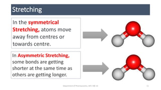
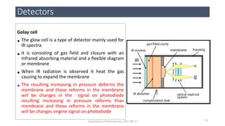
• The IR energy absorption corresponds to specific modes ; It
corresponding to combination of atomic movements.
• Vibrational force are two type
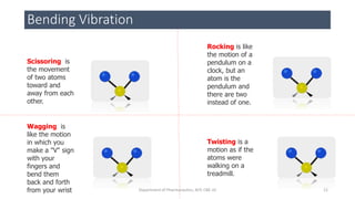
• BENDING VIBRATIONAL FORCE [change in bond angles]
• Scissoring
• Wagging
• Rocking
• Twisting
• STRETCHING VIBRATIONAL FORCE [Change in bond length]
• Symmetrical & Asymmetrical
• It corresponds to vibrational and rotational force
10Department of Pharmaceutics, KCP, CBE-32
11. Stretching
In the symmetrical
Stretching, atoms move
away from centres or
towards centre.
In Asymmetric Stretching,
some bonds are getting
shorter at the same time as
others are getting longer.
11Department of Pharmaceutics, KCP, CBE-32
12. Bending Vibration
Scissoring is
the movement
of two atoms
toward and
away from each
other.
Wagging is
like the motion
in which you
make a "V" sign
with your
fingers and
bend them
back and forth
from your wrist
Rocking is like
the motion of a
pendulum on a
clock, but an
atom is the
pendulum and
there are two
instead of one.
Twisting is a
motion as if the
atoms were
walking on a
treadmill.
12Department of Pharmaceutics, KCP, CBE-32
13. HOOKE's LAW
• Definition
Hooke’s law is a law of physics that states that the force needed to extend or
compress spring by some distance
• In Infrared spectroscopy, Hooke’s law is applied to determine the
molecules based on the peak range
• A vibrating bond in the IR can be compared to the physical model of a
vibrating spring system that can be described Hooke’s law of
harmonic oscillation.
• Using the force constant K and masses of two atoms M, then the
absorption should change as the properties of the system
13Department of Pharmaceutics, KCP, CBE-32
14. • In physics Hooke’s law used to determine force of spring
• Note : This is for understanding purpose
Cont...
14Department of Pharmaceutics, KCP, CBE-32
15. Cont ....
• In Infrared spectroscopy, Hooke's law is applied to determine the
vibrational stretching of atoms
15Department of Pharmaceutics, KCP, CBE-32
16. Cont...
• Hooke’s law given as
Where K is force constant (dynes /cm) ie bond strength
C = velocity of light
µ = m1 m2 / m1 + m2 ( where m is mass of atom )
16Department of Pharmaceutics, KCP, CBE-32
17. Determination of %Transmission
• Hooke’s law used in the determine the frequency required to stretch
the molecules
• Example:
The frequency required to stretch the C – H bond can be
calculated by using the Hooke’s formula.
k = 5x10⁵ dyne/cm
m1 = C = 12/6.02x1023 = 1.99x10⁻²³ g
m2 = H = 1/6.02x1023 = 0.167x10⁻²³ g
c = 3x1010 cm/sec
Single bond = 5x10⁵ dyne/cm
Double bond = 10x105 dyne/cm
Triple bond = 15x105 dyne/cm
17Department of Pharmaceutics, KCP, CBE-32
20. Sampling technique for Powders Sample
• Infrared spectroscopy incorporate several type of measurement
method like Diffuse reflectance method and attenuated total
reflection method
• The methods are selected according to sample form
• The KBr Method and Nujol method is classical method. How ever in
FTIR has become more common, The diffuse reflection method has
become more common and its widely used.
• The attenuated total reflection is method that allows direct
measurement of powder sample
20Department of Pharmaceutics, KCP, CBE-32
21. Cont....
The following methods are analysis the powder sample
• KBr Pellets method
• Nujol Method
• Diffuse Reflection Method
• Attenuated Total Reflection
21Department of Pharmaceutics, KCP, CBE-32
22. KBr Pellets method
• KBr Pellets Method:
• Alkali Halides are used in this method because it form a
transparent sheet when it is subject high pressure and this
transparent pellets allow the IR.
• KBr is the commonest alkali halide used in the pellets.
Cesium Iodide may be used to measure the lR region of 400
to 250 cm-1 wave number region
22Department of Pharmaceutics, KCP, CBE-32
23. Preparation Procedure
The size of pellets is
13mm in diameter
0.1 to 1.0% sample is
well mixes into 200 to
250mg fine KBr
The mixture is
pulverized and passed
into 200 mesh sieve
and dry at 100° C for
2 – 3 hours
The mixture is
compressed into
pellets
The compressional
force approx 8 tons is
applied under vacuum
for several minutes to
form a transparent
pellets
Degassing is applied
to remove all the air
and moisture
Inadequate vacuum
results is breaking and
scatter of IR
When measuring
background can be
measured with an
empty pellets without
sample and the only
analysis the pellets
containing sample
23
Department of Pharmaceutics, KCP, CBE-32
27. Diffuse Reflectance Method
• Some IR are undergoes specular reflection at the powder surface and other light enter into sample at the
surface of the powder and and reappear after repeated transmission and reflection.
• In this Spectrum is obtained from diffuse reflected light
• Procedure
KBr powder are measured first (without sample)
0.1 to 10% of KBr Powder are diluted with sample 5 to 10 ng and it is packed into micro cup
Measure under IR
Advantage
• Little sample only required
• No need to form pellets
• Fast and easy cleanup
27
Department of Pharmaceutics, KCP, CBE-32
28. Pulverized
the sample
and 1g of
sample is
taken
To this add
one or two
drops of
liquid
paraffin
Mix to
distribute a
sample in
liquid
paraffin
Apply the
Paste to
Liquid cell
Measure
with IR
Note : Since
liquid paraffin
exhibit spectra
3000 ,2800,
1460, 1375,
730 cm-1
Nujol Method
28
Department of Pharmaceutics, KCP, CBE-32
30. •This method is used to measure the IR spectra of sample is KBr or in
Liquid paraffin
•In this method the sample is pressed against the High reflective index
prism (It made up of Zinc selenide or Germanium)
•A Sample is mixed with KBr / liquid paraffin, and background data
collected without sample and then sample along KBr is analysed.
Advantages
•Faster Sampling
• Improving sample to sample reproducibility
• Minimize the spectral variation
Cont...
30
Department of Pharmaceutics, KCP, CBE-32
31. Sampling Method for Liquid Sample
• The liquid samples are measured by transmission method.
• In the transmission method two types of cells are commonly
used
1. Liquid Cell
2. Fixed Cell
31Department of Pharmaceutics, KCP, CBE-32
32. •In the liquid cell, the sample droplets are placed on to the sodium
chloride or potassium bromide the appitude plate and sandwich in a
under another appitude plate such that no gas bubbles are trapped.
•The thickness is adjusted according to sample by using the screws.
•It is demountable sales or disassemble
•This is very useful to measuring the high viscous liquid an unsuitable for
low boiling point
Liquid Cell
32Department of Pharmaceutics, KCP, CBE-32
35. •The fixed cell are used to measure a volatile sample
or a solution dissolved in appropriate solvent
•The reason furious in stopper at the sample and let
others a sample introduction and extraction from
the cell and cell washing without this disassemble.
•It is leak free
•It is used for analyser volatile sample
•the appropriate thickness is above 0.1 mm
thickness
Fixed Cell
35Department of Pharmaceutics, KCP, CBE-32
36. Sampling Method for Gas Sample
• The Gas sample are analysed by using and gas cell.
• There are two types of gas cell.
• 5cm and 10cm Gas cell
• Each cell body is consist of Glass material
• End of the cell is covered with potassium bromide window it
allow infrared radiation.
• 5 cm cell capacity is 42 ml and 10cm gas cell capacity is 98ml
36Department of Pharmaceutics, KCP, CBE-32
39. INSTRUMENTATION
• In the IR spectroscopy there are two types of instruments are
most commonly used, they are
1.Dispersive Infrared Spectrscopy
2.Fourier transform IR spectra
39Department of Pharmaceutics, KCP, CBE-32
40. Dispersive Infrared Spectrscopy
• The Dispersive infrared spectroscopy it has two type
single and double beam spectroscopy
• In the single beam Spectrscopy reference standard
cannot be analysed
• But in the double beam spectroscopy both reference
sample and standard Sample are analysed.
40Department of Pharmaceutics, KCP, CBE-32
41. Radiation source
Nerst Glower
Nichrome Wire
Globar
Monochromater
Prism
Grating
Detectors
Bolometer
Thermocouple
Golay cell
Pyroelectric detectors
Components of IR Instruments
41Department of Pharmaceutics, KCP, CBE-32
42. Nerst glower :
• It was used to provide a continuous source of IR radiation i.e near IR
• It Is Composed of
• Zirconium oxide 90% w/w
• Yttrium oxide 7% w/w
• Erbium oxide 3% w/w
• When it is heated electrically at the temperature of 2300°K (2026°C)
Radiation source
42Department of Pharmaceutics, KCP, CBE-32
44. Nichrome Wire:
• Nichrome is consist of Nickel
– Chromium wire
• It is 10 cm long wire
• When it is heated 1100°C it
can emita IR radiation ( Near
IR)
Radiation source
44Department of Pharmaceutics, KCP, CBE-32
45. GloBar:
• Globar is made up of Silicon Carbide
• It is 20-50mm in length and 5-10mm width
• It is heated by Electrically
• The silicon carbide rod is heated to temperature 1000°C to 1650°C
• Interference filter to produce a radiation between 4 to 15 micrometre
Radiation source
45Department of Pharmaceutics, KCP, CBE-32
46. Prism and Differential Grating:
• In traditional IR instruments monochromator are construct with Nacl or KBr Prism but
disadvantages of Prism is hygroscopic in Nature.
• For these reasons prism are not used instead of that diffraction grating have used
• Grating provide a Higher resolution power than Prism
• Even so most of grating operate automatically change around IR 2000 cm
Monochromator
46Department of Pharmaceutics, KCP, CBE-32
47. Working - Double beam IR Spectra
47Department of Pharmaceutics, KCP, CBE-32
48. Bolometer
•Bolometer are detectors used to measure the
Infrared Radiation
•They are very sensitive to thermal radiation
and are predominantly used in IR spectra
between 10 to 5000 micrometres
•Any thermal radiation that impinges upon the
detector will cause a temperature change. This
will cause change in resistance which is
amplified and measured as a voltage difference
Detectors
48Department of Pharmaceutics, KCP, CBE-32
49. Fields of application:
• Aerospace/astronomy
• Defense and security
• Firefighting
• Inspection/maintenance
• Laser beam profiling
• Medical
• Night vision
• Remote sensing
• Search and rescue
• Transportation
Detectors
49Department of Pharmaceutics, KCP, CBE-32
50. Thermocouple
•The thermocouple is made by welding
together of a two wires of metal 1 and
2
•The metals are Bismuth and antimony
•There are two junction hot junction
and cold junction.
•The IR radiation falls on hot zone and
the potential difference between the
hot and constant temperature zone is
measured and sent to amplifier.
Detectors
50Department of Pharmaceutics, KCP, CBE-32
51. Detectors
51Department of Pharmaceutics, KCP, CBE-32
Thermocouple Applications
Some of the applications of thermocouple include the
following.
•These are used as the temperature sensors in
thermostats in offices, homes, offices & businesses.
•These are used in industries for monitoring
temperatures of metals in iron, aluminum, and
metal.
•These are used in the food industry for cryogenic
and Low-temperature applications. Thermocouples
are used as a heat pump for performing
thermoelectric cooling.
•These are used to test temperature in the
chemical plants, petroleum plants.
•These are used in gas machines for detecting the
pilot flame
52. Golay cell
• The glow cell is a type of detector mainly used for
IR spectra
• It is consisting of gas field and closure with an
infrared absorbing material and a flexible diagram
or membrane
• When IR radiation is observed it heat the gas
causing to expand the membrane
• The resulting increasing in pressure deforms the
membrane and these reforms in the membrane
will be changes in the signal on photodiode
resulting increasing in pressure reforms than
membrane and these reforms in the membrane
will be changes engine signal on photodiode
Detectors
52Department of Pharmaceutics, KCP, CBE-32
53. 1. Infrared spectroscopy https://www.ru.nl/systemschemistry/equipment/optical-
spectroscopy/infrared/
2. Vogel’s Text book of Quantitative chemical analysis by Jerrfy et.al
3. Infrared Spectroscopy theory and Interpretation of IR https://slideplayer.com/slide/8335494/
4. Fundamental of IR spectroscopy https://www.shimadzu.com/an/ftir/support/ftirtalk/index.html
5. Attenuated total reflection sampling
https://www.google.com/url?sa=t&source=web&rct=j&url=https://www.utsc.utoronto.ca/~traceslab/
ATR_FTIR.pdf&ved=2ahUKEwirjobmuujfAhUVAXIKHefqD5YQFjAZegQIAxAB&usg=AOvVaw1p-7DG-
txypQxhy1wqJSQq&cshid=1547303582941
6. Practical Sampling Techniques for Infrared Analysis edited by Patricia B. Coleman
https://books.google.co.in/books?hl=en&lr=&id=JY_H61INJqMC&oi=fnd&pg=PA2&dq=sample+handli
ng+techniques+infrared+spectroscopy&ots=DCbrdbqsy2&sig=KkZdnDxPzvitsd4lF14pGlzhA6w#v=onep
age&q=sample%20handling%20techniques%20infrared%20spectroscopy&f=false
7. Introduction to Infrared Spectroscopy April 2012, Research gate
https://www.researchgate.net/publication/224831013
Reference
53Department of Pharmaceutics, KCP, CBE-32
54. 7. FT-IR vs. Dispersive Infrare Theory of Infrared Spectroscopy Instrumentation
https://www.google.com/url?sa=t&source=web&rct=j&url=http://www.thermo.com.cn/Resou
rces/200802/productPDF_21615.pdf&ved=2ahUKEwj8qPnNkevfAhUErI8KHUerBjYQFjAAegQIA
xAB&usg=AOvVaw2oR7zNrc-XIvb4HibRdXjc
8. Near Infrared Spectroscopy: fundamentals, practical aspects and analytical applications ,
Journal of the Brazilian Chemical Society vol 14 Sau paul April 2003
http://www.scielo.br/scielo.php?pid=S0103-50532003000200006&script=sci_arttext
9. Application of Non-Dispersive Infrared (NDIR) Spectroscopy to the Measurement of
Atmospheric Trace gases
https://www.google.com/url?sa=t&source=web&rct=j&url=https://ir.canterbury.ac.nz/bitstrea
m/handle/10092/2160/Thesis_fulltext.pdf%3Bsequence%3D1&ved=2ahUKEwj6zP7uherfAhXFL
48KHWOLCIsQFjATegQIAhAB&usg=AOvVaw06IQX-ysQupfLLm0HLASCZ
10. Monochromator
https://www.shimadzu.com/an/uv/support/fundamentals/monochromators.html
11. Golay cell https://en.m.wikipedia.org/wiki/Golay_cell
Reference
54Department of Pharmaceutics, KCP, CBE-32
55. Reference
12. Pyroelectric Detector https://www.infratec.in/sensor-division/service-
support/glossary/pyroelectric-detector/
13. Thermal detector https://www.slideshare.net/mobile/sampathramaswamy1/thermal-
detectors-of-ir
14. Introduction and calculations of IR https://www.chromacademy.com/lms/sco533/06-
infrared-spectroscopy-calculation-molecular-vibrations.html
15. Hand book of thermal detector Chapter 7
https://www.google.com/url?sa=t&source=web&rct=j&url=https://www.hamamatsu.
com/resources/pdf/ssd/e07_handbook_Thermal_detectors.pdf&ved=2ahUKEwi1gc7Y
k-vfAhWGNY8KHYQaAnkQFjAAegQIBRAB&usg=AOvVaw0NY1YMZO7MkrCMtm3-Gzek
55Department of Pharmaceutics, KCP, CBE-32