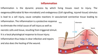
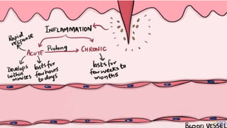
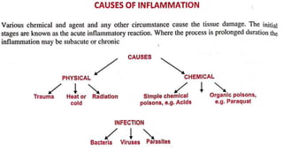
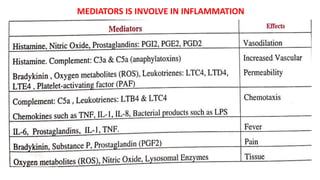
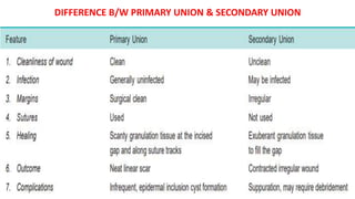
PATHOPHYSIOLOGY
b.Pharma 2nd semester unit-1ST
INFLAMMATION
Mr. Bulet Kumar Gupta
Assistant Professor
Sai College of Pharmacy, Mau
Inflammation
Inflammation is the dynamic process by which living tissues react to injury. The
exogenous(Microbial & Non-microbials) and endogenous (Cell signalling, injured tissue) stimulus
is lead to a cell injury, cause complex reactions in vascularised connective tissue leading to
inflammation. The inflammation is a protective response
to eliminate the initial cause of cell injury as well as
necrotic cells and tissue, resulting from triggered stimuli.
It is a local physiological response to tissue injury.
Inflammation thus helps in clear infection and repairs
and also does the healing of the wound.
Types of Inflammation
Inflammation is mainly two types—
A) Acute inflammation
B) Chronic inflammation
Vascular Events
1- Vasoconstriction
2- Vasodilation
3- Increase permeability
4- Stasis
Chronic inflammation
Chronic inflammation is an inflammatory response of prolonged duration: weeks, months or
even indefinitely by a continuous causative stimulus to inflammation in the tissue. The
inflammatory process causes tissue damage and is accompanied by simultaneous healing and
repair.
Chronic inflammation can be caused by one of the following 3 ways—
1. Chronic inflammation following acute inflammation- When the tissue destruction is
extensive, or the bacteria survive and persist in small numbers at the site of acute
inflammation e.g. in osteomyelitis, pneumonia terminating in lung abscess.
2. Recurrent attacks of acute inflammation-- When repeated bouts of acute inflammation
culminate in chronicity of the process e.g. in recurrent urinary tract infection leading to chronic
pyelonephritis, repeated acute infection of gallbladder leading to chronic cholecystitis.
3) Chronic inflammation starting de novo- When the infection with organisms of low
pathogenicity is chronic from the beginning e.g. infection with Mycobacterium tuberculosis.
TYPES OF CHRONIC INFLAMMATION
1) NON-SPECIFIC- When the irritant substance produces a nonspecific chronic inflammatory
reaction with formation of granulation tissue & healing by fibrosis.
e.g. Osteomyelitis & Chronic ulcer.
2) SPECIFIC- Each irritant & organism produce a characteristic microscopic picture called
granuloma. E.g. Tuberculosis & Leprosy.
GRANULOMA
Chronic specific inflammation forming a tumour like mass and characterized microscopically by
focal accumulation of large no. of chronic inflammatory cells.
ETIOLOGY OF CHRONIC INFLAMMATION
May progress from acute inflammation, if the acute process cannot be recovered because of a
persistence of the agent or interference with normal healing. However, Some specific injurious
agents which typically cause chronic inflammation, such as
(a) Persistent viral infections (e.g. caprine arthritis encephalitis virus).
(b) Persistent microbial infections (e.g. Mycobacterium spp. and fungi).
(c) Prolonged exposure to some toxic agents (e.g. asbestos).
(d) Few autoimmune diseases (e.g. lupus erythematosus).
ALTERATION IN VASCULAR PERMEABILITY AND BLOOD FLOW
In the early stages, oedema fluid, fibrin and neutrophil polymorphs accumulate in the
extracellular spaces of the damaged tissue. The presence of the cellular component, neutrophil
polymorph, is essential for a histological diagnosis of acute inflammation.
The acute inflammatory response involves three processes:
1. Changes in vessel Caliber and consequently flow.
2. Increased vascular permeability and formation of the fluid exudates.
3. Formation of the cellular exudate - emigration of the neutrophil polymorphs into the
extravascular space.
MIGRATION OF WBC'S OR LEUKOCYTES
The main inflammatory cells are polymorphonuclear leukocytes (neutrophils/heterophils,
eosinophils, basophils), mast cells, mononuclear cells (monocytes/macrophages, lymphocytes,
plasma cells), and platelets. Most cells, except for plasma cells, macrophages & mast cells, are
normal inhabitants of the circulating blood.
The total leukocyte (WBC) count in peripheral blood and the relative proportions of different
white blood cells may be greatly modified in the systemic response to inflammation and can,
therefore, be used as a diagnostic tool.
The migration of leukocytes involves in following Steps. These are---
1) Margination (2) Rolling & Adhesion
(3) Emigration (4) Chemotaxis
(5) Phagocytosis
1). Margination: In the normal blood circulation, White blood cells are travelled generally to the
central (axial) stream in blood vessels, and do not flow in the peripheral (plasmatic) zone near to
the endothelium.
2). Rolling & Adhesion: Marginated leukocytes line of the endothelium. Leukocytes start to
become adhered to the surface of endothelial cells through various adhesion molecules.
3). Emigration of leukocyte: The process by which leukocytes escape from the blood to
perivascular tissues, moving to the site of inflammation.
4). Chemotoxins: The initial margination of neutrophils and mononuclears is potentiated by
slowing of blood flow and by increased 'stickiness' of the endothelial surface. After penetration
of the vessel wall, the subsequent movement of the leucocytes is controlled by chemotaxis.
5) Phagocytosis: Phagocytosis is a process of engulf, kill and degrade foreign material; most
commonly bacteria. In this process, the neutrophils and macrophages clear the injurious agent
MEDIATORS OF INFLAMMATION
A chemical mediator is any messenger that acts on blood vessels, inflammatory cells or other
cells to contribute to an inflammatory response.
Mediator of inflammation originates from plasma or cells.
When mediators in plasma, are in an inactive state and must be activated and when in cells, they
are often within granules and need to be secreted or they are synthesized in response to a
stimulus.
The production of active mediators is triggered by microbial products or host proteins
(e.g. cytokines).
However, some inflammatory mediators have direct enzymatic activity; most require binding to
specific receptors on target cells for biologic activity.
Some mediator can' stimulate the release of other mediators by target cells.
MEDIATORS IS INVOLVE IN INFLAMMATION
BASIC PRINCIPLE OF WOUND HEALING IN THE SKIN
Injury to tissue may result in cell death and tissue destruction.
Healing is the body response to injury in an attempt to restore normal structure and function.
It involves two processes:---
A. Regeneration: When healing takes place by proliferation of parenchymal cells and usually
result in complete restoration of the original tissues.
B. Repair: When the healing takes place by proliferation of connective tissue elements resulting
in fibrosis and scarring.
 Wound healing can be accomplished in one of the following two ways:
1) Healing by first intention (primary union)
2) Healing by second intention (secondary union).
1) Healing by First Intention (Primary Union)
This is defined as healing of a wound which has the following characteristics:
i) Clean and uninfected
ii) Surgically incised
iii) Without much loss of cells and tissue
iv) Edges of wound are approximated by surgical sutures.
The sequence of events in primary union is illustrated---
1. Initial Haemorrhage- Immediately after injury, the space between the approximated surfaces
of incised wound is filled with blood which then clots and seals the wound against
dehydration and infection.
2. Acute inflammatory response- This occurs within 24 hours with appearance of polymorphs
from the margins of incision. By 3rd day, polymorphs are replaced by macrophages.
3. Epithelial changes- The basal cells of epidermis from both the cut margins start proliferating
and migrating towards incisional space in the form of epithelial spurs.
A well approximated wound is covered by a layer of epithelium in 48 hours.
4. Organisation- By 3rd day, fibroblasts also invade the wound area. By 5th day, new collagen
fibrils start forming which dominate till healing is completed.
5. Suture tracks- Each suture track is a separate wound and incites the same phenomena as in
healing of the primary wound.
2) Healing by Second Intention (Secondary Union)
This is defined as healing of a wound having the following characteristics:
i) open with a large tissue defect, at times infected
ii) having extensive loss of cells and tissues
iii) the wound is not approximated by surgical sutures but is left open.
The basic events in secondary union are similar to primary union but differ in having a larger
tissue defect which has to be bridged.
The sequence of events in secondary union is illustrated---
1. Initial Haemorrhage- As a result of injury, the wound space is filled with blood and fibrin clot
which dries.
2. Inflammatory phase- There is an initial acute inflammatory response followed by appearance
of macrophages which clear off the debris as in primary union.
3. Epithelial changes- As in primary healing, the epidermal cells from both the margins of
wound proliferate and migrate into the wound in the form of epithelial spurs till they meet in the
middle and re-epithelialise the gap completely.
4. Granulation tissue- Main bulk of secondary healing is by granulations.
5. Wound contraction- Contraction of wound is an important feature of secondary healing, not
seen in primary healing.
Complications of Wound Healing
During the course of healing, following complications may occur:
1. Infection of wound- due to entry of bacteria delays the healing.
2. Implantation (epidermal) cyst formation- may occur due to persistence of epithelial cells in
the wound after healing.
3. Pigmentation- Healed wounds may at times have rust-like colour due to staining with
haemosiderin. Some coloured particulate material left in the wound may persist and impart
colour to the healed wound.
4. Deficient scar formation- This may occur due to inadequate formation of granulation tissue
DIFFERENCE B/W PRIMARY UNION & SECONDARY UNION
inflammation ppt BKG.pptx Acute & chronic

inflammation ppt BKG.pptx Acute & chronic

  • 1.
    PATHOPHYSIOLOGY b.Pharma 2nd semesterunit-1ST INFLAMMATION Mr. Bulet Kumar Gupta Assistant Professor Sai College of Pharmacy, Mau
  • 2.
    Inflammation Inflammation is thedynamic process by which living tissues react to injury. The exogenous(Microbial & Non-microbials) and endogenous (Cell signalling, injured tissue) stimulus is lead to a cell injury, cause complex reactions in vascularised connective tissue leading to inflammation. The inflammation is a protective response to eliminate the initial cause of cell injury as well as necrotic cells and tissue, resulting from triggered stimuli. It is a local physiological response to tissue injury. Inflammation thus helps in clear infection and repairs and also does the healing of the wound.
  • 9.
    Types of Inflammation Inflammationis mainly two types— A) Acute inflammation B) Chronic inflammation
  • 10.
    Vascular Events 1- Vasoconstriction 2-Vasodilation 3- Increase permeability 4- Stasis
  • 12.
    Chronic inflammation Chronic inflammationis an inflammatory response of prolonged duration: weeks, months or even indefinitely by a continuous causative stimulus to inflammation in the tissue. The inflammatory process causes tissue damage and is accompanied by simultaneous healing and repair. Chronic inflammation can be caused by one of the following 3 ways— 1. Chronic inflammation following acute inflammation- When the tissue destruction is extensive, or the bacteria survive and persist in small numbers at the site of acute inflammation e.g. in osteomyelitis, pneumonia terminating in lung abscess. 2. Recurrent attacks of acute inflammation-- When repeated bouts of acute inflammation culminate in chronicity of the process e.g. in recurrent urinary tract infection leading to chronic pyelonephritis, repeated acute infection of gallbladder leading to chronic cholecystitis.
  • 13.
    3) Chronic inflammationstarting de novo- When the infection with organisms of low pathogenicity is chronic from the beginning e.g. infection with Mycobacterium tuberculosis. TYPES OF CHRONIC INFLAMMATION 1) NON-SPECIFIC- When the irritant substance produces a nonspecific chronic inflammatory reaction with formation of granulation tissue & healing by fibrosis. e.g. Osteomyelitis & Chronic ulcer. 2) SPECIFIC- Each irritant & organism produce a characteristic microscopic picture called granuloma. E.g. Tuberculosis & Leprosy. GRANULOMA Chronic specific inflammation forming a tumour like mass and characterized microscopically by focal accumulation of large no. of chronic inflammatory cells.
  • 15.
    ETIOLOGY OF CHRONICINFLAMMATION May progress from acute inflammation, if the acute process cannot be recovered because of a persistence of the agent or interference with normal healing. However, Some specific injurious agents which typically cause chronic inflammation, such as (a) Persistent viral infections (e.g. caprine arthritis encephalitis virus). (b) Persistent microbial infections (e.g. Mycobacterium spp. and fungi). (c) Prolonged exposure to some toxic agents (e.g. asbestos). (d) Few autoimmune diseases (e.g. lupus erythematosus).
  • 18.
    ALTERATION IN VASCULARPERMEABILITY AND BLOOD FLOW In the early stages, oedema fluid, fibrin and neutrophil polymorphs accumulate in the extracellular spaces of the damaged tissue. The presence of the cellular component, neutrophil polymorph, is essential for a histological diagnosis of acute inflammation. The acute inflammatory response involves three processes: 1. Changes in vessel Caliber and consequently flow. 2. Increased vascular permeability and formation of the fluid exudates. 3. Formation of the cellular exudate - emigration of the neutrophil polymorphs into the extravascular space.
  • 19.
    MIGRATION OF WBC'SOR LEUKOCYTES The main inflammatory cells are polymorphonuclear leukocytes (neutrophils/heterophils, eosinophils, basophils), mast cells, mononuclear cells (monocytes/macrophages, lymphocytes, plasma cells), and platelets. Most cells, except for plasma cells, macrophages & mast cells, are normal inhabitants of the circulating blood. The total leukocyte (WBC) count in peripheral blood and the relative proportions of different white blood cells may be greatly modified in the systemic response to inflammation and can, therefore, be used as a diagnostic tool. The migration of leukocytes involves in following Steps. These are--- 1) Margination (2) Rolling & Adhesion (3) Emigration (4) Chemotaxis (5) Phagocytosis
  • 21.
    1). Margination: Inthe normal blood circulation, White blood cells are travelled generally to the central (axial) stream in blood vessels, and do not flow in the peripheral (plasmatic) zone near to the endothelium. 2). Rolling & Adhesion: Marginated leukocytes line of the endothelium. Leukocytes start to become adhered to the surface of endothelial cells through various adhesion molecules. 3). Emigration of leukocyte: The process by which leukocytes escape from the blood to perivascular tissues, moving to the site of inflammation. 4). Chemotoxins: The initial margination of neutrophils and mononuclears is potentiated by slowing of blood flow and by increased 'stickiness' of the endothelial surface. After penetration of the vessel wall, the subsequent movement of the leucocytes is controlled by chemotaxis. 5) Phagocytosis: Phagocytosis is a process of engulf, kill and degrade foreign material; most commonly bacteria. In this process, the neutrophils and macrophages clear the injurious agent
  • 22.
    MEDIATORS OF INFLAMMATION Achemical mediator is any messenger that acts on blood vessels, inflammatory cells or other cells to contribute to an inflammatory response. Mediator of inflammation originates from plasma or cells. When mediators in plasma, are in an inactive state and must be activated and when in cells, they are often within granules and need to be secreted or they are synthesized in response to a stimulus. The production of active mediators is triggered by microbial products or host proteins (e.g. cytokines). However, some inflammatory mediators have direct enzymatic activity; most require binding to specific receptors on target cells for biologic activity. Some mediator can' stimulate the release of other mediators by target cells.
  • 23.
    MEDIATORS IS INVOLVEIN INFLAMMATION
  • 24.
    BASIC PRINCIPLE OFWOUND HEALING IN THE SKIN Injury to tissue may result in cell death and tissue destruction. Healing is the body response to injury in an attempt to restore normal structure and function. It involves two processes:--- A. Regeneration: When healing takes place by proliferation of parenchymal cells and usually result in complete restoration of the original tissues. B. Repair: When the healing takes place by proliferation of connective tissue elements resulting in fibrosis and scarring.  Wound healing can be accomplished in one of the following two ways: 1) Healing by first intention (primary union) 2) Healing by second intention (secondary union).
  • 25.
    1) Healing byFirst Intention (Primary Union) This is defined as healing of a wound which has the following characteristics: i) Clean and uninfected ii) Surgically incised iii) Without much loss of cells and tissue iv) Edges of wound are approximated by surgical sutures. The sequence of events in primary union is illustrated--- 1. Initial Haemorrhage- Immediately after injury, the space between the approximated surfaces of incised wound is filled with blood which then clots and seals the wound against dehydration and infection. 2. Acute inflammatory response- This occurs within 24 hours with appearance of polymorphs from the margins of incision. By 3rd day, polymorphs are replaced by macrophages.
  • 26.
    3. Epithelial changes-The basal cells of epidermis from both the cut margins start proliferating and migrating towards incisional space in the form of epithelial spurs. A well approximated wound is covered by a layer of epithelium in 48 hours. 4. Organisation- By 3rd day, fibroblasts also invade the wound area. By 5th day, new collagen fibrils start forming which dominate till healing is completed. 5. Suture tracks- Each suture track is a separate wound and incites the same phenomena as in healing of the primary wound.
  • 27.
    2) Healing bySecond Intention (Secondary Union) This is defined as healing of a wound having the following characteristics: i) open with a large tissue defect, at times infected ii) having extensive loss of cells and tissues iii) the wound is not approximated by surgical sutures but is left open. The basic events in secondary union are similar to primary union but differ in having a larger tissue defect which has to be bridged. The sequence of events in secondary union is illustrated--- 1. Initial Haemorrhage- As a result of injury, the wound space is filled with blood and fibrin clot which dries. 2. Inflammatory phase- There is an initial acute inflammatory response followed by appearance of macrophages which clear off the debris as in primary union.
  • 28.
    3. Epithelial changes-As in primary healing, the epidermal cells from both the margins of wound proliferate and migrate into the wound in the form of epithelial spurs till they meet in the middle and re-epithelialise the gap completely. 4. Granulation tissue- Main bulk of secondary healing is by granulations. 5. Wound contraction- Contraction of wound is an important feature of secondary healing, not seen in primary healing.
  • 29.
    Complications of WoundHealing During the course of healing, following complications may occur: 1. Infection of wound- due to entry of bacteria delays the healing. 2. Implantation (epidermal) cyst formation- may occur due to persistence of epithelial cells in the wound after healing. 3. Pigmentation- Healed wounds may at times have rust-like colour due to staining with haemosiderin. Some coloured particulate material left in the wound may persist and impart colour to the healed wound. 4. Deficient scar formation- This may occur due to inadequate formation of granulation tissue
  • 30.
    DIFFERENCE B/W PRIMARYUNION & SECONDARY UNION