
Recommended
Recommended
More Related Content
What's hot
What's hot (20)
Similar to unit 4 immunoblotting technique complete.pptx
Similar to unit 4 immunoblotting technique complete.pptx (20)
More from BkGupta21
More from BkGupta21 (10)
Recently uploaded
Recently uploaded (20)
unit 4 immunoblotting technique complete.pptx
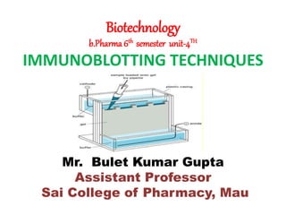
- 1. Biotechnology b.Pharma 6th semester unit-4TH IMMUNOBLOTTING TECHNIQUES Mr. Bulet Kumar Gupta Assistant Professor Sai College of Pharmacy, Mau
- 2. DEFINITION OF BLOTTING • Visualization of specific DNA , RNA & protein among many thousands of contaminating molecules requires the convergence of number of techniques which are collectively termed BLOT transfer.
- 3. IMMUNOBLOTTING • Viral antigens are detected with a polyclonal or a MAb onto nitrocellulose paper. • After incubation, the protein bands (immune complexes) are visualized with peroxides-conjugated protein and a color reagent. • A color develops in the bands where antibody binds to the antigen. • Immunoblotting assay mixture of this two technique. WESTERN BLOTTING •Western blotting is based on the principles of immunochromatography where proteins were separated into poly acryl amide gel according to the isoelectric point and molecular weight.
- 4. •A technique for detecting specific proteins separated by electrophoresis by use of labeled antibodies. •Immunoblotting is performed chiefly in diagnostic laboratories to identify the desirable protein antigens in complex mixtures. • An improved immunoblot method Zestern analysis, is able to address this issue without the electrophoresis step, thus significantly improving the efficiency of protein analysis. • Other related techniques include dot blot analysis, zestern analysis, immunohistochemistry where antibodies are used to detect proteins in tissues and cells by immunostaining and enzyme-linked immunosorbent assay (ELISA)
- 5. ELISA Enzyme-linked immunosorbent assay (ELISA): ELISA stands for "enzyme- linked immunosorbent assay." This is a rapid immunochemical test that involves an enzyme (a protein that catalyzes a biochemical reaction). It also involves an antibody or antigen (immunologic molecules). ELISA tests are utilized to detect substances that have antigenic properties, primarily proteins (as opposed to small molecules and ions such as glucose and potassium). Some of these include hormones, bacterial antigens and antibodies. TYPES OF ELISA 1- Direct ELISA 2- Indirect ELISA 3- Sandwich ELISA 4- Competitive ELISA
- 7. Diseases That Can Be Diagnosed Using ELISA ELISA can be used to detect some of these conditions: •Ebola •Pernicious anaemia •AIDS •Rotavirus •Lyme disease •Syphilis •Toxoplasmosis •Zika virus •Carcinoma of the epithelial cells
- 8. Applications of ELISA •The applications of ELISA are discussed below: •The presence of antibodies and antigens in a sample can be determined. •It is used in the food industry to detect any food allergens present. •To determine the concentration of serum antibody in a virus test. •During a disease outbreak, to evaluate the spread of the disease, e.g. during recent COVID-19 outbreak, rapid testing kits are being used to determine presence of antibodies in the blood sample.
- 10. The technique was first described by Harry Towbin in 1979 and named by W. Neal Burnette in 1981. The procedure of western blotting can be understood by the following points: Gel Electrophoresis: The protein samples are separated using gel electrophoresis. SDS-PAGE is commonly used for this step. Transfer: The separated proteins are transferred on a solid support as nitrocellulose or polyvinylidene difluoride membrane. Total Protein Staining: The proteins transferred on the membrane are visualised by staining the membrane using dyes such as Ponceau S, Coomasie R-350 or amido black.
- 11. Blocking: This step is done to block the interaction between the membrane and the antibody that will be applied in the next step. Since the target molecule and antibodies are both proteins, there are chances that the membrane might interact with the antibody. Incubation: An antibody with a reporter enzyme is probed on the membrane. A substrate that is specific to the enzyme is applied on the surface. If binding happens, a colourimetric reaction is produced. Detection and Visualization: The unbound probes are washed away and the gel is visualised for the bound proteins.
- 12. Applications •Detection of particular protein from a mixture of proteins. •Size and amount estimate of proteins in the mixture. •Verification following a high sensitivity ELISA test for diagnosis of Lyme, HIV infection, BSE, HBV and so on. •Detect condensed isoforms of proteins as well as tagged proteins. Advantage •Effective early diagnostic tool. •Detect minimal immunogenic response form virus or bacteria. •Requires fewer antibodies for testing. •Detect specific protein from a large mixture of different proteins. (Even more than 300,000).
- 13. Disadvantages •Requires specific primary antibodies to perform test on desired protein of interest. •Challenging and hence requires well trained staffs. •Poorer results as antibodies may revel off-target bindings. •Detecting and imaging the results can be expensive as equipment cost is high.
- 14. Southern blotting Southern blotting is a molecular biology technique used for DNA detection, characterization, and quantification. An example of RFLP(restriction fragment length polymorphism), southern blotting can be defined as an analytical technique for identifying specific sequences of DNA by separating fragments on a gel and transferring them to a second medium (carrier membrane) on which hybridization testing may be carried out. During southern blotting, the DNA fragments are immobilized as a result, the membrane carries a semi-permanent reproduction of the banding pattern of the gel. The DNA are then exposed to hybridization analysis allowing bands with sequence resemblance to a labeled probe to be identified.
- 16. Procedure of Southern Blotting 1. Extract and purify DNA from cells We separate the DNA to be tested from the rest of the cellular material in the nucleus. We then incubate the specimen with detergent to promote cell lysis (frees cellular proteins and DNA). Proteins are removed through organic and non-organic extraction. We then use alcohol precipitation to purify the DNA from the solution. Visible DNA fibers are removed and suspended in buffer. 2. DNA fragmentation We use restriction endonuclease enzyme to break long nucleotide sequence into smaller fragments for purification or identification process.
- 17. 3. Gel electrophoresis Sorts the complex mixture of DNA fragments according to size. The percentage and size of the gel to be used must be determined. Gels consist of microscopic pores and are solid (agarose/Polyacrylmide) Generally, 0.7 – 2% gel is considered to be adequate for most of the applications. Nucleic acids have negative charge and move from left to right. The large molecules are held up while smaller ones move faster causing separation by size. Gels are stained with ethidium bromide to permit photography under UV light. A single band form is given to intact high quality DNA.
- 18. 4. Denature DNA DNA obtained are double stranded in nature. Alkalis are used to denature the restriction fragments in the gel that makes double stranded DNA to become single stranded. To avoid re-hybridization, we use NaCl so that DNA is neutralized. 5. Blotting Transfer DNA from the gel to solid support (carrier membrane). We dry the blot (around 80°C) or use UV radiation to make it permanent. 6. Washing Despite using blocking agents, some excess probe binds to the membrane. Wash buffers containing NaCl and detergent washes away the excess probe.
- 19. Applications of Southern Blotting •Identify specific DNA from a DNA sample. •Identification of viral and bacterial infections. •Important in the study of gene mutation, deletion and rearrangements. •DNA fingerprinting (maternity and paternity analysis, forensic studies and personal identification). •Diagnosis of neonatal and genetic diseases including cancer. •Discovery of RFLP (restriction fragment length polymorphism) to map crucial genomes.
- 20. GENETIC ORGANIZATION OF EUKARYOTES AND PROKAROYOTES EUKARYOTES AND PROKAROYOTES Prokaryotes are organisms made up of cells that lack a cell nucleus or any membrane-encased organelles. Eukaryotes are organisms made up of cells that possess a membrane-bound nucleus that holds genetic material as well as membrane-bound organelles. Eukaryotic Genome The eukaryotic genome configuration consists of protein-coding regions, gene regulatory regions, gene-related sequences, and intergenic DNA or extra genic DNA, which comprises low copy number and moderate or high copy number repetitive sequences.
- 23. Bacterial Transformation Transformation is the process of DNA uptake by the bacteria from the surrounding environment. The cells that have the ability to uptake DNA are known as competent cells.
- 24. TRANSDUCTION Transduction is the process by which foreign DNA is introduced into a bacterial cell by a virus or viral vector. An example is the viral transfer of DNA from one bacterium to another and hence an example of horizontal gene transfer. Transduction is the process of transfer of genes from the recipient to the donor through bacteriophage. Transduction is of two types: 1) Generalized Transduction 2) Specialized Transduction
- 26. CONJUGATION Bacterial conjugation is the transfer of genetic material between bacterial cells by direct cell-to-cell contact or by a bridge-like connection between two cells. This takes place through a Pilus. There are various conjugal plasmids carried by various bacterial species. Conjugation is carried out in several steps: •Mating pair formation •Conjugal DNA synthesis •DNA transfer •Maturation
- 28. Mechanism of Bacterial Conjugation Bacterial conjugation involves the following steps: Pilus Formation The donor cells (F+ cells) form a sex pilus and begin contact with an F- recipient cell. Physical Contact between Donor and Recipient Cell The pilus forms a conjugation tube and enables direct contact between the donor and the recipient cells. Transfer of F-Plasmid The F-factor opens at the origin of replication. One strand is cut at the origin of replication, and the 5’ end enters the recipient cell.
- 29. Synthesis of Complementary Strand The donor and the recipient strand both contain a single strand of the F- plasmid. Thus, a complementary strand is synthesized in both the recipient and the donor. The recipient cell now contains a copy of F plasmid and becomes a donor cell.
- 30. MICROBIAL BIOTRANSFORMATION BIOTRANSFORMATION Biotransformation are structural modifications in a chemical compound by organisms /enzyme systems that lead to the formation of molecules with relatively greater polarity. This mechanism has been developed by microbes to acclimatize to environmental changes and it is useful in a wide range of biotechnological Processess. The most significant aspect of biotransformation is that it maintains the original carbon skeleton after obtaining the products.
- 31. TYPES OF BIOTRANSFORMATION Biotransformation is of two types: 1.Enzymatic: Microsomal biotransformation is caused by enzymes present within the lipophilic membranes of smooth endoplasmic reticulum . 2.Nonenzymatic: Non-Microsomal Biotransformation involves the enzymes which are present within the mitochondria. Microbial cells are ideal choice for biotransformation due to certain reasons like: I. Surface-volume ratio: Microbial biotransformation has high surface- volume ratio.
- 32. II. Growth Rate: Higher growth rate of microbial cells reduces the time of biomass transformation. III. Metabolism Rate: Higher rate of the metabolism in microbes leads to efficient transformation of substrate. IV. Sterility: It is easier to maintain sterile conditions when microbes are used APPLICATION OF MICROBIAL BIOTRANSFORMATION •Transformation of steroids and sterols. •Transformation of Pollutants. •Transformation of Antibiotics & Non-Steroid Compounds.. •Transformation of Pesticides. •Petroleum Biotransformation
- 33. Mutation Introduction • Sudden heritable change in genetic material or character of an organism is known as mutation. • Individuals showing these changes are known as mutants. • An individual showing an altered phenotype due to mutation are known as variant. • Factor or agents causing mutation are known as mutagens • Mutation which causes changes in base sequence of a gene are known as gene mutation or point mutation
- 34. Characteristics of Mutation • Generally mutant alleles are recessive to their wild type or normal alleles • Most mutations have harmful effect, but some mutations are beneficial. • Spontaneous mutations occurs at very low rate • Some genes shows high rate of mutation such genes are called as mutable gene • Highly mutable sites within a gene are known as hotspots. • Mutation can occur in any tissue/cell (somatic or germinal) of an organism.
- 35. Classification of mutation • Based on the survival of an individual 1. Lethal mutation – when mutation causes death of all individuals undergoing mutation are known as lethal 2. Sub lethal mutation - causes death of 90% individuals 3. Sub vital mutation– such mutation kills less than 90% individuals 4. Vital mutation- when mutation don’t affect the survival of an individual are known as vital 5. Supervital mutation – This kind of mutation enhances the survival of individual.
- 36. Based on causes of mutation 1. Spontaneous mutation- Spontaneous mutation occurs naturally without any cause. The rate of spontaneous mutation is very slow eg- Methylation followed by deamination of cytosine. Rate of spontaneous mutation is higher in eukaryotes than prokaryotes. Eg. UV light of sunlight causing mutation in bacteria 2. Induced Mutation- Mutations produced due to treatment with either a chemical or physical agent are called induced mutation .The agents capable of inducing such mutations are known as mutagen.use of induced mutation for crop improvement program is known as mutation breeding. Eg. X- rays causing mutation in cereals
- 37. Based on tissue of origin 1. Somatic mutation-A mutation occurring in somatic cell is called somatic mutation. In asexually reproducing species somatic mutations transmits progeny to the next progeny 2. Germinal Mutation- When mutation occur in gametic cells or reproductive cells are known as germinal mutation. In sexually reproductive species only germinal mutation are transmitted to the next generation Based on direction of mutation 1. Forward mutation- When mutation occurs from the normal/wild type allele to mutant allele are known as forward mutation
- 38. 2. Reverse mutation- When mutation occurs in reverse direction that is from mutant allele to the normal/wild type allele are known as reverse mutation. Type of trait affected 1. Visible mutation- affects on phenotypic character Those mutation and can be which detected by normal observation are known as visible mutation 2. Biochemical mutation- mutation which affect the production of biochemicals and which does not not show any phenotypic character are known as biochemical mutation
- 39. Chromosome Mutations • It May Involve: – Changing the structure loss or gain Chromosome Mutations • Five types exist: 1) Deletion 2) Inversion 3) Translocation 4) Non-disjunction 5) Duplication
- 40. 1) Deletion • Due to breakage • A piece of a chromosome is lost 2) Inversion • Chromosome segment breaks off •Segment flip around backward • Segment reattaches
- 41. 3) Duplication • Occurs when a gene sequence is repeated 4) Translocation • Involves two chromosomes that aren’t homologous • Part of one chromosome is transferred to another chromosome
- 43. 5) Nondisjunction • Failure of chromosomes to separate during meiosis • Causes gamete to have too many or too few chromosomes • Disorders: – Down Syndrome – Turner Syndrome – Klinefelter’s Syndrome Types of Gene Mutations • Include: – 1-Point Mutations 2-Substitutions 3-Insertions 4-Deletions 5-Frameshift