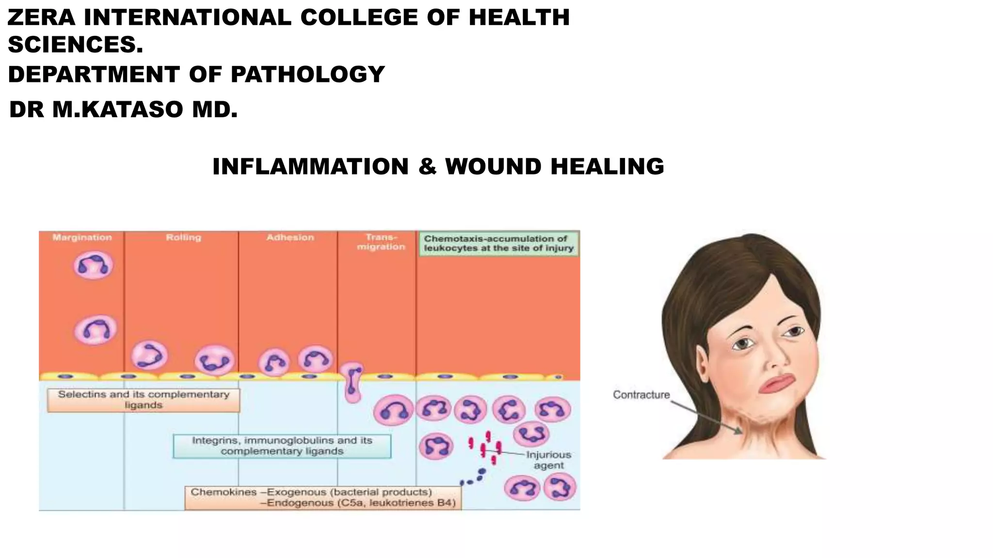
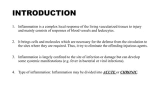
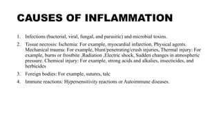
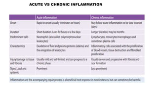
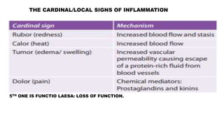
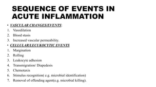
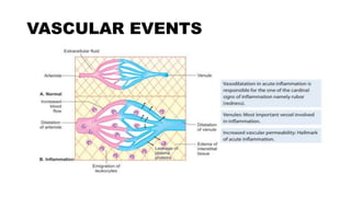
ZERA INTERNATIONAL COLLEGE OF HEALTH
SCIENCES.
DEPARTMENT OF PATHOLOGY
DR M.KATASO MD.
INFLAMMATION & WOUND HEALING
INTRODUCTION
1. Inflammation is a complex local response of the living vascularized tissues to injury
and mainly consists of responses of blood vessels and leukocytes.
2. It brings cells and molecules which are necessary for the defense from the circulation to
the sites where they are required. Thus, it try to eliminate the offending injurious agents.
3. Inflammation is largely confined to the site of infection or damage but can develop
some systemic manifestations (e.g. fever in bacterial or viral infections).
4. Type of inflammation: Inflammation may be divided into ACUTE or CHRONIC.
CAUSES OF INFLAMMATION
1. Infections (bacterial, viral, fungal, and parasitic) and microbial toxins.
2. Tissue necrosis: Ischemia: For example, myocardial infarction, Physical agents.
Mechanical trauma: For example, blunt/penetrating/crush injuries, Thermal injury: For
example, burns or frostbite ,Radiation ,Electric shock, Sudden changes in atmospheric
pressure. Chemical injury: For example, strong acids and alkalies, insecticides, and
herbicides
3. Foreign bodies: For example, sutures, talc
4. Immune reactions: Hypersensitivity reactions or Autoimmune diseases.
ACUTE VS CHRONIC INFLAMMATION
THE CARDINAL/LOCAL SIGNS OF INFLAMMATION
5TH ONE IS FUNCTIO LAESA: LOSS OF FUNCTION.
SEQUENCE OF EVENTS IN
ACUTE INFLAMMATION
• VASCULAR CHANGES/EVENTS:
1. Vasodilation
2. Blood stasis
3. Increased vascular permeability.
• CELLULAR/LECUKOCYTIC EVENTS
1. Margination
2. Rolling
3. Leukocyte adhesion
4. Transmigration/ Diapedesis
5. Chemotaxis
6. Stimulus recognition( e.g. microbial identification)
7. Removal of offending agent(e.g. microbial killing).
VASCULAR EVENTS
• Purpose: to deliver the circulating cells, fluids and plasma proteins from the circulation to
sites of infection or tissue injury.
• Vasodilatation: It is the earliest feature of acute inflammation. The vessel increases in
caliber. This results in blood stasis.
• Another feature is increased vascular permeability: this results into exudation.
Exudation is escape/extravasation of protein rich fluid into the interstitium from the
vascular space. This edema is the reason why there is edema. This edema is called
exudative edema.
• There are two main types of edema: exudative & transudative edema.
• Chemical mediators responsible for this include: Histamine, prostaglandins, platelet-
activating factor, kinins and nitric oxide (NO).
VASCULAR EVENTS
EXUDATIVE VS TRANSUDATIVE EDEMA.
EDEMA: ACCUMULATION OF FLUID IN THE INTERSTITIUM & OR BODY CAVITIES
CELLULAR EVENTS.
1. LEUCKOCYTE RECRUITMENT
2. LEUKOCYTE ACTIVATION.
Phagocytes such as neutrophils &
macrophages are involved.
CHEMICAL MEDIATORS OF INFLAMMATION.
ARACHDONIC METABOLISM
OUTCOMES OF ACUTE
INFLAMMATION
1. Resolution: Complete return of tissue architecture to normal following acute
inflammation. It occurs: When the injury is limited or short-lived, with no or minimal
tissue damage or When injured tissue is capable of regeneration.
2. Organization/healing by fibrosis: Process of replacement of dead tissue by living
tissue, which matures to form scar tissue is known as organization.
3. Progression to chronic inflammation: Chronic inflammation may follow acute
inflammation, or it may be chronic from the beginning itself. Acute progress to chronic
when the acute inflammatory response cannot be resolved. This may be due to:
Persistence of the injurious agent or Abnormality in the process of healing.
SYSTEMIC SIGNS OF
INFLAMMATION
• Systemic changes in acute inflammation are collectively known as acute-phase response,
or the systemic inflammatory response syndrome (SIRS).
1. Fever: Pyrogens: These are molecules that cause fever. It may be exogenous (bacterial
products, like LPS), which stimulate leukocytes to release endogenous pyrogens
(cytokines such as IL-1 and TNF). This stimulation cause prostaglandin release which
acts on the hypothalamic regulatory center.
2. Raised plasma levels of acute-phase proteins: These are plasma proteins synthesized in
the liver and may be markedly raised in response to inflammatory stimuli. Types of
acute-phase proteins: (1) C-reactive protein (CRP), (2) fibrinogen. The effect of this
raised levels of protein is increased erythrocyte sedimentation rate ( ESR). ESR & C-RP
are markers of inflammation.
3. Changes in the leukocytes: leukocytosis- increase in total WBC count.
4. Others: includes: Increased pulse ( tachycardia) and blood pressure. Anorexia and
malaise, probably due to cytokines acting on brain cells.
WOUND HEALING
• Healing is a process of replacement of dead tissue by living tissue.It can be broadly divided into
regeneration and repair.
• Regeneration: is a process in which lost tissue is replaced by tissue of similar type. It results in the
complete restoration of lost or damaged tissue by proliferation of residual uninjured cells and
replacement from stem cells.
• Repair: is defined as a process in which lost/damaged tissue is replaced by fibrous tissue or scar.
• FACTORS DECIDING THE PATTERN OF HEALING
1. Proliferative Capacity of the Tissue: According to proliferative capacity of the cells, the tissues
of the body can be divided into three groups:
a) Labile (continuously dividing) tissues: The cells of labile tissues proliferate throughout life,
replacing the lost cells from stem cells. Examples: Hematopoietic cells of the bone marrow
,Surface epithelia of the skin, oral cavity, vagina, and cervix ,Columnar epithelium of the
gastrointestinal tract and uterus.
PROLIFERATIVE CAPACITY
b) Stable (quiescent) tissues: Cells of stable tissue normally do not proliferate; but can
proliferate in response to injury or loss of tissue. Examples: Parenchymal cells of liver,
kidneys, and pancreas.
c) Permanent (nondividing) tissues: Cells of these tissues cannot proliferate after birth. In
these tissues, repair is by scar formation. Example: Neurons: Damaged neurons are
replaced by the proliferation of the glial cells. Skeletal muscle cells & Cardiac muscle
cells.
FACTORS DICIDING PATTERN OF
HEALING
2. Extent of Tissue Injury
a. Mild and short duration: The damaged tissue is healed by regeneration without
significant scarring.
b. Severe and chronic: Healing occurs by fibrous tissue forming scar
CUTANEOUS WOUND HEALING
• Healing by Primary Union or by First Intention: Healing of a clean, uninfected surgical
incision in the skin joined with surgical sutures is known as healing by primary union or
by first intention.
• Healing by Secondary Union or by Second Intention: When injury produces large
defects on the skin surface with extensive loss of cells and tissue, the healing process is
more complicated. Healing in such cutaneous wound is referred to as healing by
secondary union or by second intention.
FACTORS DELAYING WOUND
HEALING
• LOCAL FACTORS:
1. Infection
2. Mechanical factors: movement of wounded area may compress the blood vessels.
3. Foreign bodies
4. Poor local blood supply
5. Large & extensive wounds
6. Location of wounds: bony areas delay
7. Ionizing radiation
FACTORS DELAYING WOUND
HEALING
• SYSTEMIC FACTORS:
1. Age
2. Nutritional deficiencies: vitamin c, zinc.
3. Metabolic status: diabetes mellitus
4. Hematological abnormalities: anemia, leukemia or lylphomas.
5. Smocking
6. Steroids
7. Inadequate blood supply.
COMPLICATIONS
1. Wound dehiscence
2. Contractures
3. Keloids
4. Hypertrophic scar
5. Marjolijn's ulcer
6. Infections

INFLAMMATION & WOUD HEALING..pptx

  • 1.
    ZERA INTERNATIONAL COLLEGEOF HEALTH SCIENCES. DEPARTMENT OF PATHOLOGY DR M.KATASO MD. INFLAMMATION & WOUND HEALING
  • 2.
    INTRODUCTION 1. Inflammation isa complex local response of the living vascularized tissues to injury and mainly consists of responses of blood vessels and leukocytes. 2. It brings cells and molecules which are necessary for the defense from the circulation to the sites where they are required. Thus, it try to eliminate the offending injurious agents. 3. Inflammation is largely confined to the site of infection or damage but can develop some systemic manifestations (e.g. fever in bacterial or viral infections). 4. Type of inflammation: Inflammation may be divided into ACUTE or CHRONIC.
  • 3.
    CAUSES OF INFLAMMATION 1.Infections (bacterial, viral, fungal, and parasitic) and microbial toxins. 2. Tissue necrosis: Ischemia: For example, myocardial infarction, Physical agents. Mechanical trauma: For example, blunt/penetrating/crush injuries, Thermal injury: For example, burns or frostbite ,Radiation ,Electric shock, Sudden changes in atmospheric pressure. Chemical injury: For example, strong acids and alkalies, insecticides, and herbicides 3. Foreign bodies: For example, sutures, talc 4. Immune reactions: Hypersensitivity reactions or Autoimmune diseases.
  • 4.
    ACUTE VS CHRONICINFLAMMATION
  • 5.
    THE CARDINAL/LOCAL SIGNSOF INFLAMMATION 5TH ONE IS FUNCTIO LAESA: LOSS OF FUNCTION.
  • 6.
    SEQUENCE OF EVENTSIN ACUTE INFLAMMATION • VASCULAR CHANGES/EVENTS: 1. Vasodilation 2. Blood stasis 3. Increased vascular permeability. • CELLULAR/LECUKOCYTIC EVENTS 1. Margination 2. Rolling 3. Leukocyte adhesion 4. Transmigration/ Diapedesis 5. Chemotaxis 6. Stimulus recognition( e.g. microbial identification) 7. Removal of offending agent(e.g. microbial killing).
  • 7.
    VASCULAR EVENTS • Purpose:to deliver the circulating cells, fluids and plasma proteins from the circulation to sites of infection or tissue injury. • Vasodilatation: It is the earliest feature of acute inflammation. The vessel increases in caliber. This results in blood stasis. • Another feature is increased vascular permeability: this results into exudation. Exudation is escape/extravasation of protein rich fluid into the interstitium from the vascular space. This edema is the reason why there is edema. This edema is called exudative edema. • There are two main types of edema: exudative & transudative edema. • Chemical mediators responsible for this include: Histamine, prostaglandins, platelet- activating factor, kinins and nitric oxide (NO).
  • 8.
  • 9.
    EXUDATIVE VS TRANSUDATIVEEDEMA. EDEMA: ACCUMULATION OF FLUID IN THE INTERSTITIUM & OR BODY CAVITIES
  • 10.
    CELLULAR EVENTS. 1. LEUCKOCYTERECRUITMENT 2. LEUKOCYTE ACTIVATION. Phagocytes such as neutrophils & macrophages are involved.
  • 11.
    CHEMICAL MEDIATORS OFINFLAMMATION.
  • 12.
  • 13.
    OUTCOMES OF ACUTE INFLAMMATION 1.Resolution: Complete return of tissue architecture to normal following acute inflammation. It occurs: When the injury is limited or short-lived, with no or minimal tissue damage or When injured tissue is capable of regeneration. 2. Organization/healing by fibrosis: Process of replacement of dead tissue by living tissue, which matures to form scar tissue is known as organization. 3. Progression to chronic inflammation: Chronic inflammation may follow acute inflammation, or it may be chronic from the beginning itself. Acute progress to chronic when the acute inflammatory response cannot be resolved. This may be due to: Persistence of the injurious agent or Abnormality in the process of healing.
  • 14.
    SYSTEMIC SIGNS OF INFLAMMATION •Systemic changes in acute inflammation are collectively known as acute-phase response, or the systemic inflammatory response syndrome (SIRS). 1. Fever: Pyrogens: These are molecules that cause fever. It may be exogenous (bacterial products, like LPS), which stimulate leukocytes to release endogenous pyrogens (cytokines such as IL-1 and TNF). This stimulation cause prostaglandin release which acts on the hypothalamic regulatory center. 2. Raised plasma levels of acute-phase proteins: These are plasma proteins synthesized in the liver and may be markedly raised in response to inflammatory stimuli. Types of acute-phase proteins: (1) C-reactive protein (CRP), (2) fibrinogen. The effect of this raised levels of protein is increased erythrocyte sedimentation rate ( ESR). ESR & C-RP are markers of inflammation. 3. Changes in the leukocytes: leukocytosis- increase in total WBC count. 4. Others: includes: Increased pulse ( tachycardia) and blood pressure. Anorexia and malaise, probably due to cytokines acting on brain cells.
  • 15.
    WOUND HEALING • Healingis a process of replacement of dead tissue by living tissue.It can be broadly divided into regeneration and repair. • Regeneration: is a process in which lost tissue is replaced by tissue of similar type. It results in the complete restoration of lost or damaged tissue by proliferation of residual uninjured cells and replacement from stem cells. • Repair: is defined as a process in which lost/damaged tissue is replaced by fibrous tissue or scar. • FACTORS DECIDING THE PATTERN OF HEALING 1. Proliferative Capacity of the Tissue: According to proliferative capacity of the cells, the tissues of the body can be divided into three groups: a) Labile (continuously dividing) tissues: The cells of labile tissues proliferate throughout life, replacing the lost cells from stem cells. Examples: Hematopoietic cells of the bone marrow ,Surface epithelia of the skin, oral cavity, vagina, and cervix ,Columnar epithelium of the gastrointestinal tract and uterus.
  • 16.
    PROLIFERATIVE CAPACITY b) Stable(quiescent) tissues: Cells of stable tissue normally do not proliferate; but can proliferate in response to injury or loss of tissue. Examples: Parenchymal cells of liver, kidneys, and pancreas. c) Permanent (nondividing) tissues: Cells of these tissues cannot proliferate after birth. In these tissues, repair is by scar formation. Example: Neurons: Damaged neurons are replaced by the proliferation of the glial cells. Skeletal muscle cells & Cardiac muscle cells.
  • 17.
    FACTORS DICIDING PATTERNOF HEALING 2. Extent of Tissue Injury a. Mild and short duration: The damaged tissue is healed by regeneration without significant scarring. b. Severe and chronic: Healing occurs by fibrous tissue forming scar
  • 18.
    CUTANEOUS WOUND HEALING •Healing by Primary Union or by First Intention: Healing of a clean, uninfected surgical incision in the skin joined with surgical sutures is known as healing by primary union or by first intention. • Healing by Secondary Union or by Second Intention: When injury produces large defects on the skin surface with extensive loss of cells and tissue, the healing process is more complicated. Healing in such cutaneous wound is referred to as healing by secondary union or by second intention.
  • 19.
    FACTORS DELAYING WOUND HEALING •LOCAL FACTORS: 1. Infection 2. Mechanical factors: movement of wounded area may compress the blood vessels. 3. Foreign bodies 4. Poor local blood supply 5. Large & extensive wounds 6. Location of wounds: bony areas delay 7. Ionizing radiation
  • 20.
    FACTORS DELAYING WOUND HEALING •SYSTEMIC FACTORS: 1. Age 2. Nutritional deficiencies: vitamin c, zinc. 3. Metabolic status: diabetes mellitus 4. Hematological abnormalities: anemia, leukemia or lylphomas. 5. Smocking 6. Steroids 7. Inadequate blood supply.
  • 21.
    COMPLICATIONS 1. Wound dehiscence 2.Contractures 3. Keloids 4. Hypertrophic scar 5. Marjolijn's ulcer 6. Infections