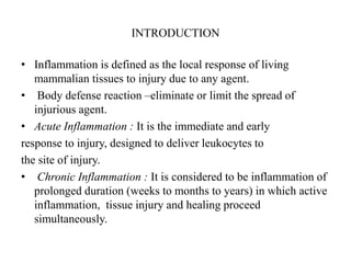
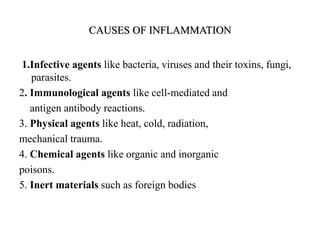
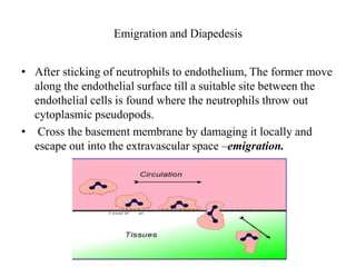
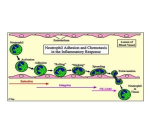
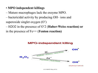
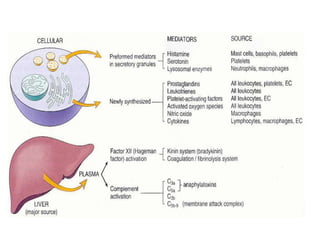
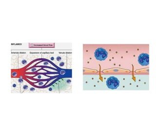
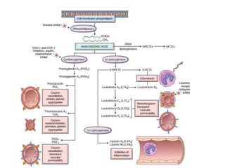
INFLAMMATION
Presented by-Dr. Honey Khatri
PG II yr
Contents
• Introduction
• Causes of inflammation
• Types of inflammation
• Acute inflammation
-Vascular events
- Haemodynamic changes
-Changes in vascular permeability
INTRODUCTION
• Inflammation is defined as the local response of living
mammalian tissues to injury due to any agent.
• Body defense reaction –eliminate or limit the spread of
injurious agent.
• Acute Inflammation : It is the immediate and early
response to injury, designed to deliver leukocytes to
the site of injury.
• Chronic Inflammation : It is considered to be inflammation of
prolonged duration (weeks to months to years) in which active
inflammation, tissue injury and healing proceed
simultaneously.
CAUSES OF INFLAMMATION
1.Infective agents like bacteria, viruses and their toxins, fungi,
parasites.
2. Immunological agents like cell-mediated and
antigen antibody reactions.
3. Physical agents like heat, cold, radiation,
mechanical trauma.
4. Chemical agents like organic and inorganic
poisons.
5. Inert materials such as foreign bodies
SIGNS OF INFLAMMATION
4 cardinal signs
(Celsus)
– rubor (redness);
– tumor (swelling);
– calor (heat);
– dolor (pain)
• 5th sign functiolaesa
(loss of function-Virchow
TYPES OF INFLAMMATION
Mainly of 2 types i.e. acute and chronic
• Acute Inflammation
– short duration.
– represents the early body reaction- followed by
Healing.
• Chronic inflammation
– longer duration.
– causative agent of acute inflammation persists
for a long time.
• Another variant, Chronic active inflammation :
stimulus is such that it induces chronic inflammation from the
beginning.
ACUTE INFLAMMATION
The main features of acute
inflammation are:
– accumulation of fluid and
plasma at the affected site;
– intravascular activation of
platelets;
– polymorphonuclear
neutrophils as inflammatory
cells.
• Acute inflammatory response is a continous process but for
understanding it is divided into
-vascular events
-cellular events.
VASCULAR EVENTS
• Alteration in microvasculature is the earliest response to the
tissue injury.
• It includes –
- haemodynamic changes
-vascular permeability
HAEMODYNAMIC CHANGES
1.Transient vasoconstriction :
immediate vascular response
irrespective of the type of
injury,
- mainly arterioles
– Mild injury - 3-5 seconds
– Severe injury - 5 minutes
2. Persistent progressive
vasodilatation : mainly
arterioles, others to a lesser
extent.
– obvious within half an hour of
injury
3.Progressive vasodilatation elevate the local hydrostatic
pressure.
– transudation of fluid into
the extracellular space
– swelling
4. Slowing or stasis increased concentration of red cells,
and thus, raised blood viscosity.
5.Leucocytic Margination peripheral orientation
of leucocytes (mainly neutrophils) along the
vascular endothelium
– stick to the vascular endothelium
– move and migrate through
the gaps between the
endothelial cells – extravascular
space.
– This is known is emigration
ALTERED VASCULAR PERMEABILITY
Accumulation of oedema fluid in the interstitial compartment
comes from blood plasma by its escape through the endothelial
wall of peripheral vascular bed.
• Escape of fluid is due to vasodilatation and consequent
elevation in hydrostatic pressure - transudate.
• Subsequently, the characteristic inflammatory oedema, appears
by increased vascular permeability of microcirculation –
exudate
STARLING HYPOTHESIS
• The appearance of inflammatory oedema is explained on the
basis of starling hypothesis
• Forces that causes the outward movement of the fluid are
-intravascular hydrostatic pressure
-colloidal osmotic pressure of interstitial fluid .
• Forces that causes inward movement of fluid are –
Intravasular colloidal osmotic pressure
-hydrostatic pressure of interstitial fluid
Mechanism of increased vascular premeability
1. Contraction of endothelial cells.
2. Retraction of endothelial cells
3. Direct injury to endothelial cells
4. Endothelial injury mediated by leucocytes
5. Leakiness and neo-vascularisation
1. CONTRACTION OF ENDOTHELIAL CELLS –
-Most common mechanism.
-Mainly affects the venules .
-temporary gap develops
between the endothelial cells.
-mediated by release of histamine,
bradykinin and other
chemical mediators .
-Short duration (15-30 mins )
Eg- immediate transient leakage
is –mild thermal injury of skin of forearm.
2.RETRACTION OF ENDOTHELIAL CELLS
Reversible mechanism
Induced by cytokine mediators (TNF & IL-1)
Structural re-organisation of
the cytoskeleton of
endothelial cells - Reversible
retraction at the intercellular
Junctions.
DIRECT INJURY TO ENDOTHELIAL CELLS
• Causes cell necrosis and
appearance of physical gaps.
• Process of thrombosis is initiated
at the site of damaged endothelial
cells.
• Affects all levels of
microvasculature.
• Either appear immediately after
injury and last for several hours or
days – severe bacterial infections
• Or delay of 2-12 hours and last for
hours or days - moderate thermal
injury and radiation injury
ENDOTHELIAL INJURY MEDIATED BY LEUKOCYTES
Adherence of leucocytes to the endothelium
at the site of inflammation.
• Activation of leucocytes
- release proteolytic
enzymes and toxic oxygen species.
• Cause endothelial injury
and increased vascular leakiness.
• Affects mostly venules
and is a late response.
NEOVASCULARISATION
Newly formed capillaries under the influence
of vascular endothelial growth factor (VEGF).
• Process of repair
excessively leaky
Presented By-
Dr.Honey Khatri
PG II Yr
CELLULAR EVENTS
• Cellular phase of inflammation consists of 2
• processes
• 1. Exudation of leucocytes
• 2. Phagocytosis.
Exudation of leucocytes
1. Changes in the formed elements of blood.
2. Rolling and adhesion
3. Emigration
4. Chemotaxis
Changes in the formed elements of the blood
Central stream of cells comprised by leucocytes and RBCs and
peripheral cell free layer of plasma close to vessel wall.
• Later, central stream of cells widens and peripheral plasma zone
becomes narrower because of loss of plasma by exudation.
This phenomenon is known as margination.
• Neutrophils of the central column come close to the vessel wall
- pavementing
Rolling and adhesion
Peripherally marginated and pavemented neutrophils slowly roll
over the endothelial cells lining the vessel wall (rolling
phase).
• Transient bond between the leucocytes and endothelial cells
becoming firmer (adhesion phase).
• The following molecules bring about rolling and adhesion
phases
– Selectins
– Integrins
– Immunoglobulin gene superfamily adhesion molecule
• E-selectins are expressed at low levels or are not present at
all on normal cells. They are upregulated after stimulation by
specific mediators. eg.IL-1 and TNF.
• P-selectins are found intracellularly,
which once stimulated by mediators such as histamine are
distributed over the cell surface.
• L-selectins interact with carbohydrate molecules known as
vascular addresins (eg.sialomucin) on the luminal surface of
endothelial cells. This brief interaction manifests itself as
rolling of the leukocyte along the luminal surface of
endothelium.
Emigration and Diapedesis
• After sticking of neutrophils to endothelium, The former move
along the endothelial surface till a suitable site between the
endothelial cells is found where the neutrophils throw out
cytoplasmic pseudopods.
• Cross the basement membrane by damaging it locally and
escape out into the extravascular space –emigration.
• Diapedesis - escape of red cells through gaps between the
endothelial cells
• – passive phenomenon.
• – raised hydrostatic pressure
• – haemorrhagic appearance to the inflammatory
• exudate
chemotaxis
After extravasating from the blood, leukocytes
migrate toward sites of injury along a chemical
gradient in a process called chemotaxis.
Soluble bacterial products : N-formylmethionine termini.
- Components of complement system : C5a
- Products of lipoxygenase pathway : leukotriene B4
- Cytokines : IL-1, IL-8
Phagocytosis
• The process of engulfment of solid particulate
material by the cells.
2 main types of phagocytic cells
– Polymorphonuclear neutrophils (PMNs) : early in acute
inflammatory response, also known as microphages
– Macrophages : Circulating monocytes and fixed tissue
mononuclear phagocytes
• This phagocytic cells releases proteolytic enzymes
- lysozyme, protease, collagenase, elastase,
lipase, proteinase, gelatinase and acid hydrolases.
• The microbe undergoes the process of
• phagocytosis in following 3 steps :
• – Recognition and attachment
• – Engulfment
• – Killing and degradation
Recognition and attachment
Phagocytosis is initiated by the expression of surface receptors
on macrophages.
• Its further enhanced when the microorganisms are coated with
specific proteins, opsonins.
– Establish a bond between bacteria and the cell membrane of
phagocytic cell.
– Major opsonins are
• IgG opsonin .
• C3b opsonin
• Lectins
Engulfment
Formation of cytoplasmic pseudopods around
the particle due to activation of actin filaments
around cell wall.
• Eventually plasma membrane
gets lysed and
fuses with nearby
lysosomes – phagolysosome.
Killing and degradation
• Killing take place by Antibacterial
substances further degraded by hydrolytic
Enzymes.
Next comes the stage of disposal of micro organisms after killing
and degradation
Disposal of micro organisms
• Intracellular mechanisms
– Oxidative bactericidal mechanism by oxygen free radicals
• MPO-dependent
• MPO-independent
– Oxidative bactericidal mechanism by lysosomal granules
– Non-oxidative bactericidal mechanism
• Extracellular mechanisms
– Granules
– Immune mechanisms
Intracellular mechanism
Kill microbes by oxidative mechanism and less often
non-oxidative pathways
• Oxidative bactericidal mechanism by oxygen free
radicals.
– production of reactive oxygen metabolites (O’2, H2O2,
OH’,HOCl, HOI, HOBr)
– activated phagocytic leucocytes requires the essential presence
of NADPH oxidase
– present in the cell membrane of phagosome reduces
oxygen to superoxide ion (O’2)
Superoxide is subsequently converted into H2O2 .
• Bactericidal activity is carried out either via enzyme
myeloperoxidase (MPO) or MPO independent.
• MPO dependent killings
– MPO acts on H2O2 in the presence of halides – form
hypohalous acid (HOCl, HOI, HOBr)
• MPO independent killings
– Mature macrophages lack the enzyme MPO.
– bactericidal activity by producing OH– ions and
superoxide singlet oxygen (O’)
– H2O2 in the presence of O’2 (Haber-Weiss reaction) or
in the presence of Fe++ (Fenton reaction)
Oxidative bactericidal mechanism by lysosomal
granules
– preformed granule-stored products of neutrophils and
macrophages.
– secreted into the phagosome and the extracellular environment.
• Non-oxidative bactericidal mechanism
– Some agents released from the granules of phagocytic cells do
not require oxygen for bactericidal activity
• Granules : cause lysis of micro organisms within phagosome
, ex: lysosomal hydrolases, permeability increasing factors,
cationic proteins (defensins), lipases, ptoteases, DNAases.
• Nitric oxide : reactive free radicals similar to oxygen free
radicals
– potent mechanism of microbial killing
– produced by endothelial cells as well as by activated
macrophages
Extra cellular mechanism
Granules
– Degranulation of macrophages and neutrophils
• Immune mechanisms
– immune-mediated lysis of microbes
– takes place outside the cells
– by mechanisms of cytolysis, antibody-mediated
lysis and by cell-mediated cytotoxicity
• mediators of inflammation
Contents
Introduction
Mediators also called as permeability factors or endogenous
mediators are the substances that initiate and regulate
inflammatory reactions.
These are:
1.cell derived
2. plasma protein derived.
• Mechanisms of action
receptor-ligand interactions (1o)
direct enzymatic activity
oxidative damage
• Network of interacting chemicals
high degree of redundancy
guarantees amplification & maintenance of inflammatory response
• Short half life
• Potentially harmful
Vasoactive Amines: Histamine and
Serotonin
Acts on blood vessels.
• First mediators
• Histamine
• Mast cells are richest source of histamine
• Histamine stored as granules and released by degranulation in
response to various stimuli
1. physical injuries
2.Antibodies mediated – hypersensitivity reaction
3. Complement products- anaphyltoxin C3a, C5a.
4. Neuropeptides (e.g., substance P) and cytokines
(IL1, IL8)
• Histamine causes dilation of arterioles and increases the
permeability of venules.
• Principal mediator of the
immediate transient phase .
• It increases vascular permeability,
producing interendothelial gaps in venules.
• Act via h1 receptors present
on microvascular
Endothelial cells.
Serotonin(5-hydroxytryptamine)
• Preformed mediator
• Present in plateletes and certain
• Neuroendocrinal cells like GIT.
• Primary function is as a neurotransmitter.
• Vasoconstriction
Arachidonic Acid Metabolites
• The lipid mediators prostaglandins and leukotrienes are
produced from arachidonic acid (AA) present in membrane
phospholipids
• Stimulate vascular and cellular reactions.
• Arachidonic acid derived from dietary sources or by
conversion from the linoleic acid.
• Mechanical, chemical, and physical stimuli or other mediators
(e.g., C5a) release AA from membrane phospholipids through
the action of cellular phospholipases, mainly phospholipase
A2.
Prostaglandins
• Produced by mast cells , macrophages, endothelial cells, .
• it involved in vascular and systemic reactions of
inflammation.
• COX-1 & COX-2 is involved in synthesis of prostaglandins.
• COX -1 is produced in response to inflammatory stimuli and
is also constitutively expressed
• COX2 is induced by inflammatory stimuli .
Throboxane A2
• derived by action of thromboxane synthase
• present in platelets.
Functions –
• platelets aggregation
• Vasoconstrictions
• it is unstable and converted to inactive forms TxB2
Prostacyclin( PGI2)
• Formed by action of prostacyclin synthase
Present in vascular endothelium.
• Functions
Increase vascular permeability
inhibit platelets aggregation
• PGD2 produced by mast cells along with PGE2.
• • it causes vasodilation and increases the
• permeability of postcapillary venules, thus
• potentiating edema formation.
• • PGF2- stimulates contraction of uterine and
• bronchial smooth muscles and small arterioles.
• • Prostaglandins involved in the pathogenesis of
• pain and fever in inflmmation.
• • PGE2 is hyperalgesic makes the skin
• hypersensitive to painful stimuli
Leukotriene
• Produced by leukocytes and mast cells by the action of
lipoxygenase.
• It involved in vascular and smooth muscle reactions and
leukocyte recruitment.
• Three types of lipoxygenase
1. - Lipoxygenase – present in neutrophils, convert
AA to 5hydroxyeicosatetraenoic acid
• Act as chemotactic for neutrophils
• LTB4- Chemotactic and neutrophil activator.
• • leads to aggregation and adhesion of the cells
• to venular endothelium.
• • Generation of ROS
• • Release of lysosomal enzymes
• LTC4, LTD4, and LTE4- causes
• • vasoconstrictions
• • Bronchospasm
• • increased permeability of venules
• • Leukotrienes are more potent than is
• histamine in increasing vascular permeability
• and causing bronchospasm.
Generated from AA by the lipoxygenase pathway.
• It suppress inflammation by inhibiting recruitment of
leukocytes.
• Inhibit neutrophil chemotaxis and adhesion to
endothelium.
Cytokines and Chemokines
• Cytokines are proteins produced by many celltypes (activated
lymphocytes, macrophages, and dendritic cells)
• mediate and regulate immune and inflammatory reactions.
Produced by activated lymphocytes and activated monocytes.
Major cytokines are
Interelukin 1
Tnf- α and β ,
interferon and
chemokines (IL-8)
Tumor Necrosis Factor (TNF) and
Interleukin-1 (IL-1)
• Roles in leukocyte recruitment and promoting adhesion of
leukocytes to endothelium and their migration through vessels.
• Produced by – macrophages and dendritic cells.
• Tnf-β is produced by activated t cells .
• Stimulus for secretion- microbial products, immune
complexes, foreign bodies, physical injury, and a variety of
other inflammatory stimuli.
Roles of cytokines in inflammation
• Endothelial activation.- by TNF and IL-1
• increased expression of adhesion molecules ( Pand
E- selectins and ligands foe leukocytes
• increased production of various mediators
• increased procoagulant activity
• Activation of leukocytes and other cells-
• stimulates microbicidal activity of macrophages by
inducing production of NO.
• IL1 activates fibroblasts to synthesize collagen
and stimulates proliferation of synovial and other
mesenchymal cells.
• stimulates TH17 responses- induce acute
inflmmation
Nitrous oxide
• nitric oxide (NO) synthesized from L-arginine.
• Effects: -
- smooth muscle relaxation
-vasodilation
- reduce platelet aggregation & adhesion
- bactericidal (forms peroxynitrite)
Plasma derived mediators
• These include the various products derived
from activation and interaction of four
interlinked system
• Kinin,
• clotting ,
• fibrinolytic and
• compliment
Kinin system
Clotting system and Fibrinolytic system
Complement System
• The complement system is a collection of soluble proteins and
membrane receptors that function mainly in host defense
against microbes and in pathologic inflammatory reactions.
• The system consists of more than 20 proteins,some of which are
numbered C1 through C9.
• Functions in both innate and adaptive immunity
• Complement proteins are present in inactive forms in the
plasma.
• Cleavage of C3 can occur by one of three pathways:
• classical pathway- triggered by fixation of C1 to antibody
(IgM or IgG) that has combined with antigen.
• Alternative pathway- triggered by microbial surface molecules
(e.g., endotoxin, or LPS), complex polysaccharides, cobra
venom, and other substances, in the absence of antibody.
• The lectin pathway- plasma mannose binding lectin binds to
carbohydrates on microbes and directly activates C1.
Other Mediators of Inflammation
• Platelet-Activating Factor (PAF)- phospholipid
derived mediator
• Secreted by platelets, basophils, mast cells,
neutrophils, macrophages, and endothelial cells.
• It causes- platelets aggregation
- vasoconstriction
- bronchoconstiction
- at low concentrations it induces vasodilation
and increased venular permeability.
Products of coagulation
• Kinins- vasoactive peptides derived from plasma
• proteins, called kininogens, by the action of
• specific proteases called kallikreins.
• • Bradykinin- increases vascular permeability, and
• smooth muscles contractions,
• • Dilatation of blood vessels and pain
• • Neuropeptides-secreted by sensory nerves and
• various leukocytes.
• • substance P and neurokinin A
Outcomes of inflammation
• Thankyou
Inflammation [autosaved]

Inflammation [autosaved]

  • 1.
  • 2.
    Contents • Introduction • Causesof inflammation • Types of inflammation • Acute inflammation -Vascular events - Haemodynamic changes -Changes in vascular permeability
  • 3.
    INTRODUCTION • Inflammation isdefined as the local response of living mammalian tissues to injury due to any agent. • Body defense reaction –eliminate or limit the spread of injurious agent. • Acute Inflammation : It is the immediate and early response to injury, designed to deliver leukocytes to the site of injury. • Chronic Inflammation : It is considered to be inflammation of prolonged duration (weeks to months to years) in which active inflammation, tissue injury and healing proceed simultaneously.
  • 4.
    CAUSES OF INFLAMMATION 1.Infectiveagents like bacteria, viruses and their toxins, fungi, parasites. 2. Immunological agents like cell-mediated and antigen antibody reactions. 3. Physical agents like heat, cold, radiation, mechanical trauma. 4. Chemical agents like organic and inorganic poisons. 5. Inert materials such as foreign bodies
  • 5.
    SIGNS OF INFLAMMATION 4cardinal signs (Celsus) – rubor (redness); – tumor (swelling); – calor (heat); – dolor (pain) • 5th sign functiolaesa (loss of function-Virchow
  • 6.
    TYPES OF INFLAMMATION Mainlyof 2 types i.e. acute and chronic • Acute Inflammation – short duration. – represents the early body reaction- followed by Healing. • Chronic inflammation – longer duration. – causative agent of acute inflammation persists for a long time. • Another variant, Chronic active inflammation : stimulus is such that it induces chronic inflammation from the beginning.
  • 7.
    ACUTE INFLAMMATION The mainfeatures of acute inflammation are: – accumulation of fluid and plasma at the affected site; – intravascular activation of platelets; – polymorphonuclear neutrophils as inflammatory cells.
  • 8.
    • Acute inflammatoryresponse is a continous process but for understanding it is divided into -vascular events -cellular events.
  • 9.
    VASCULAR EVENTS • Alterationin microvasculature is the earliest response to the tissue injury. • It includes – - haemodynamic changes -vascular permeability
  • 10.
    HAEMODYNAMIC CHANGES 1.Transient vasoconstriction: immediate vascular response irrespective of the type of injury, - mainly arterioles – Mild injury - 3-5 seconds – Severe injury - 5 minutes 2. Persistent progressive vasodilatation : mainly arterioles, others to a lesser extent. – obvious within half an hour of injury
  • 11.
    3.Progressive vasodilatation elevatethe local hydrostatic pressure. – transudation of fluid into the extracellular space – swelling 4. Slowing or stasis increased concentration of red cells, and thus, raised blood viscosity.
  • 12.
    5.Leucocytic Margination peripheralorientation of leucocytes (mainly neutrophils) along the vascular endothelium – stick to the vascular endothelium – move and migrate through the gaps between the endothelial cells – extravascular space. – This is known is emigration
  • 14.
    ALTERED VASCULAR PERMEABILITY Accumulationof oedema fluid in the interstitial compartment comes from blood plasma by its escape through the endothelial wall of peripheral vascular bed. • Escape of fluid is due to vasodilatation and consequent elevation in hydrostatic pressure - transudate. • Subsequently, the characteristic inflammatory oedema, appears by increased vascular permeability of microcirculation – exudate
  • 16.
    STARLING HYPOTHESIS • Theappearance of inflammatory oedema is explained on the basis of starling hypothesis • Forces that causes the outward movement of the fluid are -intravascular hydrostatic pressure -colloidal osmotic pressure of interstitial fluid . • Forces that causes inward movement of fluid are – Intravasular colloidal osmotic pressure -hydrostatic pressure of interstitial fluid
  • 18.
    Mechanism of increasedvascular premeability 1. Contraction of endothelial cells. 2. Retraction of endothelial cells 3. Direct injury to endothelial cells 4. Endothelial injury mediated by leucocytes 5. Leakiness and neo-vascularisation
  • 20.
    1. CONTRACTION OFENDOTHELIAL CELLS – -Most common mechanism. -Mainly affects the venules . -temporary gap develops between the endothelial cells. -mediated by release of histamine, bradykinin and other chemical mediators . -Short duration (15-30 mins ) Eg- immediate transient leakage is –mild thermal injury of skin of forearm.
  • 21.
    2.RETRACTION OF ENDOTHELIALCELLS Reversible mechanism Induced by cytokine mediators (TNF & IL-1) Structural re-organisation of the cytoskeleton of endothelial cells - Reversible retraction at the intercellular Junctions.
  • 22.
    DIRECT INJURY TOENDOTHELIAL CELLS • Causes cell necrosis and appearance of physical gaps. • Process of thrombosis is initiated at the site of damaged endothelial cells. • Affects all levels of microvasculature. • Either appear immediately after injury and last for several hours or days – severe bacterial infections • Or delay of 2-12 hours and last for hours or days - moderate thermal injury and radiation injury
  • 23.
    ENDOTHELIAL INJURY MEDIATEDBY LEUKOCYTES Adherence of leucocytes to the endothelium at the site of inflammation. • Activation of leucocytes - release proteolytic enzymes and toxic oxygen species. • Cause endothelial injury and increased vascular leakiness. • Affects mostly venules and is a late response.
  • 24.
    NEOVASCULARISATION Newly formed capillariesunder the influence of vascular endothelial growth factor (VEGF). • Process of repair excessively leaky
  • 25.
    Presented By- Dr.Honey Khatri PGII Yr CELLULAR EVENTS
  • 26.
    • Cellular phaseof inflammation consists of 2 • processes • 1. Exudation of leucocytes • 2. Phagocytosis.
  • 27.
    Exudation of leucocytes 1.Changes in the formed elements of blood. 2. Rolling and adhesion 3. Emigration 4. Chemotaxis
  • 29.
    Changes in theformed elements of the blood Central stream of cells comprised by leucocytes and RBCs and peripheral cell free layer of plasma close to vessel wall. • Later, central stream of cells widens and peripheral plasma zone becomes narrower because of loss of plasma by exudation. This phenomenon is known as margination. • Neutrophils of the central column come close to the vessel wall - pavementing
  • 30.
    Rolling and adhesion Peripherallymarginated and pavemented neutrophils slowly roll over the endothelial cells lining the vessel wall (rolling phase). • Transient bond between the leucocytes and endothelial cells becoming firmer (adhesion phase).
  • 31.
    • The followingmolecules bring about rolling and adhesion phases – Selectins – Integrins – Immunoglobulin gene superfamily adhesion molecule • E-selectins are expressed at low levels or are not present at all on normal cells. They are upregulated after stimulation by specific mediators. eg.IL-1 and TNF. • P-selectins are found intracellularly, which once stimulated by mediators such as histamine are distributed over the cell surface.
  • 32.
    • L-selectins interactwith carbohydrate molecules known as vascular addresins (eg.sialomucin) on the luminal surface of endothelial cells. This brief interaction manifests itself as rolling of the leukocyte along the luminal surface of endothelium.
  • 33.
    Emigration and Diapedesis •After sticking of neutrophils to endothelium, The former move along the endothelial surface till a suitable site between the endothelial cells is found where the neutrophils throw out cytoplasmic pseudopods. • Cross the basement membrane by damaging it locally and escape out into the extravascular space –emigration.
  • 34.
    • Diapedesis -escape of red cells through gaps between the endothelial cells • – passive phenomenon. • – raised hydrostatic pressure • – haemorrhagic appearance to the inflammatory • exudate
  • 36.
    chemotaxis After extravasating fromthe blood, leukocytes migrate toward sites of injury along a chemical gradient in a process called chemotaxis.
  • 37.
    Soluble bacterial products: N-formylmethionine termini. - Components of complement system : C5a - Products of lipoxygenase pathway : leukotriene B4 - Cytokines : IL-1, IL-8
  • 38.
    Phagocytosis • The processof engulfment of solid particulate material by the cells. 2 main types of phagocytic cells – Polymorphonuclear neutrophils (PMNs) : early in acute inflammatory response, also known as microphages – Macrophages : Circulating monocytes and fixed tissue mononuclear phagocytes • This phagocytic cells releases proteolytic enzymes - lysozyme, protease, collagenase, elastase, lipase, proteinase, gelatinase and acid hydrolases.
  • 39.
    • The microbeundergoes the process of • phagocytosis in following 3 steps : • – Recognition and attachment • – Engulfment • – Killing and degradation
  • 40.
    Recognition and attachment Phagocytosisis initiated by the expression of surface receptors on macrophages. • Its further enhanced when the microorganisms are coated with specific proteins, opsonins. – Establish a bond between bacteria and the cell membrane of phagocytic cell. – Major opsonins are • IgG opsonin . • C3b opsonin • Lectins
  • 41.
    Engulfment Formation of cytoplasmicpseudopods around the particle due to activation of actin filaments around cell wall. • Eventually plasma membrane gets lysed and fuses with nearby lysosomes – phagolysosome.
  • 42.
    Killing and degradation •Killing take place by Antibacterial substances further degraded by hydrolytic Enzymes. Next comes the stage of disposal of micro organisms after killing and degradation
  • 43.
    Disposal of microorganisms • Intracellular mechanisms – Oxidative bactericidal mechanism by oxygen free radicals • MPO-dependent • MPO-independent – Oxidative bactericidal mechanism by lysosomal granules – Non-oxidative bactericidal mechanism • Extracellular mechanisms – Granules – Immune mechanisms
  • 44.
    Intracellular mechanism Kill microbesby oxidative mechanism and less often non-oxidative pathways • Oxidative bactericidal mechanism by oxygen free radicals. – production of reactive oxygen metabolites (O’2, H2O2, OH’,HOCl, HOI, HOBr) – activated phagocytic leucocytes requires the essential presence of NADPH oxidase – present in the cell membrane of phagosome reduces oxygen to superoxide ion (O’2)
  • 45.
    Superoxide is subsequentlyconverted into H2O2 . • Bactericidal activity is carried out either via enzyme myeloperoxidase (MPO) or MPO independent. • MPO dependent killings – MPO acts on H2O2 in the presence of halides – form hypohalous acid (HOCl, HOI, HOBr)
  • 46.
    • MPO independentkillings – Mature macrophages lack the enzyme MPO. – bactericidal activity by producing OH– ions and superoxide singlet oxygen (O’) – H2O2 in the presence of O’2 (Haber-Weiss reaction) or in the presence of Fe++ (Fenton reaction)
  • 47.
    Oxidative bactericidal mechanismby lysosomal granules – preformed granule-stored products of neutrophils and macrophages. – secreted into the phagosome and the extracellular environment. • Non-oxidative bactericidal mechanism – Some agents released from the granules of phagocytic cells do not require oxygen for bactericidal activity
  • 48.
    • Granules :cause lysis of micro organisms within phagosome , ex: lysosomal hydrolases, permeability increasing factors, cationic proteins (defensins), lipases, ptoteases, DNAases. • Nitric oxide : reactive free radicals similar to oxygen free radicals – potent mechanism of microbial killing – produced by endothelial cells as well as by activated macrophages
  • 49.
    Extra cellular mechanism Granules –Degranulation of macrophages and neutrophils • Immune mechanisms – immune-mediated lysis of microbes – takes place outside the cells – by mechanisms of cytolysis, antibody-mediated lysis and by cell-mediated cytotoxicity
  • 50.
    • mediators ofinflammation
  • 51.
  • 52.
    Introduction Mediators also calledas permeability factors or endogenous mediators are the substances that initiate and regulate inflammatory reactions. These are: 1.cell derived 2. plasma protein derived.
  • 53.
    • Mechanisms ofaction receptor-ligand interactions (1o) direct enzymatic activity oxidative damage • Network of interacting chemicals high degree of redundancy guarantees amplification & maintenance of inflammatory response • Short half life • Potentially harmful
  • 55.
    Vasoactive Amines: Histamineand Serotonin Acts on blood vessels. • First mediators • Histamine • Mast cells are richest source of histamine • Histamine stored as granules and released by degranulation in response to various stimuli 1. physical injuries 2.Antibodies mediated – hypersensitivity reaction 3. Complement products- anaphyltoxin C3a, C5a. 4. Neuropeptides (e.g., substance P) and cytokines (IL1, IL8)
  • 56.
    • Histamine causesdilation of arterioles and increases the permeability of venules. • Principal mediator of the immediate transient phase . • It increases vascular permeability, producing interendothelial gaps in venules. • Act via h1 receptors present on microvascular Endothelial cells.
  • 58.
    Serotonin(5-hydroxytryptamine) • Preformed mediator •Present in plateletes and certain • Neuroendocrinal cells like GIT. • Primary function is as a neurotransmitter. • Vasoconstriction
  • 59.
    Arachidonic Acid Metabolites •The lipid mediators prostaglandins and leukotrienes are produced from arachidonic acid (AA) present in membrane phospholipids • Stimulate vascular and cellular reactions. • Arachidonic acid derived from dietary sources or by conversion from the linoleic acid. • Mechanical, chemical, and physical stimuli or other mediators (e.g., C5a) release AA from membrane phospholipids through the action of cellular phospholipases, mainly phospholipase A2.
  • 61.
    Prostaglandins • Produced bymast cells , macrophages, endothelial cells, . • it involved in vascular and systemic reactions of inflammation. • COX-1 & COX-2 is involved in synthesis of prostaglandins. • COX -1 is produced in response to inflammatory stimuli and is also constitutively expressed • COX2 is induced by inflammatory stimuli .
  • 62.
    Throboxane A2 • derivedby action of thromboxane synthase • present in platelets. Functions – • platelets aggregation • Vasoconstrictions • it is unstable and converted to inactive forms TxB2
  • 63.
    Prostacyclin( PGI2) • Formedby action of prostacyclin synthase Present in vascular endothelium. • Functions Increase vascular permeability inhibit platelets aggregation
  • 64.
    • PGD2 producedby mast cells along with PGE2. • • it causes vasodilation and increases the • permeability of postcapillary venules, thus • potentiating edema formation. • • PGF2- stimulates contraction of uterine and • bronchial smooth muscles and small arterioles. • • Prostaglandins involved in the pathogenesis of • pain and fever in inflmmation. • • PGE2 is hyperalgesic makes the skin • hypersensitive to painful stimuli
  • 65.
    Leukotriene • Produced byleukocytes and mast cells by the action of lipoxygenase. • It involved in vascular and smooth muscle reactions and leukocyte recruitment. • Three types of lipoxygenase 1. - Lipoxygenase – present in neutrophils, convert AA to 5hydroxyeicosatetraenoic acid • Act as chemotactic for neutrophils
  • 66.
    • LTB4- Chemotacticand neutrophil activator. • • leads to aggregation and adhesion of the cells • to venular endothelium. • • Generation of ROS • • Release of lysosomal enzymes
  • 67.
    • LTC4, LTD4,and LTE4- causes • • vasoconstrictions • • Bronchospasm • • increased permeability of venules • • Leukotrienes are more potent than is • histamine in increasing vascular permeability • and causing bronchospasm.
  • 68.
    Generated from AAby the lipoxygenase pathway. • It suppress inflammation by inhibiting recruitment of leukocytes. • Inhibit neutrophil chemotaxis and adhesion to endothelium.
  • 69.
    Cytokines and Chemokines •Cytokines are proteins produced by many celltypes (activated lymphocytes, macrophages, and dendritic cells) • mediate and regulate immune and inflammatory reactions. Produced by activated lymphocytes and activated monocytes. Major cytokines are Interelukin 1 Tnf- α and β , interferon and chemokines (IL-8)
  • 70.
    Tumor Necrosis Factor(TNF) and Interleukin-1 (IL-1) • Roles in leukocyte recruitment and promoting adhesion of leukocytes to endothelium and their migration through vessels. • Produced by – macrophages and dendritic cells. • Tnf-β is produced by activated t cells . • Stimulus for secretion- microbial products, immune complexes, foreign bodies, physical injury, and a variety of other inflammatory stimuli.
  • 72.
    Roles of cytokinesin inflammation • Endothelial activation.- by TNF and IL-1 • increased expression of adhesion molecules ( Pand E- selectins and ligands foe leukocytes • increased production of various mediators • increased procoagulant activity • Activation of leukocytes and other cells- • stimulates microbicidal activity of macrophages by inducing production of NO. • IL1 activates fibroblasts to synthesize collagen and stimulates proliferation of synovial and other mesenchymal cells. • stimulates TH17 responses- induce acute inflmmation
  • 74.
    Nitrous oxide • nitricoxide (NO) synthesized from L-arginine. • Effects: - - smooth muscle relaxation -vasodilation - reduce platelet aggregation & adhesion - bactericidal (forms peroxynitrite)
  • 75.
    Plasma derived mediators •These include the various products derived from activation and interaction of four interlinked system • Kinin, • clotting , • fibrinolytic and • compliment
  • 77.
  • 78.
    Clotting system andFibrinolytic system
  • 79.
    Complement System • Thecomplement system is a collection of soluble proteins and membrane receptors that function mainly in host defense against microbes and in pathologic inflammatory reactions. • The system consists of more than 20 proteins,some of which are numbered C1 through C9. • Functions in both innate and adaptive immunity
  • 80.
    • Complement proteinsare present in inactive forms in the plasma. • Cleavage of C3 can occur by one of three pathways: • classical pathway- triggered by fixation of C1 to antibody (IgM or IgG) that has combined with antigen. • Alternative pathway- triggered by microbial surface molecules (e.g., endotoxin, or LPS), complex polysaccharides, cobra venom, and other substances, in the absence of antibody. • The lectin pathway- plasma mannose binding lectin binds to carbohydrates on microbes and directly activates C1.
  • 82.
    Other Mediators ofInflammation • Platelet-Activating Factor (PAF)- phospholipid derived mediator • Secreted by platelets, basophils, mast cells, neutrophils, macrophages, and endothelial cells. • It causes- platelets aggregation - vasoconstriction - bronchoconstiction - at low concentrations it induces vasodilation and increased venular permeability.
  • 83.
    Products of coagulation •Kinins- vasoactive peptides derived from plasma • proteins, called kininogens, by the action of • specific proteases called kallikreins. • • Bradykinin- increases vascular permeability, and • smooth muscles contractions, • • Dilatation of blood vessels and pain • • Neuropeptides-secreted by sensory nerves and • various leukocytes. • • substance P and neurokinin A
  • 84.
  • 85.

Editor's Notes

  • #23 Immediated sustained leakage Delayed prolonged leakage