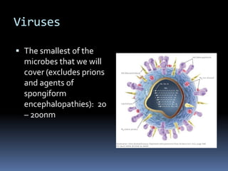
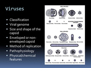
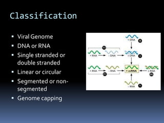
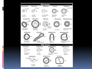
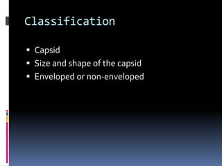
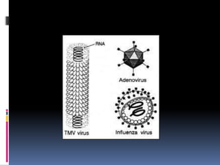
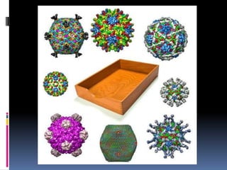
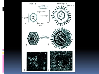
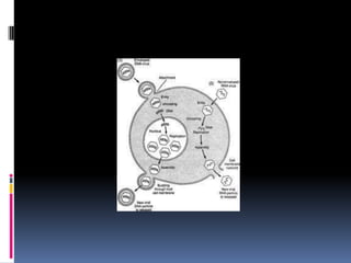
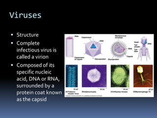
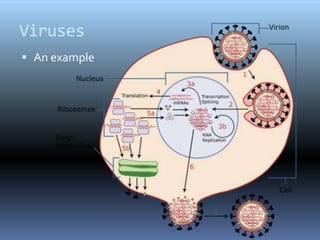
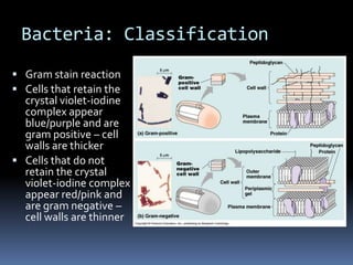
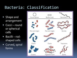
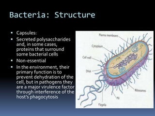
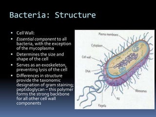
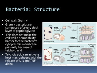
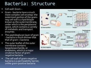
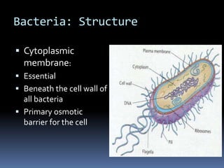
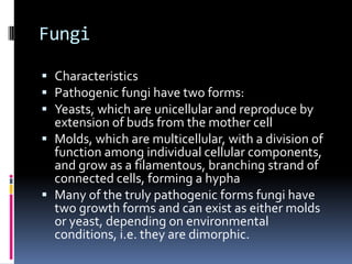
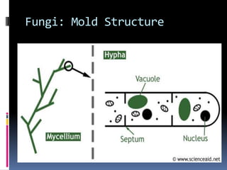
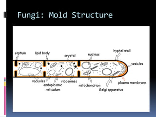
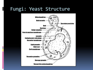
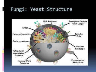
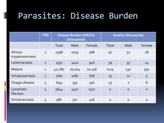
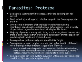
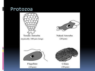
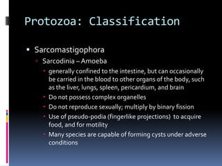
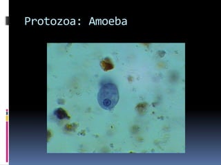
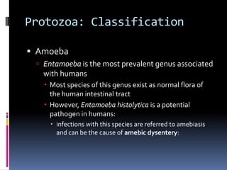
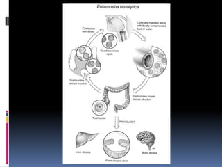
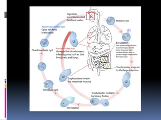
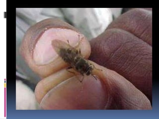
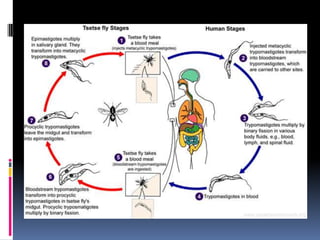
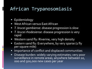
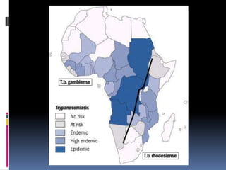
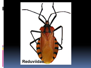
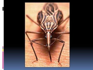
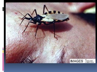
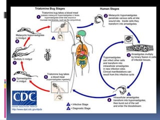
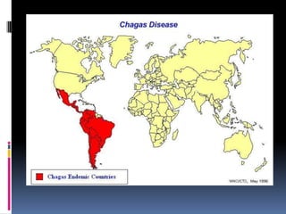
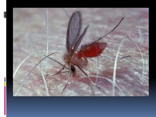
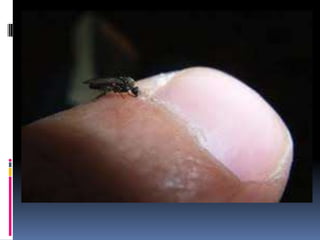
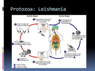
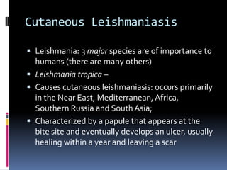
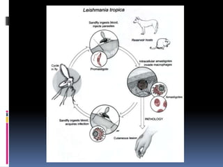
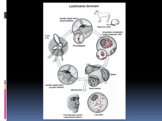
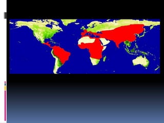
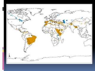
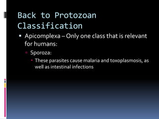
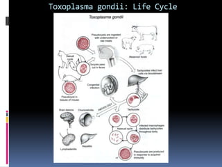
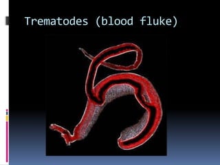
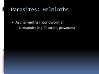
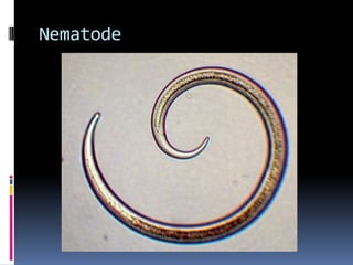
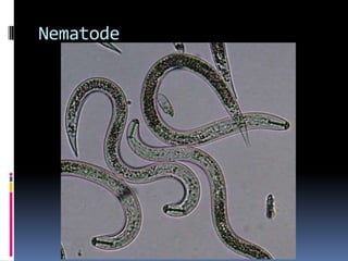
Infectious disease epidemiologyInfectious Organisms
VirusesThe smallest of the microbes that we will cover (excludes prions and agents of spongiform encephalopathies):  20 – 200nm
VirusesClassificationViral genomeSize and shape of the capsidEnveloped or non-enveloped capsidMethod of replicationPathophysiologyPhysical/chemical features
ClassificationViral GenomeDNA or RNASingle stranded or double strandedLinear or circularSegmented or non-segmentedGenome capping
ClassificationCapsidSize and shape of the capsidEnveloped or non-enveloped
Classification Method of replication
ClassificationPathophysiology of the virus such as host range, antigenic composition, vectors, and tissue tropism Physical/chemical features, such as susceptibility to acid or lipid solvents
VirusesStructureComplete infectious virus is called a virionComposed of its specific nucleic acid, DNA or RNA, surrounded by a protein coat known as the capsid
VirusesAn example
BacteriaClassification: Morphologic classification based on 1) staining characteristics, 2) shape and size of the organismBacteria: ClassificationGram stain reactionCells that retain the crystal violet-iodine complex appear blue/purple and are gram positive – cell walls are thicker Cells that do not retain the crystal violet-iodine complex appear red/pink and are gram negative – cell walls are thinner
Bacteria: ClassificationShape and arrangementCocci – round or spherical cellsBacilli – rod-shaped cellsCurved, spiral forms
Bacteria: ClassificationOther classificationmetabolic and physiologic differences, as measured by the presence or absence of specific enzymes these identify bacteria that are able to use particular nutrients for growth, or metabolize particular substratesGenetic methods
BacteriaStructure
Bacteria: StructureFlagella: Long, complex structures that are important for motility Non-essentialAn important antigen for identification and classification among those bacteria that possess them
Bacteria: StructurePili or fimbriae: Short, non-flexible structures that surround the surface of the cell  Responsible for adherence to host cell membranes through a very specific interaction that frequently determines the organotropism of a particular pathogen Non-essential Conjugation – the attachment and transfer of DNA between similar species of bacteria by way of the sex or F pili – this is process is the most common method for acquisition of antibiotic resistance determinants  by bacteria
Bacteria: StructureCapsules: Secreted polysaccharides and, in some cases, proteins that surround some bacterial cells Non-essential In the environment, their primary function is to prevent dehydration of the cell, but in pathogens they are a major virulence factor through interference of the host’s phagocytosis
Bacteria: StructureCell Wall: Essential component to all bacteria, with the exception of the mycoplasmaDetermines the size and shape of the cell Serves as an exoskeleton, preventing lysis of the cell Differences in structure provide the taxonomic designation of gram staining; peptidoglycan – this polymer forms the strong backbone for all other cell wall components
Bacteria: StructureCell wall: Gram +Gram + bacteria are composed of a very thick layer of peptidoglycan This does not make the cell wall a permeability barrier for the bacteria’s cytoplasmic membrane, primarily because of teichoic acid Teichoic acid can activate host macrophages with the release of IL-1 and TNF-alpha
Bacteria: StructureCell wall: Gram –
Gram - bacteria have a much more complex cell envelop: the outermost portion of the gram-neg cell wall is a lipid bilayer known as the outermembrane, under which is the periplasmic space, which contains a variety of metabolic and transport enzymes
The peptidoglycan layer of gram - bacteria is much thinner that that of gram + bacteria
The outer leaflet of the outer membrane contains lipopolysaccharide, or endotoxin, which is a major virulence factor of gram-negative bacteria
 The cell wall of gram-negative bacteria is a permeability barrier, unlike gram-positive bacteriaBacteria: StructureCytoplasmic membrane:EssentialBeneath the cell wall of all bacteriaPrimary osmotic barrier for the cell
FungiCharacteristicsPathogenic fungi have two forms: Yeasts, which are unicellular and reproduce by extension of buds from the mother cell Molds, which are multicellular, with a division of function among individual cellular components, and grow as a filamentous, branching strand of connected cells, forming a hyphaMany of the truly pathogenic forms fungi have two growth forms and can exist as either molds or yeast, depending on environmental conditions, i.e. they are dimorphic.
Fungi: Mold Structure
Fungi: Yeast Structure
FungiFungi of medical importance can be grouped according to the type or location of infection they cause:Superficial mycoses or infections that involve only the outermost layers of the skin and hairCutaneous mycoses that involve primarily the epidermisSubcutaneous mycoses that cause infections of the dermis and subcutaneous tissueSystemic mycoses, which are infections of internal organ systems
FungiSystemic MycosesThere are those systemic mycoses that are caused by true, or primary, pathogens, which are capable of causing disease in healthy peopleThere are those systemic mycoses that are opportunistic pathogens, which are marginally pathogenic and cause disseminated or deep-tissue infection in immunocompromised or debilitated hosts
ParasitesParasites: parasitic diseases are spread in conditions of poverty – substandard housing, poor water treatment, and crowding facilitate their transmission
Parasites: Disease Burden
Parasites: ProtozoaBelong to a subkingdom (Protozoa) as they are neither plant nor animalOval, spherical, or elongated cells that range in size from 1-300µm to 1-2 mmCytoplasmic membrane that encloses cytoplasm containing membrane-bound nuclei, mitochondria, 80S ribosomes, and a variety of specialized organelles associated with higher life formsMajority of protozoa are aquatic, living in soil water, rivers, oceans, etc; there is a small subset that are obligate parasites of animals capable of producing both acute and chronic diseaseCan reproduce both asexually and sexually (like fungi)Some protozoa have complex reproductive cycles, in which different hosts are required for different stages of the life cycleHosts in which sexual reproduction occurs is called the definitive host, while asexual multiplication occurs in the intermediate host, among Protozoa requiring multiple hosts to complete their life cyclesSome protozoa can form cysts to survive in unfavorable environmental conditions (like spore-forming bacteria)
Protozoa
Protozoa: ClassificationSarcomastigophoraSarcodinia – Amoebagenerally confined to the intestine, but can occasionally be carried in the blood to other organs of the body, such as the liver, lungs, spleen, pericardium, and brainDo not possess complex organellesDo not reproduce sexually; multiply by binary fissionUse of pseudo-podia (fingerlike projections)  to acquire food, and for motilityMany species are capable of forming cysts under adverse conditions
Protozoa: Amoeba
Protozoa: ClassificationAmoebaEntamoeba is the most prevalent genus associated with humansMost species of this genus exist as normal flora of the human intestinal tractHowever, Entamoebahistolytica is a potential pathogen in humans: infections with this species are referred to amebiasis and can be the cause of amebic dysentery:
ProtozoaAmoebic dysenteryActively growing organisms (trophozoites) invade the intestinal mucosa, resulting in lesions that can range in symptoms (few daily loose stools with small amounts of blood and mucous to numerous intestinal ulcers causing severe diarrhea and substantial amounts of blood and mucous; in some individuals these intestinal ulcers may erode into adjoining blood vessels, allowing the amoeba to spread to other organs, especially the liver and lungs, and eventually leading to abscess formationMore prevalent in tropical and subtropical regions than in temperate regionsOften associated with poor sanitary conditionsIn most cases, transmission is the result of ingestion of cysts from chronic carriers who shed the cyst form of the organism in their fecesUnlike acute cases, which tend to shed the trophozoite forms in feces, cysts are relatively resistant to harsh environmental conditions and therefore survive long enough to establish new infections
Protozoa: ClassificationMastigophora: flagellated protozoa commonly divided into those that cause intestinal or genital tract disease (intestinal flagellates) and those transmitted by blood-sucking insects (hemoflagellates)
Flagellated Protozoa
Flagellate Life Cycle
Protozoa: ClassificationMastigophora:Intestinal (genital) flagellatesGiardialamblia – causes giardiasisTrichomonasvaginalis – trichomoniasisHemoflagellatesTrypanosomaLeishmania
Example: TrypanosomiasisHemoflagellatesTrypanosomaTrypanosomes are the causative agents for: West and East African trypanosomiasis (Sleeping Sickness)  American trypanosomiasis (Chagas’s disease)
Example: TrypanosomiasisAfrican trypanosomiasis (Sleeping sickness)Tsetse fly (Glossinia) is the vectorThe trypanosomes migrate through the blood to lymph nodes This triggers ongoing attacks of fever The attacks can be intermittent and recur over a period of weeks to months These sustained attacks can often result in heart damage  As the disease progresses trypanosomes invade the central nervous system, causing meningoencephalitis
African TrypanosomiasisEpidemiologyWest African versus East AfricanT. bruceigambiense: disease progression is slow T. brucei rh0desiense: disease progression is very rapidWestern sand fly: Riverine, very high densityEastern sand fly: Everywhere, by very sparse (1 fly per square mile) Importance of conflict and displaced communitiesDisease burden: widely varying estimates; very poor surveillance in remote areas; anywhere between 10, 000 and 500,000 new cases per year
American TrypanosomiasisAmerican trypanosomiasis (Chagas’ Disease)Reduviid bug is the vectorThe trypanosome is unable to multiply outside the cell of its vertebrate host, and therefore undergoes a change to the amastigote form and can be found multiplying in virtually every cell in every organ of the body Hollow organsThe organ most often effected is the heart, wherein inflammation causes an enlargement of the heart
American TrypanosomiasisEpidemiologyHousing is a critical characteristic for reduviid bug control: thatched roofs; dogs and pigs close to or in the homeDisease Burden: prevalence ~ 8 million across all endemic countries; incidence ~ 40 – 50,000 per yearChronic disease lasting decades is quite common, so humans are a critical reservoir as well
Example: LeishmaniasisLeishmania spp.Causative agent of leishmaniasisTransmitted by the vector sand fly (Phlebotomusgenus in the Old World, Lutzomyia in the New World)The flagellated promastigote is the form that is in the vector gut and the form that is transferred to the human hostThe flagellated promastigotes then transform into nonmotileamastigotes that then proliferate in cells of the reticuloendothelial system, specifically, macrophages and endothelial cellsLeishmania cause disease by co-opting the immune response
Protozoa: Leishmania
CutaneousLeishmaniasisLeishmania: 3 major species are of importance to humans (there are many others)Leishmaniatropica – Causes cutaneousleishmaniasis: occurs primarily in the Near East, Mediterranean, Africa, Southern Russia and South Asia; Characterized by a papule that appears at the bite site and eventually develops an ulcer, usually healing within a year and leaving a scar
Cutaneousleishmaniasis
MucocutaneousLeishmaniasisL. braziliensis – Causes mucocutaneousleishmaniasis: a variant of cutaneous form that involves the mucous membranes of the nasopharyngeal area; If untreated the nasal septum, lips, and soft palate may be destroyed, resulting asphyxiation due to airway collapse or secondary bacterial infection
Mucocutaneousleishmaniasis
Visceral LeishmaniasisL. donovani – Causes visceral leishmaniasis: the leishmania are able to invade the reticuloendothelial system throughout the body, especially the liver and spleen As a result, these organs become enlarged causing abdominal swelling and often result in death an average of two years after onset of initial symptoms
Visceral leishmaniasis
Leishmaniasis EpidemiologyAgain, poor surveillance, especially for VLHundreds of millions are at genuine risk for infection. ~10 million infected, with ~1-2 million incident cases per year (includes all forms)Vector control has figured prominently: insecticide impregnated bed nets (similar to malaria)
Back to ProtozoanClassificationApicomplexa – Only one class that is relevant for humans:Sporoza: These parasites cause malaria and toxoplasmosis, as well as intestinal infections
Protozoa: ClassificationSporoza:Plasmodium vivax, P. ovale, P. malariae, and P. falciparum:Cause malariaTransmission by vector of the female mosquito (Anopheles)Clinical symptoms are variable across the different species, but in general include chills and fever at intermittent, regular intervals followed by profuse sweating
Protozoa: ClassificationSporozaToxoplasmagondiiCryptosporidium
Toxoplasmagondii: Life Cycle
Parasites: HelminthsPlatyhelminthes (flatworms): Typically no digestive tract, or only a rudimentary one Typically flat Most are hermaphroditic Humans are often the definitive hosts for adult worms, though there are also often intermediate hosts in other animals in different developmental stages of the worm
Parasites: HelminthsPlatyhelminthsCestodes (tapeworms)Trematodes (flukes)
Cestodes
Cestodes
Trematodes (liver fluke)
Trematodes (blood fluke)
Parasites: HelminthsAschelminths (roundworms)Nematodes (e.g. Toxocara, pinworms)

Infectious Organisms

  • 1.
  • 2.
    VirusesThe smallest ofthe microbes that we will cover (excludes prions and agents of spongiform encephalopathies): 20 – 200nm
  • 3.
    VirusesClassificationViral genomeSize andshape of the capsidEnveloped or non-enveloped capsidMethod of replicationPathophysiologyPhysical/chemical features
  • 4.
    ClassificationViral GenomeDNA orRNASingle stranded or double strandedLinear or circularSegmented or non-segmentedGenome capping
  • 7.
    ClassificationCapsidSize and shapeof the capsidEnveloped or non-enveloped
  • 11.
  • 13.
    ClassificationPathophysiology of thevirus such as host range, antigenic composition, vectors, and tissue tropism Physical/chemical features, such as susceptibility to acid or lipid solvents
  • 14.
    VirusesStructureComplete infectious virusis called a virionComposed of its specific nucleic acid, DNA or RNA, surrounded by a protein coat known as the capsid
  • 15.
  • 16.
    BacteriaClassification: Morphologic classificationbased on 1) staining characteristics, 2) shape and size of the organismBacteria: ClassificationGram stain reactionCells that retain the crystal violet-iodine complex appear blue/purple and are gram positive – cell walls are thicker Cells that do not retain the crystal violet-iodine complex appear red/pink and are gram negative – cell walls are thinner
  • 17.
    Bacteria: ClassificationShape andarrangementCocci – round or spherical cellsBacilli – rod-shaped cellsCurved, spiral forms
  • 18.
    Bacteria: ClassificationOther classificationmetabolicand physiologic differences, as measured by the presence or absence of specific enzymes these identify bacteria that are able to use particular nutrients for growth, or metabolize particular substratesGenetic methods
  • 19.
  • 20.
    Bacteria: StructureFlagella: Long,complex structures that are important for motility Non-essentialAn important antigen for identification and classification among those bacteria that possess them
  • 21.
    Bacteria: StructurePili orfimbriae: Short, non-flexible structures that surround the surface of the cell Responsible for adherence to host cell membranes through a very specific interaction that frequently determines the organotropism of a particular pathogen Non-essential Conjugation – the attachment and transfer of DNA between similar species of bacteria by way of the sex or F pili – this is process is the most common method for acquisition of antibiotic resistance determinants by bacteria
  • 22.
    Bacteria: StructureCapsules: Secretedpolysaccharides and, in some cases, proteins that surround some bacterial cells Non-essential In the environment, their primary function is to prevent dehydration of the cell, but in pathogens they are a major virulence factor through interference of the host’s phagocytosis
  • 23.
    Bacteria: StructureCell Wall:Essential component to all bacteria, with the exception of the mycoplasmaDetermines the size and shape of the cell Serves as an exoskeleton, preventing lysis of the cell Differences in structure provide the taxonomic designation of gram staining; peptidoglycan – this polymer forms the strong backbone for all other cell wall components
  • 24.
    Bacteria: StructureCell wall:Gram +Gram + bacteria are composed of a very thick layer of peptidoglycan This does not make the cell wall a permeability barrier for the bacteria’s cytoplasmic membrane, primarily because of teichoic acid Teichoic acid can activate host macrophages with the release of IL-1 and TNF-alpha
  • 25.
  • 26.
    Gram - bacteriahave a much more complex cell envelop: the outermost portion of the gram-neg cell wall is a lipid bilayer known as the outermembrane, under which is the periplasmic space, which contains a variety of metabolic and transport enzymes
  • 27.
    The peptidoglycan layerof gram - bacteria is much thinner that that of gram + bacteria
  • 28.
    The outer leafletof the outer membrane contains lipopolysaccharide, or endotoxin, which is a major virulence factor of gram-negative bacteria
  • 29.
    The cellwall of gram-negative bacteria is a permeability barrier, unlike gram-positive bacteriaBacteria: StructureCytoplasmic membrane:EssentialBeneath the cell wall of all bacteriaPrimary osmotic barrier for the cell
  • 30.
    FungiCharacteristicsPathogenic fungi havetwo forms: Yeasts, which are unicellular and reproduce by extension of buds from the mother cell Molds, which are multicellular, with a division of function among individual cellular components, and grow as a filamentous, branching strand of connected cells, forming a hyphaMany of the truly pathogenic forms fungi have two growth forms and can exist as either molds or yeast, depending on environmental conditions, i.e. they are dimorphic.
  • 31.
  • 32.
  • 33.
  • 34.
  • 35.
    FungiFungi of medicalimportance can be grouped according to the type or location of infection they cause:Superficial mycoses or infections that involve only the outermost layers of the skin and hairCutaneous mycoses that involve primarily the epidermisSubcutaneous mycoses that cause infections of the dermis and subcutaneous tissueSystemic mycoses, which are infections of internal organ systems
  • 36.
    FungiSystemic MycosesThere arethose systemic mycoses that are caused by true, or primary, pathogens, which are capable of causing disease in healthy peopleThere are those systemic mycoses that are opportunistic pathogens, which are marginally pathogenic and cause disseminated or deep-tissue infection in immunocompromised or debilitated hosts
  • 37.
    ParasitesParasites: parasitic diseasesare spread in conditions of poverty – substandard housing, poor water treatment, and crowding facilitate their transmission
  • 38.
  • 39.
    Parasites: ProtozoaBelong toa subkingdom (Protozoa) as they are neither plant nor animalOval, spherical, or elongated cells that range in size from 1-300µm to 1-2 mmCytoplasmic membrane that encloses cytoplasm containing membrane-bound nuclei, mitochondria, 80S ribosomes, and a variety of specialized organelles associated with higher life formsMajority of protozoa are aquatic, living in soil water, rivers, oceans, etc; there is a small subset that are obligate parasites of animals capable of producing both acute and chronic diseaseCan reproduce both asexually and sexually (like fungi)Some protozoa have complex reproductive cycles, in which different hosts are required for different stages of the life cycleHosts in which sexual reproduction occurs is called the definitive host, while asexual multiplication occurs in the intermediate host, among Protozoa requiring multiple hosts to complete their life cyclesSome protozoa can form cysts to survive in unfavorable environmental conditions (like spore-forming bacteria)
  • 40.
  • 41.
    Protozoa: ClassificationSarcomastigophoraSarcodinia –Amoebagenerally confined to the intestine, but can occasionally be carried in the blood to other organs of the body, such as the liver, lungs, spleen, pericardium, and brainDo not possess complex organellesDo not reproduce sexually; multiply by binary fissionUse of pseudo-podia (fingerlike projections) to acquire food, and for motilityMany species are capable of forming cysts under adverse conditions
  • 42.
  • 43.
    Protozoa: ClassificationAmoebaEntamoeba isthe most prevalent genus associated with humansMost species of this genus exist as normal flora of the human intestinal tractHowever, Entamoebahistolytica is a potential pathogen in humans: infections with this species are referred to amebiasis and can be the cause of amebic dysentery:
  • 46.
    ProtozoaAmoebic dysenteryActively growingorganisms (trophozoites) invade the intestinal mucosa, resulting in lesions that can range in symptoms (few daily loose stools with small amounts of blood and mucous to numerous intestinal ulcers causing severe diarrhea and substantial amounts of blood and mucous; in some individuals these intestinal ulcers may erode into adjoining blood vessels, allowing the amoeba to spread to other organs, especially the liver and lungs, and eventually leading to abscess formationMore prevalent in tropical and subtropical regions than in temperate regionsOften associated with poor sanitary conditionsIn most cases, transmission is the result of ingestion of cysts from chronic carriers who shed the cyst form of the organism in their fecesUnlike acute cases, which tend to shed the trophozoite forms in feces, cysts are relatively resistant to harsh environmental conditions and therefore survive long enough to establish new infections
  • 47.
    Protozoa: ClassificationMastigophora: flagellatedprotozoa commonly divided into those that cause intestinal or genital tract disease (intestinal flagellates) and those transmitted by blood-sucking insects (hemoflagellates)
  • 48.
  • 49.
  • 50.
    Protozoa: ClassificationMastigophora:Intestinal (genital)flagellatesGiardialamblia – causes giardiasisTrichomonasvaginalis – trichomoniasisHemoflagellatesTrypanosomaLeishmania
  • 51.
    Example: TrypanosomiasisHemoflagellatesTrypanosomaTrypanosomes arethe causative agents for: West and East African trypanosomiasis (Sleeping Sickness) American trypanosomiasis (Chagas’s disease)
  • 52.
    Example: TrypanosomiasisAfrican trypanosomiasis(Sleeping sickness)Tsetse fly (Glossinia) is the vectorThe trypanosomes migrate through the blood to lymph nodes This triggers ongoing attacks of fever The attacks can be intermittent and recur over a period of weeks to months These sustained attacks can often result in heart damage As the disease progresses trypanosomes invade the central nervous system, causing meningoencephalitis
  • 56.
    African TrypanosomiasisEpidemiologyWest Africanversus East AfricanT. bruceigambiense: disease progression is slow T. brucei rh0desiense: disease progression is very rapidWestern sand fly: Riverine, very high densityEastern sand fly: Everywhere, by very sparse (1 fly per square mile) Importance of conflict and displaced communitiesDisease burden: widely varying estimates; very poor surveillance in remote areas; anywhere between 10, 000 and 500,000 new cases per year
  • 58.
    American TrypanosomiasisAmerican trypanosomiasis(Chagas’ Disease)Reduviid bug is the vectorThe trypanosome is unable to multiply outside the cell of its vertebrate host, and therefore undergoes a change to the amastigote form and can be found multiplying in virtually every cell in every organ of the body Hollow organsThe organ most often effected is the heart, wherein inflammation causes an enlargement of the heart
  • 64.
    American TrypanosomiasisEpidemiologyHousing isa critical characteristic for reduviid bug control: thatched roofs; dogs and pigs close to or in the homeDisease Burden: prevalence ~ 8 million across all endemic countries; incidence ~ 40 – 50,000 per yearChronic disease lasting decades is quite common, so humans are a critical reservoir as well
  • 66.
    Example: LeishmaniasisLeishmania spp.Causativeagent of leishmaniasisTransmitted by the vector sand fly (Phlebotomusgenus in the Old World, Lutzomyia in the New World)The flagellated promastigote is the form that is in the vector gut and the form that is transferred to the human hostThe flagellated promastigotes then transform into nonmotileamastigotes that then proliferate in cells of the reticuloendothelial system, specifically, macrophages and endothelial cellsLeishmania cause disease by co-opting the immune response
  • 69.
  • 70.
    CutaneousLeishmaniasisLeishmania: 3 majorspecies are of importance to humans (there are many others)Leishmaniatropica – Causes cutaneousleishmaniasis: occurs primarily in the Near East, Mediterranean, Africa, Southern Russia and South Asia; Characterized by a papule that appears at the bite site and eventually develops an ulcer, usually healing within a year and leaving a scar
  • 72.
  • 73.
    MucocutaneousLeishmaniasisL. braziliensis –Causes mucocutaneousleishmaniasis: a variant of cutaneous form that involves the mucous membranes of the nasopharyngeal area; If untreated the nasal septum, lips, and soft palate may be destroyed, resulting asphyxiation due to airway collapse or secondary bacterial infection
  • 75.
  • 76.
    Visceral LeishmaniasisL. donovani– Causes visceral leishmaniasis: the leishmania are able to invade the reticuloendothelial system throughout the body, especially the liver and spleen As a result, these organs become enlarged causing abdominal swelling and often result in death an average of two years after onset of initial symptoms
  • 78.
  • 79.
    Leishmaniasis EpidemiologyAgain, poorsurveillance, especially for VLHundreds of millions are at genuine risk for infection. ~10 million infected, with ~1-2 million incident cases per year (includes all forms)Vector control has figured prominently: insecticide impregnated bed nets (similar to malaria)
  • 82.
    Back to ProtozoanClassificationApicomplexa– Only one class that is relevant for humans:Sporoza: These parasites cause malaria and toxoplasmosis, as well as intestinal infections
  • 83.
    Protozoa: ClassificationSporoza:Plasmodium vivax,P. ovale, P. malariae, and P. falciparum:Cause malariaTransmission by vector of the female mosquito (Anopheles)Clinical symptoms are variable across the different species, but in general include chills and fever at intermittent, regular intervals followed by profuse sweating
  • 84.
  • 85.
  • 86.
    Parasites: HelminthsPlatyhelminthes (flatworms):Typically no digestive tract, or only a rudimentary one Typically flat Most are hermaphroditic Humans are often the definitive hosts for adult worms, though there are also often intermediate hosts in other animals in different developmental stages of the worm
  • 87.
  • 88.
  • 89.
  • 90.
  • 91.
  • 92.