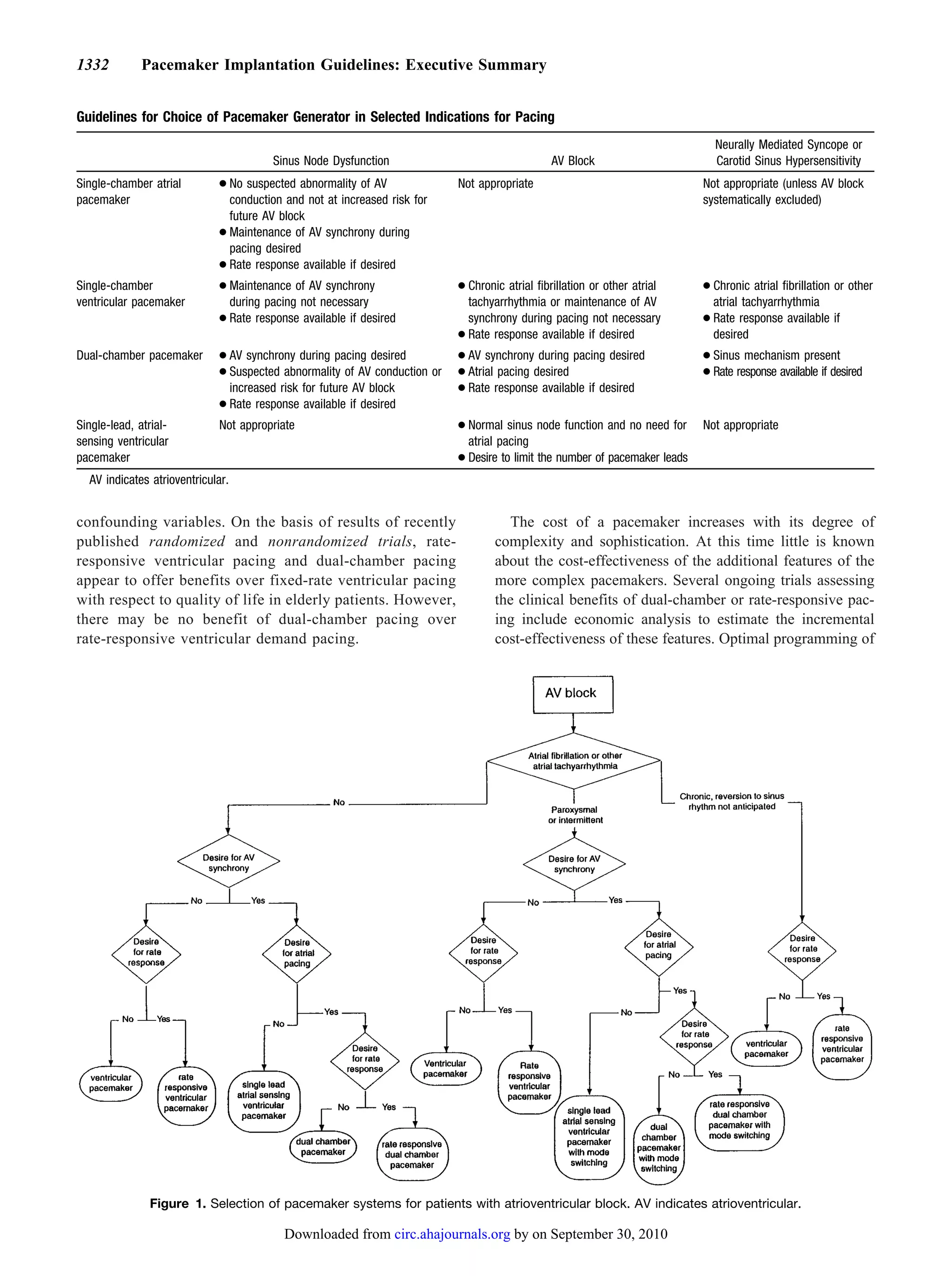
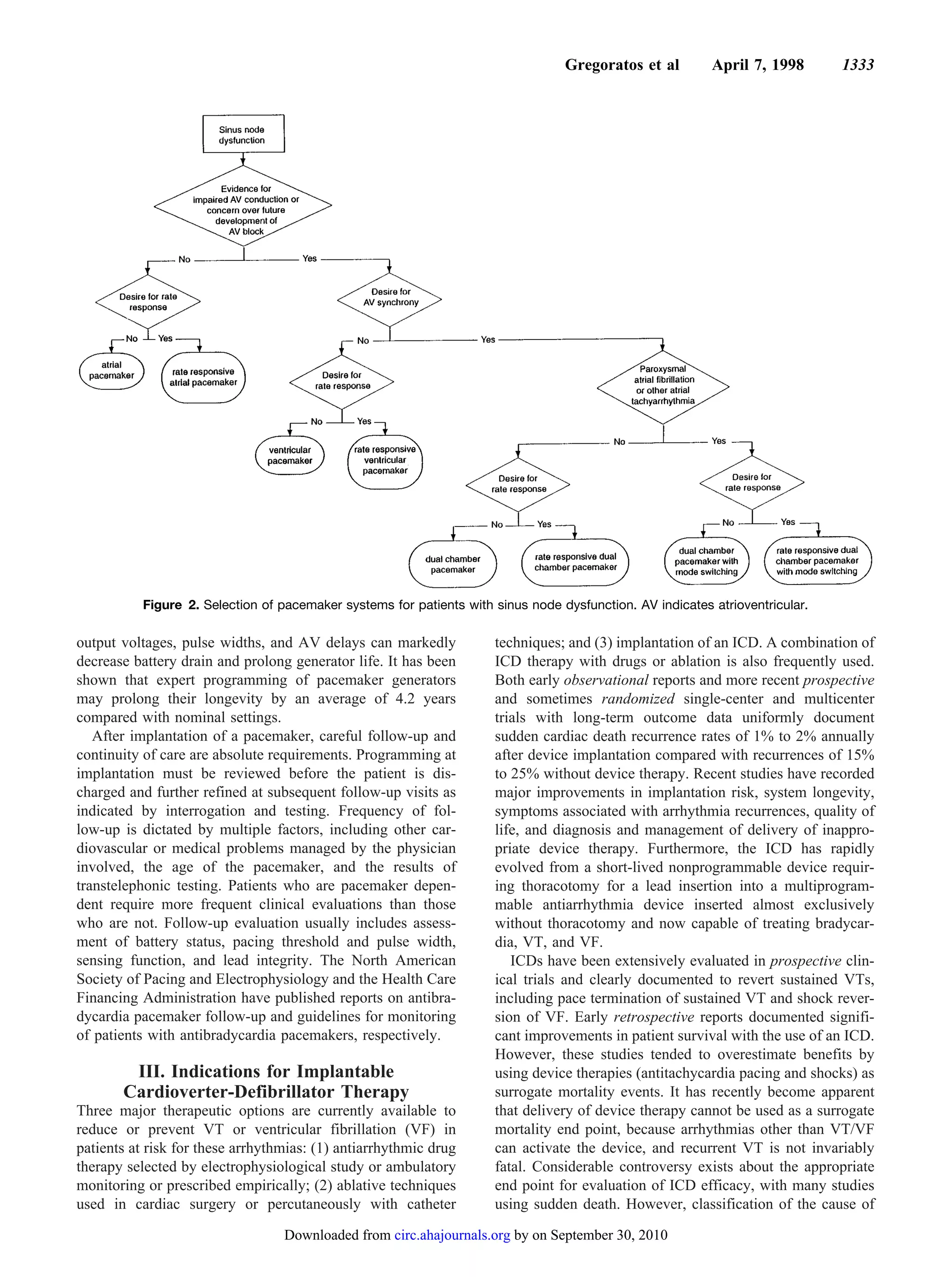
3. 1326 Pacemaker Implantation Guidelines: Executive Summary
when it can be expected to be transient. The statement block, particularly if syncope has occurred. It is now recog-
“incidental finding at electrophysiological study” is used nized that marked first-degree AV block can lead to symp-
several times in this document and does not imply such a toms even in the absence of higher degrees of AV conduction
study is indicated. Appropriate indications for electrophysio- disturbance and may be associated with a “pseudopacemaker
logical studies have been previously published. syndrome” because of close proximity of atrial systole to the
The term “symptomatic bradycardia” is used frequently preceding ventricular systole. Small uncontrolled trials have
throughout the guidelines and is defined as a documented suggested some symptomatic and functional improvement
bradyarrhythmia that is directly responsible for the develop- with pacing in patients with PR intervals 0.30 second,
ment of frank syncope or near-syncope, transient dizziness or especially those with left ventricular (LV) dysfunction, some
light-headedness, and confusional states resulting from cere- of whom may benefit from dual-chamber pacing with short
bral hypoperfusion attributable to slow heart rate. Fatigue, AV delay.
Type I second-degree AV block is unlikely to progress to
exercise intolerance, and frank congestive heart failure may
advanced AV block when the delay is within the AV node.
also result from bradycardia. These symptoms may occur at
Consequently, pacing is not usually indicated in this situation.
rest or with exertion. Definite correlation of symptoms with a
However, in patients with type II second-degree AV block
bradyarrhythmia is a requirement to fulfill the criteria of
(either intra- or infra-His), symptoms are frequent, prognosis
symptomatic bradycardia. Caution should be exercised not to is compromised, and progression to third-degree AV block is
confuse physiological sinus bradycardia (as occurs in highly common.
trained athletes) with pathological bradyarrhythmias. Physiological AV block in the presence of supraventricular
The section on indications for ICDs has been extensively tachyarrhythmias is not an indication for pacemaker implan-
revised and enlarged to reflect emerging developments in this tation except as specifically defined in the recommendations
field and the voluminous literature attesting to the efficacy of below. Similarly, neurally mediated mechanisms in young
these devices in the treatment of sudden cardiac death and patients with AV block should be assessed before proceeding
malignant ventricular arrhythmias. Indications for ICDs are with permanent pacing. Finally, permanent pacing for AV
constantly changing and can be expected to change further as block after valve surgery follows a variable natural history;
ongoing large-scale trials are reported. In these guidelines the therefore, the decision for permanent pacing is at the physi-
term “mortality” is used to indicate “all-cause” mortality cian’s discretion.
unless otherwise specified. The committee elected to use
“all-cause” mortality because of the variable definition of Indications for Permanent Pacing in Acquired
“sudden death” and the developing consensus to use “all- Atrioventricular Block in Adults
cause” mortality as the most appropriate end point of clinical Class I
trials. 1. Third-degree AV block at any anatomic level associ-
The final recommendations for indications for device ated with any one of the following conditions:
therapy are expressed in the standard ACC/AHA format: a. Bradycardia with symptoms presumed to be due to
Class I: Conditions for which there is evidence and/or AV block. (Level of evidence: C)
general agreement that a given procedure or treatment is b. Arrhythmias and other medical conditions that
beneficial, useful, and effective. require drugs that result in symptomatic bradycar-
Class II: Conditions for which there is conflicting dia. (Level of evidence: C)
evidence and/or a divergence of opinion about the useful- c. Documented periods of asystole >3.0 seconds or any
ness/efficacy of a procedure or treatment. escape rate <40 beats per minute (bpm) in awake,
Class IIa: Weight of evidence/opinion is in favor of symptom-free patients. (Level of evidence: B, C)
usefulness/efficacy. d. After catheter ablation of the AV junction. (Level of
Class IIb: Usefulness/efficacy is less well established evidence: B, C) There are no trials to assess outcome
by evidence/opinion. without pacing, and pacing is virtually always
Class III: Conditions for which there is evidence and/or planned in this situation unless the operative pro-
general agreement that a procedure/treatment is not cedure is AV junction modification.
useful/effective and in some cases may be harmful. e. Postoperative AV block that is not expected to
resolve. (Level of evidence: C)
II. Indications for Permanent Pacing f. Neuromuscular diseases with AV block such as
myotonic muscular dystrophy, Kearns-Sayre syn-
A. Pacing for Acquired Atrioventricular Block drome, Erb’s dystrophy (limb-girdle), and peroneal
in Adults muscular atrophy. (Level of evidence: B)
Patients with abnormalities of atrioventricular (AV) conduc- 2. Second-degree AV block regardless of type or site of
tion may be asymptomatic or may experience serious symp- block, with associated symptomatic bradycardia.
toms related to bradycardia, ventricular arrhythmias, or both. (Level of evidence: B)
Decisions about the need for a pacemaker are necessarily
influenced by the presence or absence of symptoms that are Class IIa
directly attributable to bradycardia. 1. Asymptomatic third-degree AV block at any anatomic
Nonrandomized studies strongly suggest that permanent site with average awake ventricular rates of 40 bpm or
pacing improves survival in patients with third-degree AV faster. (Level of evidence: B, C)
Downloaded from circ.ahajournals.org by on September 30, 2010
4. Gregoratos et al April 7, 1998 1327
2. Asymptomatic type II second-degree AV block. (Level Indications for Permanent Pacing in Chronic
of evidence: B) Bifascicular and Trifascicular Block
3. Asymptomatic type I second-degree AV block at intra- or Class I
infra-His levels found incidentally at electrophysiological
1. Intermittent third-degree AV block. (Level of evidence: B)
study for other indications. (Level of evidence: B)
2. Type II second-degree AV block. (Level of evidence: B)
4. First-degree AV block with symptoms suggestive of
pacemaker syndrome and documented alleviation of Class IIa
symptoms with temporary AV pacing. (Level of evi-
1. Syncope not proved to be due to AV block when other
dence: B) likely causes have been excluded, specifically ventric-
ular tachycardia (VT). (Level of evidence: B)
Class IIb 2. Incidental finding at electrophysiological study of
1. Marked first-degree AV block (>0.30 second) in pa- markedly prolonged HV interval (>100 milliseconds)
tients with LV dysfunction and symptoms of conges- in asymptomatic patients. (Level of evidence: B)
tive heart failure in whom a shorter AV interval 3. Incidental finding at electrophysiological study of pac-
results in hemodynamic improvement, presumably by ing-induced infra-His block that is not physiological.
(Level of evidence: B)
decreasing left atrial filling pressure. (Level of evi-
dence: C) Class IIb
None.
Class III
Class III
1. Asymptomatic first-degree AV block. (Level of evi-
1. Fascicular block without AV block or symptoms.
dence: B) (See “Pacing for Chronic Bifascicular and (Level of evidence: B)
Trifascicular Block.”) 2. Fascicular block with first-degree AV block without
2. Asymptomatic type I second-degree AV block at the symptoms. (Level of evidence: B)
supra-His (AV node) level or not known to be intra- or
infra-Hisian. (Level of evidence: B, C) C. Pacing for Atrioventricular Block Associated
3. AV block expected to resolve and unlikely to recur (eg, With Acute Myocardial Infarction
drug toxicity, Lyme disease). (Level of evidence: B) The long-term prognosis of survivors of acute myocardial
infarction (AMI) who develop AV block is related primarily
B. Pacing for Chronic Bifascicular and to the extent of myocardial damage and the character of
Trifascicular Block intraventricular conduction disturbances rather than the AV
Symptomatic advanced AV block that develops in patients block itself. Indications for permanent pacing in this setting
do not necessarily depend on the presence of symptoms.
with underlying bifascicular and trifascicular block is asso-
Patients with AMI who develop intraventricular conduction
ciated with a high mortality rate and a significant incidence of
defects (with the exception of isolated left anterior fascicular
sudden death. However, there is considerable evidence that block) have an unfavorable short- and long-term prognosis
the rate of progression of bifascicular block to third-degree and an increased incidence of sudden death.
AV block is slow. Syncope is common in patients with The decision to implant a permanent pacemaker for AV or
bifascicular block, and there is evidence that syncope in this intraventricular conduction block complicating AMI will
setting is associated with an increased incidence of sudden depend on the type of conduction disturbance, location of the
cardiac death. Therefore, if the cause of syncope in the infarction, and relation of the electrical disturbance to infarct
presence of bifascicular or trifascicular block cannot be time. Thrombolytic therapy has decreased the incidence of
determined with certainty, prophylactic permanent pacing is high-grade AV block in AMI, but mortality remains high in
indicated. this group of patients.
The PR and HV intervals have been identified as possible The impact of preexisting bundle branch block on mortal-
predictors of third-degree AV block and sudden death in the ity after AMI is uncertain. However, left bundle branch block
presence of underlying bifascicular block. However, the combined with advanced or third-degree AV block and right
prolongation is often at the level of the AV node, and bundle branch block combined with left anterior or left
posterior fascicular block carry a particularly ominous
frequently there is no correlation between the PR and HV
prognosis.
intervals and progression to third-degree AV block and
incidence of sudden cardiac death. Some investigators have Indications for Permanent Pacing After the Acute
suggested that asymptomatic patients with bifascicular block Phase of Myocardial Infarction*
and a prolonged HV interval ( 100 milliseconds) should be
Class I
considered for permanent pacing. However, the incidence of
progression to third-degree AV block is low, even in the 1. Persistent second-degree AV block in the His-Purkinje
setting of prolonged HV interval. Death is often not sudden or system with bilateral bundle branch block or third-
due to advanced AV block but rather due to the underlying *These recommendations generally follow the ACC/AHA Guidelines
heart disease itself and nonarrhythmic cardiac causes. for the Management of Patients with Acute Myocardial Infarction.
Downloaded from circ.ahajournals.org by on September 30, 2010
5. 1328 Pacemaker Implantation Guidelines: Executive Summary
degree AV block within or below the His-Purkinje genic and will occur as a consequence of essential long-
system after AMI. (Level of evidence: B) term drug therapy of a type and dose for which there are
2. Transient advanced (second- or third-degree) infran- no acceptable alternatives. (Level of evidence: C)
odal AV block and associated bundle branch block. If 2. Symptomatic chronotropic incompetence. (Level of
the site of block is uncertain, an electrophysiological evidence: C)
study may be necessary. (Level of evidence: B)
Class IIa
3. Persistent and symptomatic second- or third-degree
AV block. (Level of evidence: C) 1. Sinus node dysfunction occurring spontaneously or as
a result of necessary drug therapy with heart rate <40
Class IIa bpm when a clear association between significant
None. symptoms consistent with bradycardia and the actual
presence of bradycardia has not been documented.
Class IIb (Level of evidence: C)
1. Persistent second- or third-degree AV block at the AV
node level. (Level of evidence: B) Class IIb
1. In minimally symptomatic patients, chronic heart rate
Class III <30 bpm while awake. (Level of evidence: C)
1. Transient AV block in the absence of intraventricular Class III
conduction defects. (Level of evidence: B)
2. Transient AV block in the presence of isolated left 1. Sinus node dysfunction in asymptomatic patients, in-
cluding those in whom substantial sinus bradycardia
anterior fascicular block. (Level of evidence: B)
(heart rate <40 bpm) is a consequence of long-term
3. Acquired left anterior fascicular block in the absence
drug treatment.
of AV block. (Level of evidence: B)
2. Sinus node dysfunction in patients with symptoms
4. Persistent first-degree AV block in the presence of
suggestive of bradycardia that are clearly documented
bundle branch block that is old or age indeterminate. as not associated with a slow heart rate.
(Level of evidence: B) 3. Sinus node dysfunction with symptomatic bradycardia
due to nonessential drug therapy.
D. Pacing in Sinus Node Dysfunction
Correlation of symptoms with arrhythmias resulting from E. Prevention and Termination of
sinus node dysfunction (eg, sinus bradycardia, sinus arrest, Tachyarrhythmias by Pacing
paroxysmal supraventricular tachycardia alternating with pe- Pacing can be useful in terminating a variety of
riods of bradycardia or even asystole) is essential in deciding tachyarrhythmias, including atrial flutter, paroxysmal reen-
whether a permanent pacemaker is indicated. This correlation trant supraventricular tachycardia, and VT. A variety of
may be difficult because of the intermittent nature of the pacing patterns have been used, including programmed stim-
episodes. Sinus node dysfunction may also express itself as ulation and short bursts of rapid pacing. Antitachyarrhythmia
chronotropic incompetence. Rate-responsive pacemakers devices may detect the tachycardia and automatically activate
have clinically benefited patients by restoring physiological a pacing sequence or may respond to an external instruction
heart rate during physical activity in this setting. (eg, application of a magnet). Similarly, prevention of
Trained athletes may have a physiological sinus bradycar- tachyarrhythmias by pacing has been demonstrated in several
dia of 40 to 50 bpm while awake and at rest and a sleeping situations (eg, patients with the long QT syndrome and
heart rate as low as 30 bpm with sinus pauses producing recurrent pause-dependent VT). Combined therapy of pacing
asystolic intervals as long as 2.8 seconds. These findings are and -blockade has been reported to shorten the QT interval
due to increased vagal tone and are not an indication for and help prevent sudden cardiac death. Atrial synchronous
ventricular pacing may prevent recurrences of reentrant
permanent pacing.
supraventricular tachycardia, but this technique is rarely used
Permanent pacing in patients with sinus node dysfunction
today, given the availability of catheter ablation and other
will frequently relieve symptoms but may not necessarily
alternative therapies. In some patients with bradycardia-
result in improved survival. Whether dual-chamber pacing
dependent atrial fibrillation, atrial pacing may also be effec-
improves survival compared with ventricular pacing remains tive in reducing the frequency of recurrence. Dual-site and
controversial. Multiple prospective trials are ongoing to biatrial pacing are actively being investigated as therapies for
assess the superiority of dual-chamber versus ventricular- symptomatic drug-refractory atrial fibrillation with concom-
based pacing systems in patients with sinus node dysfunction. itant bradyarrhythmias.
Indications for Permanent Pacing in Sinus Indications for Permanent Pacemakers That
Node Dysfunction Automatically Detect and Pace to
Class I Terminate Tachycardias
1. Sinus node dysfunction with documented symptomatic Class I
bradycardia, including frequent sinus pauses that pro- 1. Symptomatic recurrent supraventricular tachycardia
duce symptoms. In some patients, bradycardia is iatro- that is reproducibly terminated by pacing after drugs
Downloaded from circ.ahajournals.org by on September 30, 2010
6. Gregoratos et al April 7, 1998 1329
and catheter ablation fail to control the arrhythmia or important vasodepressor component in their reflex re-
produce intolerable side effects. (Level of evidence: C) sponse, this component must be addressed as well.
2. Symptomatic recurrent sustained VT as part of an Ten to forty percent of syncopal episodes are due to a
automatic defibrillator system. (Level of evidence: B) variety of neurally mediated syndromes, the most common
being vasovagal syncope. Considerable controversy exists
Class IIa concerning the role of permanent pacing in refractory neu-
None. rally mediated syncope associated with significant bradycar-
Class IIb dia or asystole because approximately 25% of patients have a
predominant vasodepressor reaction without significant bra-
1. Recurrent supraventricular tachycardia or atrial flut- dycardia. There is conflicting evidence in the literature
ter that is reproducibly terminated by pacing as an regarding the efficacy of permanent pacing in neurally
alternative to drug therapy or ablation. (Level of mediated syncope, although a recent randomized trial in
evidence: C) highly symptomatic patients with bradycardia demonstrated
that permanent pacing increased the time to the first syncopal
Class III
event.
1. Tachycardias frequently accelerated or converted to
fibrillation by pacing. Indications for Permanent Pacing in
2. The presence of accessory pathways with the capacity Hypersensitive Carotid Sinus Syndrome and
for rapid anterograde conduction whether or not the Neurally Mediated Syncope
pathways participate in the mechanism of the Class I
tachycardia.
1. Recurrent syncope caused by carotid sinus stimula-
Pacing Indications to Prevent Tachycardia tion; minimal carotid sinus pressure induces ventric-
ular asystole of >3 seconds’ duration in the absence of
Class I any medication that depresses the sinus node or AV
1. Sustained pause-dependent VT, with or without pro- conduction. (Level of evidence: C)
longed QT, in which the efficacy of pacing is thor-
oughly documented. (Level of evidence: C) Class IIa
1. Recurrent syncope without clear, provocative events
Class IIa and with a hypersensitive cardioinhibitory response.
1. High-risk patients with congenital long QT syndrome. (Level of evidence: C)
(Level of evidence: C) 2. Syncope of unexplained origin when major abnormal-
ities of sinus node function or AV conduction are
Class IIb discovered or provoked in electrophysiological studies.
1. AV reentrant or AV node reentrant supraventricular (Level of evidence: C)
tachycardia not responsive to medical or ablative
therapy. (Level of evidence: C) Class IIb
2. Prevention of symptomatic, drug-refractory, recur- 1. Neurally mediated syncope with significant bradycar-
rent atrial fibrillation. (Level of evidence: C) dia reproduced by a head-up tilt with or without
isoproterenol or other provocative maneuvers. (Level
Class III of evidence: B)
1. Frequent or complex ventricular ectopic activity with-
out sustained VT in the absence of the long QT Class III
syndrome. 1. A hyperactive cardioinhibitory response to carotid
2. Long QT syndrome due to reversible causes. sinus stimulation in the absence of symptoms.
2. A hyperactive cardioinhibitory response to carotid
F. Pacing in Hypersensitive Carotid Sinus and sinus stimulation in the presence of vague symptoms
Neurally Mediated Syndromes such as dizziness, light-headedness, or both.
Hypersensitive carotid sinus syndrome is an uncommon 3. Recurrent syncope, light-headedness, or dizziness in
cause of syncope or presyncope. Symptoms in this setting the absence of a hyperactive cardioinhibitory
are mediated through both cardioinhibitory and vasode- response.
pressor reflexes. It is necessary to ascertain the relative 4. Situational vasovagal syncope in which avoidance be-
contribution of these two components of carotid sinus havior is effective.
stimulation before concluding that permanent pacing is
clinically indicated. Patients with symptoms due entirely G. Pacing in Children and Adolescents
to the cardioinhibitory response of carotid sinus stimula- Permanent pacing in children or adolescents is generally
tion can be effectively treated with permanent pacing. indicated in (1) symptomatic sinus bradycardia, (2) recurrent
However, because 10% to 20% of patients also have an bradycardia-tachycardia syndromes, (3) congenital AV block,
Downloaded from circ.ahajournals.org by on September 30, 2010
7. 1330 Pacemaker Implantation Guidelines: Executive Summary
and (4) advanced second- or third-degree surgically induced 2. Congenital third-degree AV block beyond the first
or acquired AV block. Important differences between indica- year of life with an average heart rate <50 bpm or
tions for permanent pacing in children and adults include (1) abrupt pauses in ventricular rate that are two or three
age dependency of physiological heart rate and (2) impact of times the basic cycle length. (Level of evidence: B)
residual ventricular dysfunction and abnormal circulatory 3. Long QT syndrome with 2:1 AV or third-degree AV
physiology after surgical palliation of complex congenital block. (Level of evidence: B)
cardiac defects. 4. Asymptomatic sinus bradycardia in the child with
Symptomatic bradycardia is an indication for pacemaker complex congenital heart disease with resting heart
implantation, provided other causes of symptoms have been rate <35 bpm or pauses in ventricular rate >3 sec-
excluded. The bradycardia-tachycardia syndrome is an in- onds. (Level of evidence: C)
creasingly frequent problem in young patients after surgery
for congenital heart disease. Recurrent or chronic atrial flutter Class IIb
is responsible for substantial morbidity and mortality in 1. Transient postoperative third-degree AV block that
young patients, and long-term atrial pacing at physiological reverts to sinus rhythm with residual bifascicular
rates and atrial antitachycardia pacing have been used in this block. (Level of evidence: C)
setting. However, the results of permanent pacing to date 2. Congenital third-degree AV block in the asymptomat-
have been equivocal and the source of considerable ic neonate, child, or adolescent with an acceptable rate,
controversy. narrow QRS complex, and normal ventricular func-
The indications for permanent pacing in congenital third- tion. (Level of evidence: B)
degree AV block have evolved with some studies suggesting 3. Asymptomatic sinus bradycardia in the adolescent
improved long-term survival and prevention of syncopal with congenital heart disease with resting heart rate
episodes in asymptomatic patients with congenital complete <35 bpm or pauses in ventricular rate >3 seconds.
heart block who meet specific criteria. High-grade second- or (Level of evidence: C)
third-degree AV block persisting for 7 to 14 days after cardiac
surgery is an indication for permanent pacing. The need for Class III
permanent pacing in patients with transient postoperative AV 1. Transient postoperative AV block with return of normal
block and residual bifascicular block has not been estab- AV conduction within 7 days. (Level of evidence: B)
lished, whereas patients with AV conduction that returns to 2. Asymptomatic postoperative bifascicular block with
normal have a favorable prognosis. or without first-degree AV block. (Level of evidence: C)
3. Asymptomatic type I second-degree AV block. (Level
Indications for Permanent Pacing in Children of evidence: C)
and Adolescents 4. Asymptomatic sinus bradycardia in the adolescent
Class I when the longest RR interval is <3 seconds and the
minimum heart rate is >40 bpm. (Level of evidence: C)
1. Advanced second- or third-degree AV block associated
with symptomatic bradycardia, congestive heart fail-
H. Pacing in Specific Conditions
ure, or low cardiac output. (Level of evidence: C) In patients with severely symptomatic hypertrophic cardio-
2. Sinus node dysfunction with correlation of symptoms myopathy, early nonrandomized studies demonstrated that
during age-inappropriate bradycardia. The definition implantation of a dual-chamber pacemaker with a short AV
of bradycardia varies with the patient’s age and delay decreased the magnitude of LV outflow obstruction and
expected heart rate. (Level of evidence: B) improved symptoms. However, recent observational studies
3. Postoperative advanced second- or third-degree AV suggest that a decrease in LV outflow gradient produced by a
block that is not expected to resolve or persists at least temporary dual chamber may adversely affect ventricular
7 days after cardiac surgery. (Level of evidence: B, C) filling and cardiac output. Recent randomized trials have
4. Congenital third-degree AV block with a wide QRS yielded variable results: one study demonstrated that DDD
escape rhythm or ventricular dysfunction. (Level of pacing reduced outflow tract gradient and improved New
evidence: B) York Heart Association (NYHA) functional class, whereas
5. Congenital third-degree AV block in the infant with another randomized double-blind study demonstrated no sig-
a ventricular rate <50 to 55 bpm or with congenital nificant subjective or exercise capacity improvement in the
heart disease and a ventricular rate <70 bpm. (Level paced versus nonpaced patient group at 2 to 3 months of
of evidence: B, C) follow-up, despite a significant decrease in LV outflow
6. Sustained pause-dependent VT, with or without pro- gradient. As a result, pacing indications for hypertrophic
longed QT, in which the efficacy of pacing is thor- cardiomyopathy remain controversial.
oughly documented. (Level of evidence: B)
Pacing Indications for
Class IIa Hypertrophic Cardiomyopathy
1. Bradycardia-tachycardia syndrome with the need for Class I
long-term antiarrhythmic treatment other than digi- Class I indications for sinus node dysfunction or AV block
talis. (Level of evidence: C) as previously described. (Level of evidence: C)
Downloaded from circ.ahajournals.org by on September 30, 2010
8. Gregoratos et al April 7, 1998 1331
Class IIa with previously stated Class I indications should have per-
None. manent pacing.
Class IIb
Pacing Indications After Cardiac Transplantation
1. Medically refractory, symptomatic hypertrophic car-
Class I
diomyopathy with significant resting or provoked LV
outflow obstruction. (Level of evidence: C) 1. Symptomatic bradyarrhythmias/chronotropic incompe-
tence not expected to resolve and other Class I indica-
Class III tions for permanent pacing. (Level of evidence: C)
1. Patients who are asymptomatic or medically
controlled.
Class IIa
None.
2. Symptomatic patients without evidence of LV outflow
obstruction. Class IIb
Several nonrandomized trials of patients with symptomatic 1. Symptomatic bradyarrhythmias/chronotropic incom-
dilated cardiomyopathy refractory to medical therapy have petence that, although transient, may persist for
reported limited improvement of symptoms with dual-cham- months and require intervention. (Level of evidence: C)
ber pacing with a short AV delay. However, at this time no
long-term data are available, and there is no consensus of Class III
opinion for this indication. It has been hypothesized that a 1. Asymptomatic bradyarrhythmias after cardiac trans-
well-timed atrial contraction primes the ventricles and de- plantation.
creases mitral regurgitation, thus augmenting stroke volume
and arterial pressure. However, one randomized controlled I. Selection and Follow-up of Pacemaker Devices
trial showed no significant benefit of DDD pacing through a Once a decision has been reached to implant a pacemaker,
range of PR intervals despite the presence of both tricuspid the clinician may choose from a large number of pace-
and mitral regurgitation. Preliminary data suggest that simul- maker generators and leads. Generator choices include
taneous biventricular pacing may improve cardiac hemody- single- versus dual-chamber devices, unipolar versus bi-
namics and lead to subjective and objective symptom im- polar configuration, presence of rate responsiveness and
provement. This technique must still be considered type of sensor used, advanced features such as automatic
investigational at this time. mode switching, generator size, battery capacity, and cost.
Lead choices include polarity, type of insulation material,
Pacing Indications for Dilated Cardiomyopathy active versus passive fixation mechanism, presence of
Class I steroid elution, and typical pacing impedance. Other fac-
Class I indications for sinus node dysfunction or AV block tors that frequently influence the choice of a pacemaker
as previously described. (Level of evidence: C) system include the capabilities of the pacemaker program-
mer and local availability of technical support. Current
Class IIa single-chamber pacemakers incorporate a number of pro-
None. gramming features such as pacing mode, lower rate, pulse
Class IIb width and amplitude, sensitivity, and refractory period.
Additional features of current dual-chamber pacemakers
1. Symptomatic, drug-refractory dilated cardiomyop-
include maximum tracking rate and AV delays. Rate-
athy with prolonged PR interval when acute hemo-
responsive pacemakers require programmable features to
dynamic studies have demonstrated hemodynamic
regulate the relation between sensor output and pacing rate
benefit of pacing. (Level of evidence: C)
and to limit the maximum sensor-driven pacing rate. These
Class III programmable parameters must be individualized for each
patient. Many of these considerations are beyond the scope
1. Asymptomatic dilated cardiomyopathy. of this document. The Table presents brief guidelines for
2. Symptomatic dilated cardiomyopathy when patients selecting the appropriate pacemaker for the most com-
are rendered asymptomatic by drug therapy. monly encountered indications for pacing. Fig 1 depicts a
3. Symptomatic ischemic cardiomyopathy. decision tree for selecting a pacing system for a patient
Bradyarrhythmias after cardiac transplantation are common, with AV block. Fig 2 depicts a decision tree for selecting
occurring in 8% to 23% of patients with transplantation and a pacing system for a patient with sinus node dysfunction.
are usually associated with sinus node dysfunction. Although It has been suggested that less sophisticated devices, eg,
some investigators have recommended more liberal use of single-chamber ventricular pacemakers or non–rate-re-
cardiac pacing for persistent postoperative bradycardia, ap- sponsive pacemakers, should be considered for elderly
proximately 50% of these patients demonstrate significant patients who require pacing. However, a large retrospec-
improvement within 6 to 12 months after transplantation, and tive analysis of elderly Medicare patients suggested that
therefore long-term pacing is often unnecessary. However, dual-chamber pacing is associated with improved survival
patients with irreversible sinus node dysfunction or AV block compared with ventricular pacing even after correction for
Downloaded from circ.ahajournals.org by on September 30, 2010
9. 1332 Pacemaker Implantation Guidelines: Executive Summary
Guidelines for Choice of Pacemaker Generator in Selected Indications for Pacing
Neurally Mediated Syncope or
Sinus Node Dysfunction AV Block Carotid Sinus Hypersensitivity
Single-chamber atrial ● No suspected abnormality of AV Not appropriate Not appropriate (unless AV block
pacemaker conduction and not at increased risk for systematically excluded)
future AV block
● Maintenance of AV synchrony during
pacing desired
● Rate response available if desired
Single-chamber ● Maintenance of AV synchrony ● Chronic atrial fibrillation or other atrial ● Chronic atrial fibrillation or other
ventricular pacemaker during pacing not necessary tachyarrhythmia or maintenance of AV atrial tachyarrhythmia
● Rate response available if desired synchrony during pacing not necessary ● Rate response available if
● Rate response available if desired desired
Dual-chamber pacemaker ● AV synchrony during pacing desired ● AV synchrony during pacing desired ● Sinus mechanism present
● Suspected abnormality of AV conduction or ● Atrial pacing desired ● Rate response available if desired
increased risk for future AV block ● Rate response available if desired
● Rate response available if desired
Single-lead, atrial- Not appropriate ● Normal sinus node function and no need for Not appropriate
sensing ventricular atrial pacing
pacemaker ● Desire to limit the number of pacemaker leads
AV indicates atrioventricular.
confounding variables. On the basis of results of recently The cost of a pacemaker increases with its degree of
published randomized and nonrandomized trials, rate- complexity and sophistication. At this time little is known
responsive ventricular pacing and dual-chamber pacing about the cost-effectiveness of the additional features of the
appear to offer benefits over fixed-rate ventricular pacing more complex pacemakers. Several ongoing trials assessing
with respect to quality of life in elderly patients. However, the clinical benefits of dual-chamber or rate-responsive pac-
there may be no benefit of dual-chamber pacing over ing include economic analysis to estimate the incremental
rate-responsive ventricular demand pacing. cost-effectiveness of these features. Optimal programming of
Figure 1. Selection of pacemaker systems for patients with atrioventricular block. AV indicates atrioventricular.
Downloaded from circ.ahajournals.org by on September 30, 2010
10. Gregoratos et al April 7, 1998 1333
Figure 2. Selection of pacemaker systems for patients with sinus node dysfunction. AV indicates atrioventricular.
output voltages, pulse widths, and AV delays can markedly techniques; and (3) implantation of an ICD. A combination of
decrease battery drain and prolong generator life. It has been ICD therapy with drugs or ablation is also frequently used.
shown that expert programming of pacemaker generators Both early observational reports and more recent prospective
may prolong their longevity by an average of 4.2 years and sometimes randomized single-center and multicenter
compared with nominal settings. trials with long-term outcome data uniformly document
After implantation of a pacemaker, careful follow-up and sudden cardiac death recurrence rates of 1% to 2% annually
continuity of care are absolute requirements. Programming at after device implantation compared with recurrences of 15%
implantation must be reviewed before the patient is dis- to 25% without device therapy. Recent studies have recorded
charged and further refined at subsequent follow-up visits as major improvements in implantation risk, system longevity,
indicated by interrogation and testing. Frequency of fol- symptoms associated with arrhythmia recurrences, quality of
low-up is dictated by multiple factors, including other car- life, and diagnosis and management of delivery of inappro-
diovascular or medical problems managed by the physician priate device therapy. Furthermore, the ICD has rapidly
involved, the age of the pacemaker, and the results of evolved from a short-lived nonprogrammable device requir-
transtelephonic testing. Patients who are pacemaker depen- ing thoracotomy for a lead insertion into a multiprogram-
dent require more frequent clinical evaluations than those mable antiarrhythmia device inserted almost exclusively
who are not. Follow-up evaluation usually includes assess- without thoracotomy and now capable of treating bradycar-
ment of battery status, pacing threshold and pulse width, dia, VT, and VF.
sensing function, and lead integrity. The North American ICDs have been extensively evaluated in prospective clin-
Society of Pacing and Electrophysiology and the Health Care ical trials and clearly documented to revert sustained VTs,
Financing Administration have published reports on antibra- including pace termination of sustained VT and shock rever-
dycardia pacemaker follow-up and guidelines for monitoring sion of VF. Early retrospective reports documented signifi-
of patients with antibradycardia pacemakers, respectively. cant improvements in patient survival with the use of an ICD.
However, these studies tended to overestimate benefits by
III. Indications for Implantable using device therapies (antitachycardia pacing and shocks) as
Cardioverter-Defibrillator Therapy surrogate mortality events. It has recently become apparent
Three major therapeutic options are currently available to that delivery of device therapy cannot be used as a surrogate
reduce or prevent VT or ventricular fibrillation (VF) in mortality end point, because arrhythmias other than VT/VF
patients at risk for these arrhythmias: (1) antiarrhythmic drug can activate the device, and recurrent VT is not invariably
therapy selected by electrophysiological study or ambulatory fatal. Considerable controversy exists about the appropriate
monitoring or prescribed empirically; (2) ablative techniques end point for evaluation of ICD efficacy, with many studies
used in cardiac surgery or percutaneously with catheter using sudden death. However, classification of the cause of
Downloaded from circ.ahajournals.org by on September 30, 2010
11. 1334 Pacemaker Implantation Guidelines: Executive Summary
death is often difficult and imprecise; therefore, a consensus Sudden death has been estimated to occur in 1.5% to 2.5%
has emerged that “all-cause” mortality is the appropriate of pediatric patients after repair of tetralogy of Fallot, and the
primary end point for judging ICD efficacy. Total mortality risk is even higher for patients with transposition of the great
has varied significantly between reports due to differences in arteries and aortic stenosis. Most cases are presumed to be
disease status of the population under study and LV function. due to a malignant ventricular arrhythmia associated either
Survival of ICD recipients is greatly influenced by LV with ischemia, ventricular dysfunction, or rapid ventricular
function. Patients with an LV ejection fraction 30% have response to atrial flutter. The risk of sudden cardiac death
reduced survival rates compared with those with higher may be greatest in young patients with diseases such as
ejection fractions. However, both populations appear to hypertrophic cardiomyopathy or the long QT syndrome.
derive a significant survival benefit from ICD implantation. Family history of sudden cardiac death may be an important
A significant body of information is now available com- indication for implantation of an ICD in a pediatric patient
paring the efficacy of antiarrhythmic drug therapy and ICDs with these conditions. Limited experience with ICDs in
for the secondary prevention of cardiac arrest and sustained young patients with hypertrophic cardiomyopathy after resus-
VT. Evidence from both early retrospective nonrandomized citation has been encouraging.
reports and more recent prospective randomized studies ICD therapy is also used in patients with coronary artery
comparing ICD therapy with Class III antiarrhythmic drug disease for the “primary prevention” of sudden cardiac death:
therapy indicates a significant relative risk reduction with nonsustained VT in patients with prior MI and LV dysfunc-
ICD therapy at 1 and 3 years of follow-up. In a recently tion carries a 2-year mortality estimate of 30%. Approxi-
reported large prospective trial, 98% of randomly selected mately half of this mortality is thought to be due to malignant
patients could be maintained on ICD therapy, with 25.4% ventricular arrhythmias. In general, improved patient survival
requiring the addition of drug therapy by 2 years. Therefore, with conventional antiarrhythmic drug therapy has not been
the addition of an antiarrhythmic drug for selected patients shown in this setting. Empiric amiodarone therapy has also
with ICDs may improve their quality of life by reducing shown inconsistent survival benefit, although a recent meta-
recurrence of arrhythmias and the need for defibrillation. analysis suggests that total mortality may be reduced when
Patients with coronary artery disease constitute the major- amiodarone is compared with other medical therapies. A
ity of patients receiving devices in most reports. Device recent prospective randomized trial has documented im-
implantation is widely accepted today as improving the proved survival of patients with inducible and nonsuppress-
outcome of these patients. It has been reported that patients ible ventricular tachyarrhythmias treated with ICDs when
with impaired LV function may obtain greater benefit with compared with antiarrhythmic drug therapy, including ami-
ICDs than with drug therapy. Optimal anti-ischemic therapy odarone. However, routine insertion of ICDs in patients
including (when possible) a -blocker should be used con- thought to be at high risk of sudden death who are undergoing
comitantly with an ICD. In patients with marked elevation of aortocoronary bypass graft surgery has not improved
LV filling pressures, abbreviated defibrillation threshold survival.
testing is desirable. ICDs are not recommended for a number of patients,
Patients with idiopathic dilated cardiomyopathy have a including those with ventricular tachyarrhythmias in evolving
high mortality rate within 2 years of diagnosis. Approxi- AMI or with electrolyte abnormalities, those without induc-
ible or spontaneous VT undergoing coronary bypass surgery,
mately half die suddenly and unexpectedly, and it has been
and those with preexcitation syndrome presenting with VF as
shown that the combination of poor LV function and frequent
a result of atrial fibrillation. Similarly, patients with terminal
episodes of nonsustained VT is associated with an increased
illnesses or drug-refractory NYHA Class IV congestive heart
risk of sudden death. In a recently published large prospective
failure who are not candidates for cardiac transplantation are
trial, patients with idiopathic dilated cardiomyopathy consti-
likely to obtain limited benefit from ICD therapy. A history of
tuted 10% of the study group and showed similar survival
psychiatric disorders, including severe depression and sub-
benefits with ICD therapy compared with empiric amiod-
stance abuse, that interfere with the meticulous care and
arone therapy as the entire cohort. Similarly, ICD therapy has
follow-up needed is also a relative contraindication to device
been shown to confer a significant survival benefit in selected
therapy.
patients with the long QT syndrome, hypertrophic cardiomy-
In appropriately selected patients, ICDs have been found to
opathy, arrhythmogenic right ventricular dysplasia, idiopathic
be cost-effective and comparable to other widely accepted
VF, and syncope with inducible sustained VT.
noncardiac therapies such as hemodialysis. A preliminary
Pediatric patients represent 1% of persons with ICDs.
analysis of a recent randomized clinical trial indicates that in
Nevertheless, ICD therapy is an important treatment option
this group of patients, ICDs had a cost-effectiveness ratio of
for young patients, given the problems of noncompliance and
$27,000 per life-year gained.
drug-induced side effects with lifelong pharmacological treat-
ment. Sudden cardiac death is uncommon in childhood and is Indications for ICD Therapy
mainly associated with three forms of heart disease: (1)
congenital heart disease, (2) cardiomyopathy, and (3) primary Class I
electrical disease. It is noteworthy that a lower percentage of 1. Cardiac arrest due to VF or VT not due to a transient
children who undergo resuscitation survive to hospital dis- or reversible cause. (Level of evidence: A)
charge compared with adults. 2. Spontaneous sustained VT. (Level of evidence: B)
Downloaded from circ.ahajournals.org by on September 30, 2010
12. Gregoratos et al April 7, 1998 1335
3. Syncope of undetermined origin with clinically rele- Class III
vant, hemodynamically significant sustained VT or VF
induced at electrophysiological study when drug ther- 1. Syncope of undetermined cause in a patient without
apy is ineffective, not tolerated, or not preferred. inducible ventricular tachyarrhythmias. (Level of evi-
(Level of evidence: B) dence: C)
4. Nonsustained VT with coronary disease, prior MI, LV 2. Incessant VT or VF. (Level of evidence: C)
dysfunction, and inducible VF or sustained VT at 3. VF or VT resulting from arrhythmias amenable to
electrophysiological study that is not suppressible by a surgical or catheter ablation; for example, atrial ar-
Class I antiarrhythmic drug. (Level of evidence: B) rhythmias associated with the Wolff-Parkinson-White
syndrome, right ventricular outflow tract VT, idio-
Class IIa pathic left ventricular tachycardia, or fascicular VT.
None. (Level of evidence: C)
Class IIb 4. Ventricular tachyarrhythmias due to a transient or
reversible disorder (eg, AMI, electrolyte imbalance,
1. Cardiac arrest presumed to be due to VF when
drugs, trauma). (Level of evidence: C)
electrophysiological testing is precluded by other med-
5. Significant psychiatric illnesses that may be aggra-
ical conditions. (Level of evidence: C)
2. Severe symptoms attributable to sustained ventricular vated by device implantation or may preclude system-
tachyarrhythmias while awaiting cardiac transplanta- atic follow-up. (Level of evidence: C)
tion. (Level of evidence: C) 6. Terminal illnesses with projected life expectancy <6
3. Familial or inherited conditions with a high risk for months. (Level of evidence: C)
life-threatening ventricular tachyarrhythmias such as 7. Patients with coronary artery disease with LV dys-
long QT syndrome or hypertrophic cardiomyopathy. function and prolonged QRS duration in the absence
(Level of evidence: B) of spontaneous or inducible sustained or nonsus-
4. Nonsustained VT with coronary artery disease, prior MI, tained VT who are undergoing coronary bypass
and LV dysfunction, and inducible sustained VT or VF surgery. (Level of evidence: B)
at electrophysiological study. (Level of evidence: B) 8. NYHA Class IV drug-refractory congestive heart fail-
5. Recurrent syncope of undetermined etiology in the ure in patients who are not candidates for cardiac
presence of ventricular dysfunction and inducible ven- transplantation. (Level of evidence: C)
tricular arrhythmias at electrophysiological study
when other causes of syncope have been excluded. KEY WORDS: AHA Medical/Scientific Statements pacemakers pacing
(Level of evidence: C) arrhythmia
Downloaded from circ.ahajournals.org by on September 30, 2010