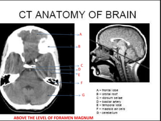
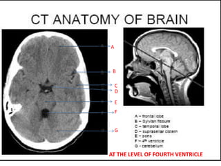
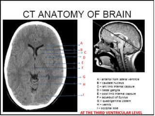
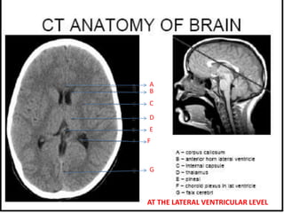
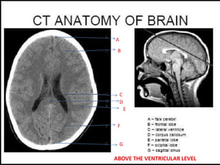
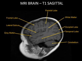
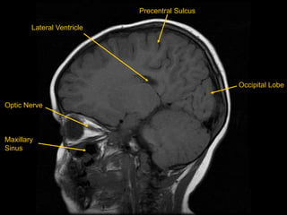
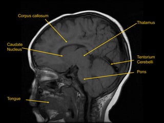
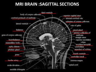
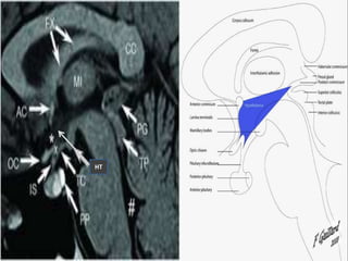
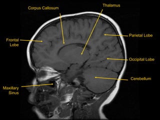
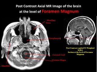
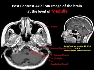
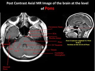
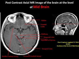
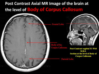
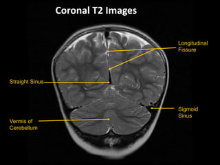
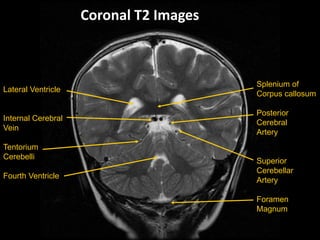
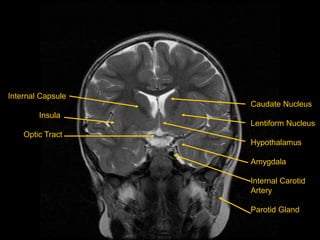
RADIOLOGICAL ANATOMY OF
NORMAL BRAIN
DR. Rahi Kiran.B
SR Neurology
GMC, KOTA
A
B
C
D
E
F
G
ABOVE THE LEVEL OF FORAMEN MAGNUM
A
B
C
D
E
F
G
AT THE LEVEL OF FOURTH VENTRICLE
A
B
C
D
E
F
G
H
I
AT THE THIRD VENTRICULAR LEVEL
A
B
C
D
E
F
G
AT THE LATERAL VENTRICULAR LEVEL
B
A
C
D
E
F
G
ABOVE THE VENTRICULAR LEVEL
MRI BRAIN – T1 SAGITTAL
White Matter
Occipital Lobe
Cerebellum
Grey Matter
Frontal Lobe
Lateral Sulcus
Parietal Lobe
Temporal Lobe
Optic Nerve
Maxillary
Sinus
Precentral Sulcus
Lateral Ventricle
Occipital Lobe
Caudate
Nucleus
Tongue
Corpus callosum
Thalamus
Tentorium
Cerebelli
Pons
MRI BRAIN :SAGITTAL SECTIONS
HT
Frontal
Lobe
Maxillary
Sinus
Parietal Lobe
Occipital Lobe
Corpus Callosum
Thalamus
Cerebellum
Frontal Lobe
Lateral Sulcus
Superior Temporal
Gyrus
Inferior Temporal
Gyrus
Parietal Lobe
Middle
Temporal
Gyrus
External
Auditory
Meatus
Post Contrast Axial MR Image of the brain
at the level of Foramen Magnum
Post Contrast Axial MR Image of the brain
at the level of Medulla
Post Contrast Axial MR Image of the brain at the level
of Pons
Post Contrast Axial MR Image of the brain at the level
of Mid Brain
Post Contrast Axial MR Image of the brain at the level
of Thalamus
Pineal gland
insular cortex
Post Contrast Axial MR Image of the brain at
the level of Body of Corpus Callosum
Longitudinal
Fissure
Sigmoid
Sinus
Straight Sinus
Vermis of
Cerebellum
Coronal T2 Images
Splenium of
Corpus callosum
Posterior
Cerebral
Artery
Superior
Cerebellar
Artery
Foramen
Magnum
Lateral Ventricle
Internal Cerebral
Vein
Tentorium
Cerebelli
Fourth Ventricle
Coronal T2 Images
Cingulate Gyrus
Choroid Plexus
Superior Colliculus
Cerebral Aqueduct
Corpus Callosum
Thalamus
Pineal Gland
Vertebral Artery
Insula
Lateral Sulcus
Hippocampus
Parahippocampal
gyrus
Olive
Crus of Fornix
Cerebral Peduncle
Middle Cerebellar
Peduncle
Internal Capsule
Insula
Optic Tract
Caudate Nucleus
Lentiform Nucleus
Hypothalamus
Amygdala
Internal Carotid
Artery
Parotid Gland
Ethmoid Sinus
Nasal Septum
Nasal Cavity
Tongue
Frontal Lobe
Nasal Turbinate
Masseter Muscle
Thank you

Imaging in neurology - normal anatomy