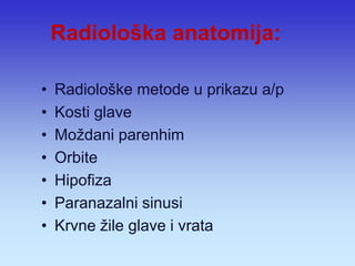
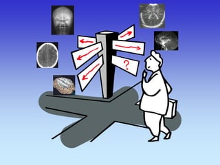
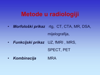
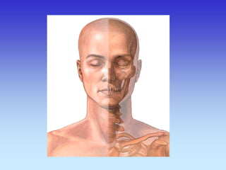
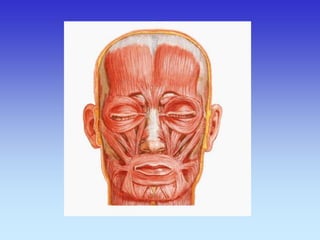
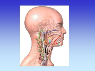
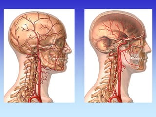
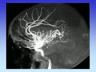
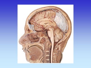
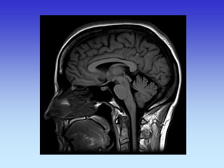
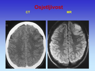
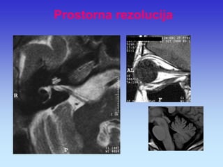
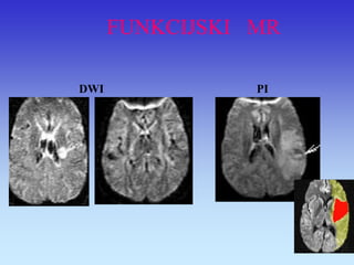
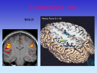
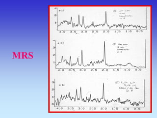
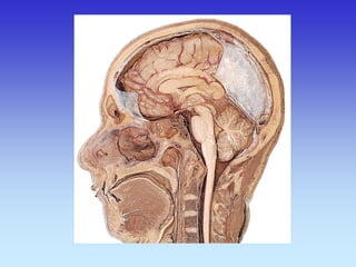
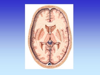
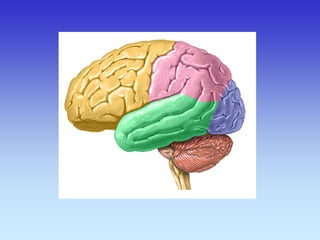
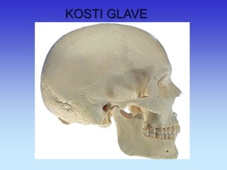
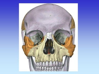
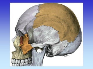
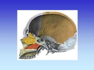
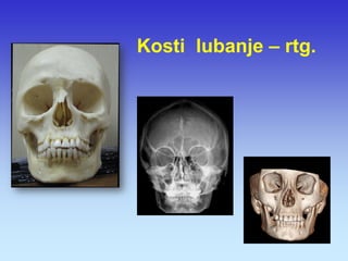
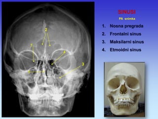
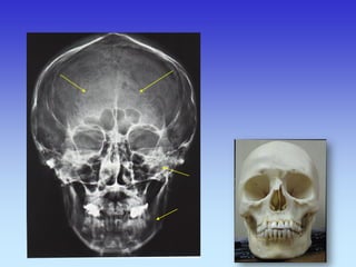
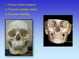
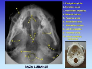
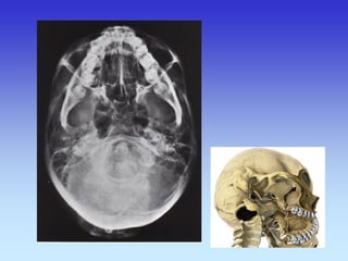
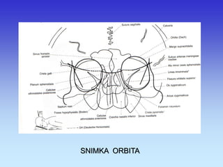
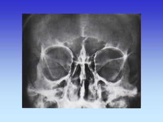
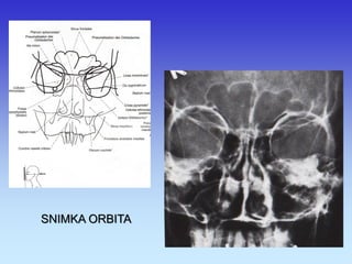
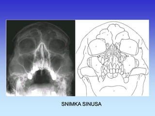
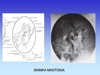
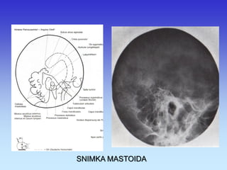
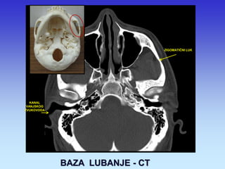
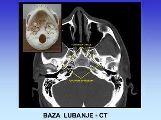
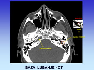
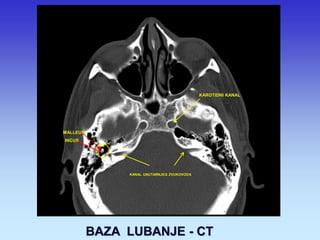
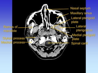
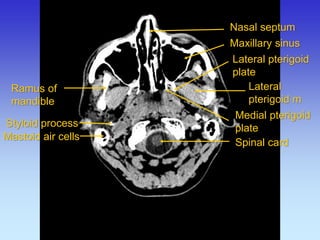
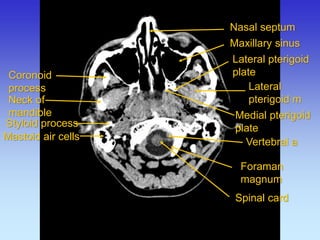
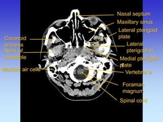
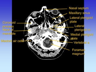
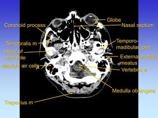
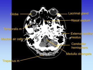
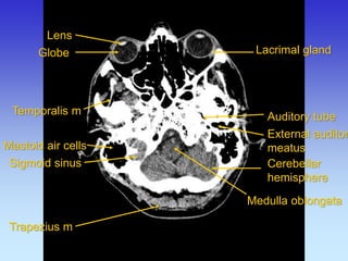
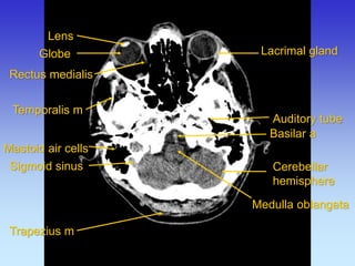
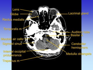
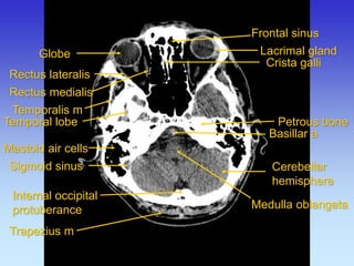
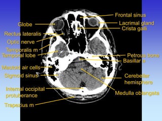
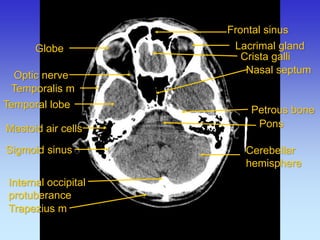
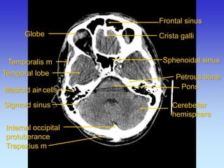
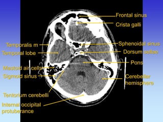
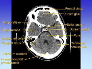
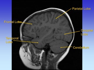
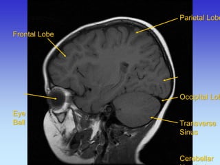
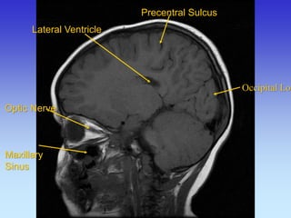
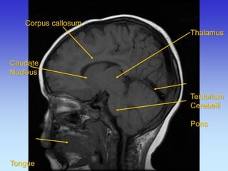
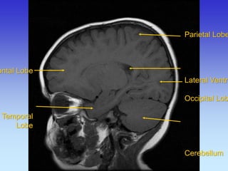
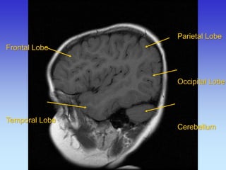
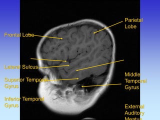
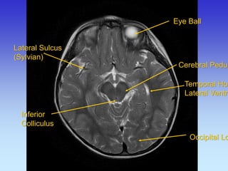
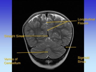
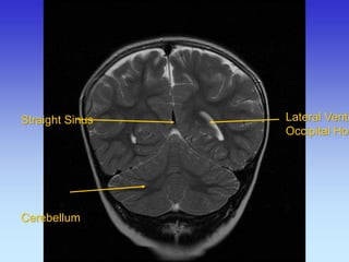
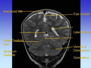
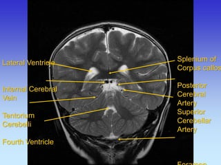
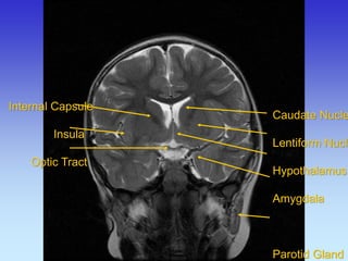
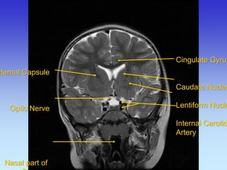
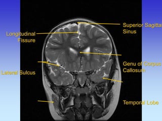
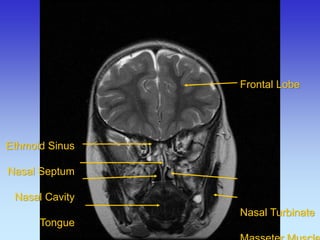
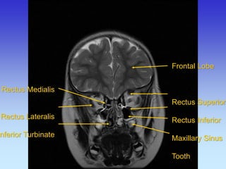
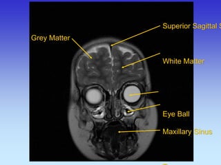
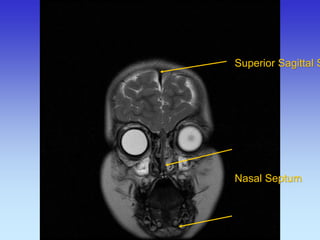
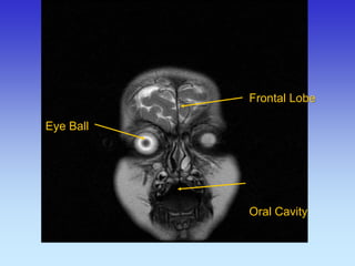
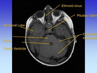
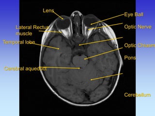
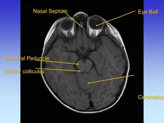
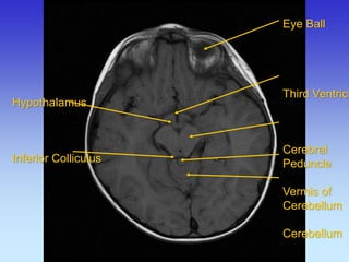
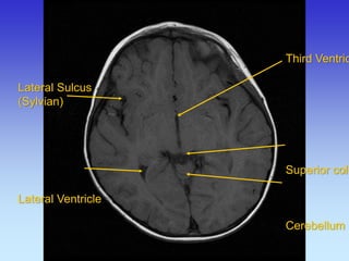
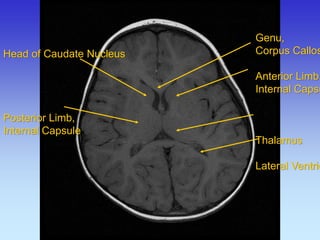
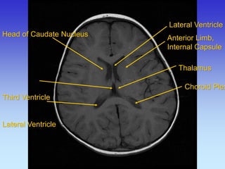
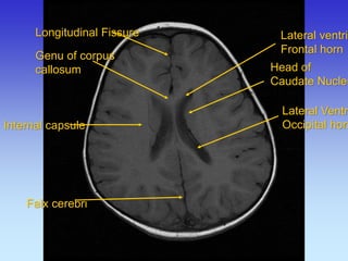
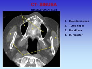
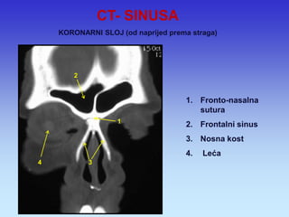
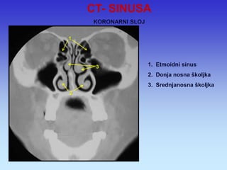
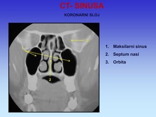
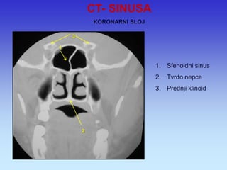
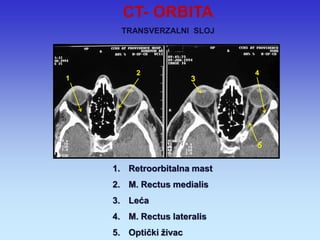
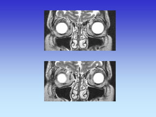
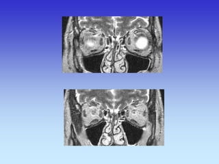
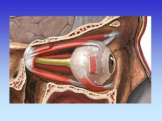
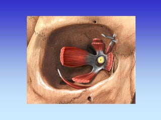
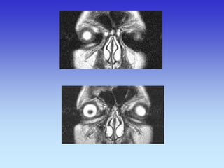
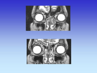
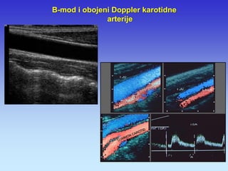
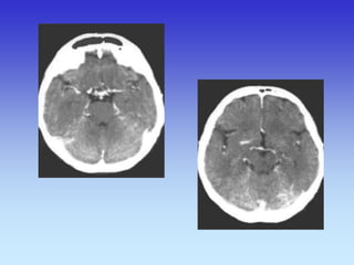
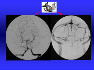
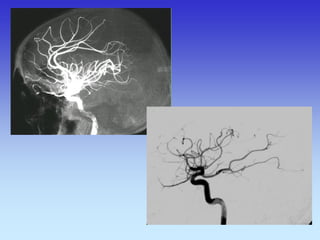
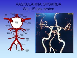
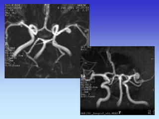
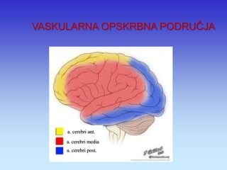
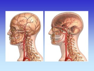
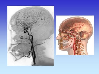
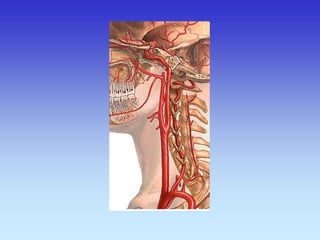
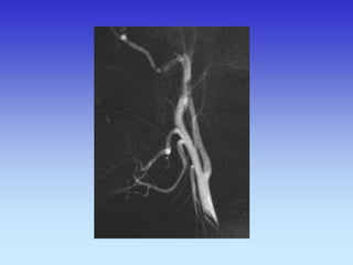
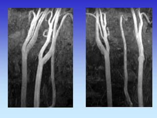
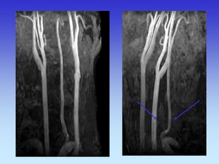
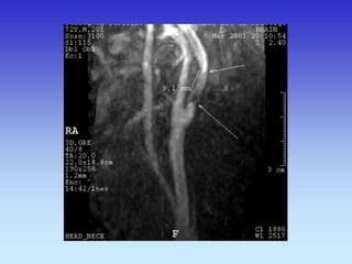
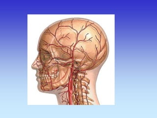
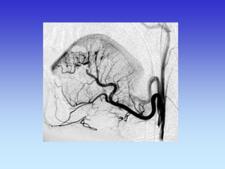
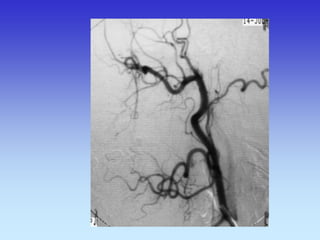
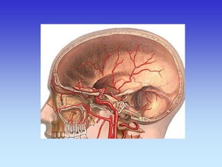
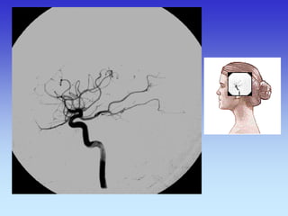
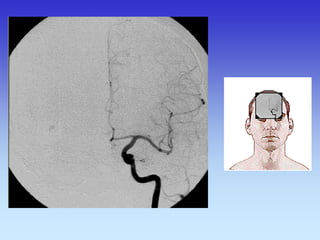
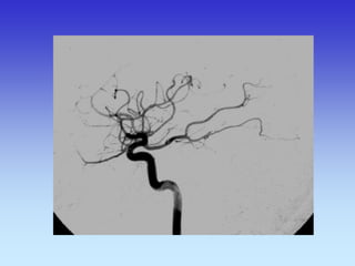
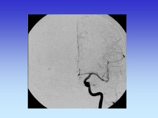
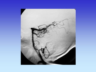
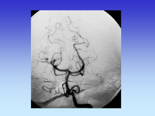
This document provides an overview of radiological anatomy and pathology of the head. It discusses various radiological imaging methods used to visualize anatomy such as bones, brain parenchyma, orbits, sinuses and blood vessels of the head and neck. Specific sections are dedicated to describing cross-sectional anatomy as visualized by CT and MRI of different regions including the skull base, orbits, sinuses and mastoids. Functional MRI techniques are also mentioned.


![Radiological anatomy of_temporal_bone[1]](https://cdn.slidesharecdn.com/ss_thumbnails/radiologicalanatomyoftemporalbone1-171112100915-thumbnail.jpg?width=640&height=640&fit=bounds)


