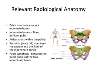
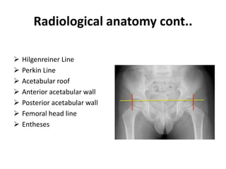
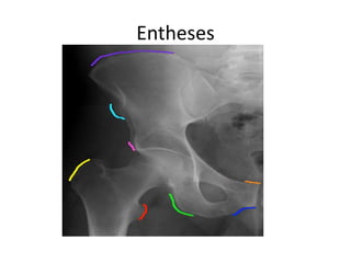
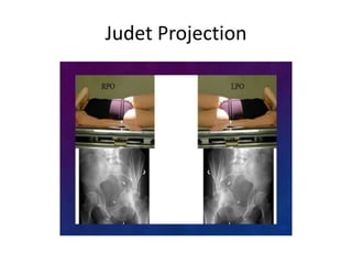
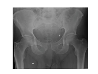
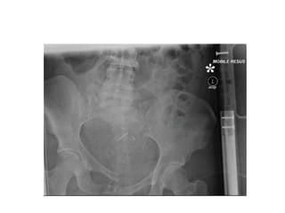
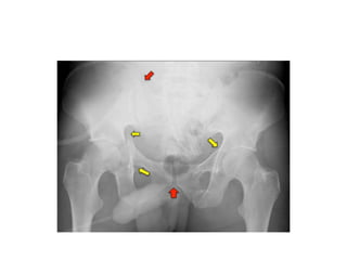
This document provides an overview of how to read pelvic and hip radiographs. It discusses the relevant radiological anatomy of the pelvis, including important bones, lines, and articulations. It describes common radiographic views used to image the hip and pelvis, such as AP, lateral, and Judet projections. It outlines how to systematically read a pelvic radiograph, including assessing adequacy, soft tissues, joint space, and bones. Examples of normal anatomy and pathological findings are shown, such as fractures, infections, and developmental dysplasia. The conclusion emphasizes understanding normal anatomy and disease patterns aids accurate diagnosis.
2. HOW TO READ PELVIC/HIP
RADIOGRAPHS
• PRESENTER:
DR JOHN ABUTU JOB
Resident doctor dept of surgery FMC bida.
Nigeria
3. OBJECTIVES
• To review the radiographic anatomy of the
pelvis placing emphasis on bones, body
landmarks and other anatomy.
• Identify Basic radiographic views of the hip
and pelvis.
• To systematically read a hip/pelvic radiographs
• Depict pathologies that disrupt the normal
contours of the pelvic reference lines, stripes
and arcs.
4. OUTLINES
• Introduction
• Relevant Radiological Anatomy
• Radiological Views/projections
• How to read hip/pelvic radiographs
• Normal pelvic radiograph
• Pathological pelvic radiograph
• Conclusion
5. INTRODUCTION
• The bony pelvis consists of the sacrum, coccyx
and a pair of innominate bones, which are
part of the appendicular skeleton.
• Pelvic Fractures account for 2-3 % of all
fractures, but the mortality rate is up to 20%
• The pelvic x-ray also forms an integral part in
trauma imaging and as part of ATLS
assessment.
6. Relevant Radiological Anatomy
• Pelvis = sacrum, coccyx +
inominate bones
• Inominate bones = ilium,
ischium, pubis
• Articulations within the pelvis:
• Sacroiliac joints (x2) - between
the sacrum and the ilium of
the innominate bones
• Pubic symphysis - between the
pubic bodies of the two
innominate bones
9. Radiological anatomy cont..
• Numerous lines, arcs and stripes
make up the familiar image in the
pelvic radiograph.
• These include:
Iliopectineal line
Ilioischial line
Sacral arcuate line
Shenton arc
Fat stripe
Line of klein
Radiographic teardrop
10. Radiological anatomy cont..
Hilgenreiner Line
Perkin Line
Acetabular roof
Anterior acetabular wall
Posterior acetabular wall
Femoral head line
Entheses
16. Male vs female pelvis
Feature Male Female
Shape Narrow, deep Wide, shallow
Superior
aperture (inlet)
Round Oval
Inferior
Aperture
(outlet)
Narrow Wide
17.
18.
19. Radiological views
• AP projections
• Lateral Hip projection
• Anxiolateral cross table Hip projection
• Inlet/outlet Projection
• Judet Projection
• Frog leg lateral
25. Reading hip/pelvic radiographs
• 1. Adequacy of film
-Penetration
-Right patient
-Right part of the body
-Positioning
-View of structure you want to see
- Atleast 2 views
28. Examples of pathological pelvic
radiograph
• A. Congenital/Inherited -Developmental
dysplasia/dislocation of the hip, achondroplasia
• B. Trauma: pelvic fractures
• -Can be simple or complex and can involve any
part of the bony pelvis.
- Anteroposterior compression - result in open
book or sprung pelvic fractures
Lateral compression
Vertical shear
Combined mechanical - complex fracture pattern
29. Examples of pathological pelvic
radiograph
• C. Infection -septic arthritis
-osteomyelitis
• D. Metabolic – Osteonecrosis(AVN femoral
head) -Paget diseease
• E. others eg protrucio Acetabuli
53. CONCLUSION
• The pelvis has complex and subtle anatomy,
important radiographic findings can be
challenging to the clinician. A good
understanding of the normal anatomy and the
patterns of disease can be useful for accurate
diagnosis.
54. References
• 1. Campbell S.E Radiography of the Hip:Lines, Signs and Patterns of
Disease. Semin
• Roentgenol 2005; 40: 290-319. DOI:10.1053/j.ro.2005.01.016.
• 2. Frank Gaillard, 'Radiopedia', http://www.radiopedia.org, 2005-
2014, (accessed 6
• January 2015).
• 3. Young JWR, Resnik CS. Fracture of the pelvis: current concepts of
classification. AJR
• 1990; 155: 1169-75.
• 4. Leunig M, Beaule PE, Ganz R. The Concept of Femoroacetabular
Impingement. Clin
• Orthop Relat Res 2009; 467: 616-22.
• 5. Blausen.com staff. "Blausen gallery 2014" Wikiversity Journal of
Medicine.
Each innominate bone consists of three separate bones (Figure 1):
• ilium
• ischium
• pubis
1: Iliac crest
2: Posterior superior iliac spine
3: Wing of ilium
4: Posterior inferior iliac spine
5: Anterior superior iliac spine
6: Arcuate line of ilium
7: Acetabular rim
8: Acetabular fossa
9: Ischial spine
10: Ischial tuberosity
11: Superior ramus of pubis
12: Inferior ramus of pubis
13: Ala of sacrum
14: Pelvic sacral foramina
15: Sacro-iliac joint
16: Intrauterine contraceptive device
(IUD)
17: Lunate surface of acetabulum
18: Coccyx
19: Obturator foramen
20: Body of pubis
21: Pubic symphysis
1: Acetabular rim
2: Femoral head
3: Femoral neck
4: Greater trochanter
5: Lesser trochanter
6: Lunate surface
7: Acetabular fossa
8: Ilio-ischial line (radiology term)
9: Fovea of femoral head
10: Acetabular rim (anterior lip)
11: Acetabular rim (posterior)
12: Acetabular notch
13: Shenton’s line (radiology term
Sacroiliac join 2-4mm,Pubic symphisis less than 5mm
The teardrop is inspected. This is a radiological feature
which correlates to the non-articular floor of
the acetabulum, and close inspection in this area
will reveal any acetabular fracture. The 'tear drop' to evaluate the anteroinferior portion of the acetabular fossa.
Hilgenreiner line: This is a line drawn horizontally through the superior aspect of both triradiate cartilages. It should be horizontal and is mainly used as a measurement of the acetabular angle and as a reference for Perkin line.
Perkin Line This is the line drawn perpendicular to the Hilgenreiner line, intersecting the lateral most aspect of the acetabular roof. The upper femoral epiphysis should be in the inferomedial quadrant: it should lie below the Hilgenreiner line and medial to Perkin line. These are used to diagnose DDH(dev displassia or dislocation of the hip)
Iliopectineal line (red); Ilioischial line (light green); Sacral arcuate lines (yellow); Shenton arc (light blue); Line of Klein (white); Gluteal fat stripe (purple); Acetabular roof (pink); Medial acetabular wall (dark green); Anterior acetabular wall (orange); Posterior acetabular wall (dark blue); Femoral head line (black).
Arcuate line
Ileoischial line
Radiographic U (teardrop)
Acetabular roof
Anterior lip of acetabulum
Posterior lip of acetabulum
Anterior and posterior acetabular rim
Entheses: Within the pelvis and hips are multiple osseous projections which serve as tendon origins/insertions
Tendon origins/insertion sites: Abdominal muscles (Iiac crest purple); Sartorius muscle (ASIS light blue); Rectus femoris muscle (pink); Rectus abdominis muscle (orange); Adductor muscles (dark blue); Hamstring muscles (light green); Gluteus medius and minimus, gemellus and piriformis muscles (yellow); Iliopsoas muscles (red).
Female pelvis is broader, bones are slender, greater breath and capacity
The sides of the male pelvis converge from the inlet to the outlet, whereas the sides of the female pelvis are wider apart. The angle between the inferior pubic rami is acute (70 degrees) in men, but obtuse (90–100 degrees) in women
Obique view. To show acetabular #.
Flamingo projection: specialised orthopaedic series consisting of 2 separate projections, used to asses stability of pubic symphysis
AP errect
Left foot raised
Right foot raised
Medially rotate toes (15-20 degrees). The anteroposterior pelvic radiograph should be made with the patient supine on the x-ray table with both lower extremities orientated in 15° of internal rotation in order to maximise the length of the femoral neck.
Adequacy: Include the anatomy from the top of both iliac crests superiorly through the ischial tuberosities including both lesser trochanters of the femurs, both anterior portions of the iliac wings should be visible.
View femoral neck
Obique view. To show acetabular #.
Supine, leg abducted and externally rotated, knee flex.
Paed age grp for DDH and SCFE. Contraindicated in acute injury
Narrow joint space in cartilage loss arthritis. Wide in dislocatiion
Fat strip- fascia planes covering muscles, directly overlie hip joint capsule. Gluteal muscle and ileosoas. Soft tissue bulge out in effusion
Four main forces in high energy blunt force trauma result in unstable pelvic fractures:
Developmental dysplasia of the left hip with the femoral epiphysis in the superolateral quadrant (lateral to the Perkin line and above the Hilgenreiner line). It should normally be in the inferomedial quadrant.
Bilateral dysplastic hips with disruption of the Shenton arcs.
SCFE. In normal hip a line drawn tangentia to superior femoral neck(klein line) intersects small portion of lateral capital epiphyseal
Apc vertical #, lateral horiontal #. LC1-rami#+insp sacral alar #, LC2 rami #+ insp posterior iliac dislocation. LC3
APC1 widen pubi symphisis less 2.5cm, APC2 greater than 2.5cm +widen ant SIJ APC 3 widen greater 2.5cm+SIJ dislocation
Vertical share fall from hight
APC II pelvic fracture A diastasis (widening of the pubic symphysis) anteriorly and widening of the left SIJ (arrow) can be seen.
Right-sided superior and inferior pubic ramus fracture. LC1
Vertical share. Vertical shear fracture (a) Note how the right hemipelvis is higher than the left.
Complex pelvic fractures with anterior and posterior columns fracture of the right acetabulum and anterior column fracture of the left acetabulum. There is widening if the right sacroiliac joint (upper red arrow) and minor widening of the pubic symphysis (lower red arrow).
The iliopectineal lines on both sides are disrupted indicating anterior column fractures of the bilateral acetabuli (arrows).
There is disruption of the right ilioischial line (arrows) suggestive of posterior acetabular fracture.
Pubic rami #
ischial tuberosity avulsion fracture right. Hamstring muscle in sport
Bilateral sacroiliac joint diastasis with verical right superior and inferior pubic rami fractures- vertical shear 'open book' injury. Combine injury
Anterior dislocation
Posterior hip dislocation
Avulsion fracture of the greater trochanter at the insertion site of gluteal tendon, with superomedial displacement of fracture fragment.
Acetabular protrusio (also known as acetabular protrusion) is intrapelvic displacement of the acetabulum and femoral head, so that the femoral head projects medial to the ischioilial line. It should be differentiated from coxa profunda.
Primary Characterized by progressive protrusio in middle aged women. The condition may be associated with osteoarthritis and may be familial.
Secondary
Paget disease
psoriatic arthropathy
rheumatoid arthritis
ankylosing spondylitis
osteomalacia / rickets
osteogenesis imperfecta
Marfan syndrome
trauma
hemophilia (in advanced arthropathy) 5
Displaced femoral neck fracture
Two-part intertrochanteric fracture
Disruption of Shenton's line in subcapital neck of femmur fracture.
avascular necrosis of right femoral head- subchondral sclerosis, fragmentation and collapse of femoral head.
Septic arthritis. Joint effusion, narowing of joint space, gass within the joint, soft tissue swelling
Fig right hip septic arthritis with complete loss of right superior femoral acetabular joint space and mild destruction of the femorl head
Previous pelvic radiograph of the same patient (Figure 36) shows focus of osteomyelitis in the left femoral neck. The femoral head appears normal. Periosteal reaction.