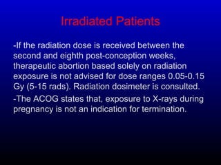
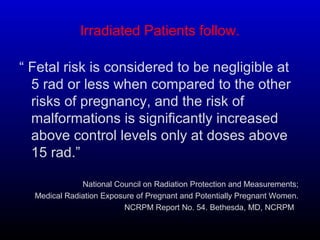
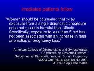
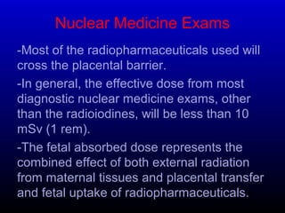
1. Imaging And Radiation HazardsImaging And Radiation Hazards
During PregnancyDuring Pregnancy
Almataria Teaching Hospital, Nasser InstituteAlmataria Teaching Hospital, Nasser Institute
Cairo, EgyptCairo, Egypt
Dr. Mamdouh SabryDr. Mamdouh Sabry
MD. Ain Shams, PhD. FranceMD. Ain Shams, PhD. France
Consultant Ob. & Gyn.Consultant Ob. & Gyn.
2. Introduction
-The application of X-ray, U.S, CT, Nuclear medicine
and MRI is unquestionable in diagnosis of acute and
chronic diseases.
-Its use during pregnancy and lactation is confusing
to most physicians and all patients.
-Women may be exposed to many sources of
radiation without knowing, as any energy-carrying
waveform is considered as a kind of radiation.
-Exposure to ionizing rays includes diagnostic rays,
nuclear medicine (radiopharmaceuticals), Therapeutic
rad., workplace exposure and environmental
exposure.
3. Data Collection
Data from most groups and societies of diagnostic and
therapeutic radiology.
Up to date.com.
Evaluation of 35 publications, obser., case rep. and
many follow up studies.
Medline & Medscape.
Our experience in N. In. , Hema. On. Gp., Ped. On. Gp.,
and organ transplant group.
4. X-rays
-Measured in several types of units.
-Radiation-absorbed dose (rad), US measurement.
-Gray (Gy) international measurement.
-Both represent a single-time exposure and radiation
absorbed dose (as diagnostic procedures).
-The measurement units; roentgen equivalent man
(rem) and Sievert (Sv) used to quantify radiation
exposure over time and radiologic effective dose (as
environmental releases).
-1 Gy equals 100 rad & 1 Sv equals 100 rem.
-Dose calculation depends on study type, machine
type, distance from machine, organ penetrated and
method of study.
5. X-ray, follow.
-X-rays have deterministic and stochastic effects.
-Deterministic effects are usually intrauterine, involving
damage to growing cells, it has a dose response curve
and a threshold effect, either all or non, the damage
threshold begins at 0.1-o.15 Gy which causes abortion
or no implantation and extends to 1Gy, which is
associated fetal death in-utero at term.
-Stochastic effects appears in the later years of
exposed persons, not needing threshold? (0.05 Gy)
relative risk documented in multiple case-control
studies, others considered this to be theoretical.
-Dose < 5 rad not associated with fetal hazards.
6. X-ray follow.
CT scanning
-New generations more
sensitive and accurate.
-0.17-0.35 rad are fetal
exposure in abdominal
or pelvic CT.
Fluoroscopy/Angiogra.
-Cerebral angiography
exposes to <10 mrad
-Risk benefit ratio is
evaluated
Plain Films
-In chest radiograph,
exposure for 2 views
<0.07 mrad
-Trauma to bone, ribs,
skull and lower limb
deliver lower doses due
to fetal distance from
target organ, but hip
and pelvis X-ray deliver
about 200 mrad.
7. X-ray Follow
SO, Concern about possible
effects of high-dose ionizing radiation
exposure should not prevent medically
indicated diagnostic x-ray procedures
from being performed on a pregnant
woman.
During pregnancy, other imaging
procedures not associated with ionizing
radiation (ultrasonography and MRI)
should be considered instead of x-rays
when appropriate.
8. Irradiated Patients
-If the radiation dose is received between the
second and eighth post-conception weeks,
therapeutic abortion based solely on radiation
exposure is not advised for dose ranges 0.05-0.15
Gy (5-15 rads). Radiation dosimeter is consulted.
-The ACOG states that, exposure to X-rays during
pregnancy is not an indication for termination.
9. Irradiated Patients follow.
“ Fetal risk is considered to be negligible at
5 rad or less when compared to the other
risks of pregnancy, and the risk of
malformations is significantly increased
above control levels only at doses above
15 rad.”
National Council on Radiation Protection and Measurements;
Medical Radiation Exposure of Pregnant and Potentially Pregnant Women.
NCRPM Report No. 54. Bethesda, MD, NCRPM, .
10. Irradiated patients follow
“Women should be counseled that x-ray
exposure from a single diagnostic procedure
does not result in harmful fetal effects.
Specifically, exposure to less than 5 rad has
not been associated with an increase in fetal
anomalies or pregnancy loss.”
American College of Obstetricians and Gynecologists,
Committee on Obstetric Practice;
Guidelines for Diagnostic Imaging During Pregnancy.
ACOG Committee Opinion No. 299.
ACOG, September 2004.
11. Nuclear Medicine Exams
-Most of the radiopharmaceuticals used will
cross the placental barrier.
-In general, the effective dose from most
diagnostic nuclear medicine exams, other
than the radioiodines, will be less than 10
mSv (1 rem).
-The fetal absorbed dose represents the
combined effect of both external radiation
from maternal tissues and placental transfer
and fetal uptake of radiopharmaceuticals.
12. Magnetic Resonance Imaging (MRI)
“Although there have been no documented
adverse fetal effects reported, the National
Radiological Protection Board arbitrarily advises
against its use in the first trimester.”
Guidelines for Diagnostic Imaging During Pregnancy.
ACOG Committee Opinion
-USA, FDA labeling MRI devices to indicate, safety of
MRI with respect to the fetus not yet confirmed.
-Gadolinium is teratogenic in animals and does cross
the placenta.
-
.
13. Ultrasound Imaging
“There have been no reports of documented adverse
fetal effects for diagnostic ultrasound procedures,
including duplex Doppler imaging.”
“There are no contraindications to ultrasound
procedures during pregnancy, and this modality
has largely replaced x-ray as the primary method
of fetal imaging during pregnancy.”
American College of Obstetricians and Gynecologists,
Committee on Obstetric Practice;
Guidelines for Diagnostic Imaging During Pregnancy.
ACOG Committee Opinion No. 299.
ACOG, September, 2004.
14. 0ccuptional Hazards
Health Care Providers
-Complete occupational and
exposure history is
mandatory before any plan
for pregnancy.
-Exclusion of pregnant
females from certain jobs.
-Contraception.
-Accurate exposure
evaluation techniques.
Aviation Workers
-Few studies on human
exposure to cosmic rad.
-It is ionizing radiation by
heavy particles such as
protons and helium nuclei in
high altitudes.
-Airplane crew and pilots
are in risk.
-Computer systems are
available to calculate
exposure
15. Electromagnetic Fields
-Very large studies about the low-energy
electromagnetic fields generated by power lines, video
displays and other electric and electronic devices,
found no demonstrable effect, it is conducted in
response to increase abortion rates in some areas.
-Microwaves raises temp. within cells, cytotoxic only
at high exposure, no DNA-damage effect.
-Short-wave and microwave therapists have a
remarkable higher rates of infertility, sp. Abortion,
altered gender ratio, cong. Malformations and low
birth weight, i.e, some groups may show worsened
preg. Outcome due to many types of exposure.
16. To Conclude
-What is the 10 days rule?
-What rad. tests contraindicated at pregnancy?
-Is 0.5 rad, & 0.05 Gy exposure safe at preg. ?
-Best time for pregnancy after radiotherapy?
-Exposure in early pregnancy dictates abortion?
-Coronary cath. May be done during pregnancy?
-Pregnant air crew may continue work?
-What imaging techniques are safe at pregnancy?