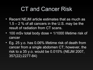
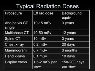
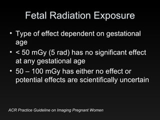
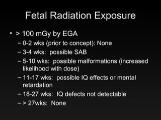
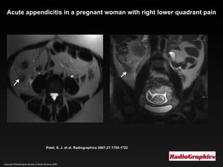
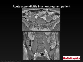
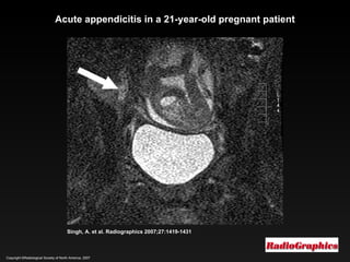
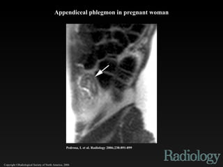
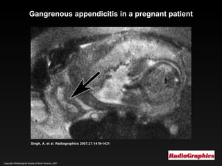
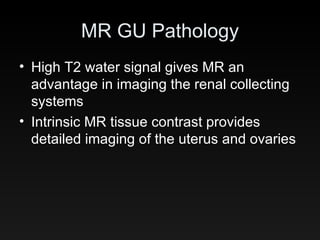
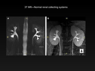
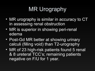
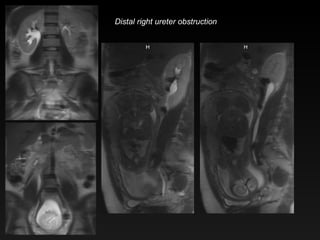
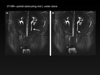
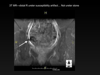
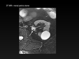
Maternal Imaging in Pregnancy Lloyd Stambaugh, M.D. Radia
CT and Cancer Risk CT scans/year 1980 3 million 2007 62 million (4M in children) Radiation dose estimate CT 10-20 mSv (per scan) 30-60 mSv for multi-phase CXR 0.2 mSv
Radiation Physics—the one slide version,   Gray—unit measures physical quantity of radiation Sievert—unit of biological radiation exposure Sievert = Gray * bio factor (1 for X-ray) So 1 Sievert = 1 Gray for X-rays
CT and Cancer Risk There is no known lower threshold for cancer induction from radiation Cancer risk from radiation is cumulative Although there is “no safe level”, potential benefits from the exam often outweigh potential risks
CT and Cancer Risk Recent NEJM article estimates that as much as 1.5 – 2 % of all cancers in the U.S. may be the result of radiation from CT scans. 100 mSv total body dose = 1/1000 lifetime risk of cancer Eg. 25 y.o. has 0.06% lifetime risk of death from cancer from a single abdomen CT; however, the risk to a 35 y.o. would be 0.015% (NEJM 2007, 357(22):2277-84)
Typical Radiation Doses 12 years 40-50 mSv Multiphase CT 150-200 days per view 1.5-2 mSv per view L-spine xrays 10 days 0.1 mSv Hand x-rays 3 months 0.7 mSv Mammogram 20 days 0.2 mSv Chest x-ray 3 years 10 mSv Spine CT 3 years 10-15 mSv Abd/pelvis CT single Background equiv Eff rad dose Procedure
Radiation and the Fetus
Fetal Radiation Exposure Type of effect dependent on gestational age < 50 mGy (5 rad) has no significant effect at any gestational age 50 – 100 mGy has either no effect or potential effects are scientifically uncertain ACR Practice Guideline on Imaging Pregnant Women
Fetal Radiation Exposure > 100 mGy by EGA 0-2 wks (prior to concept): None 3-4 wks:  possible SAB 5-10 wks:  possible malformations (increased likelihood with dose) 11-17 wks:  possible IQ effects or mental retardation 18-27 wks:  IQ defects not detectable > 27wks:  None
Fetal Radiation Exposure However, fetal exposure at any level does carry with it the small risk of cancer induction later in life.
Fetal Radiation Exposure How many fetuses are exposed ICRP indicates thousands of pregnant women are exposed to some level of medically indicated radiation each year One study reports 1% of women receiving abdominal radiographs were unknowingly pregnant in their 1 st  trimester (Mossman, Obst Gyn 1982) ICRP—Interntl Comm Radiological Protection
Fetal Radiation Exposure Study in trauma patients showed 2.9% were pregnant and unidentified pregnancy rate was 0.3% (Bochicchio, J Am Coll Surg 2001)
Fetal Doses from CT Hurwitz et al, constructed an anthropomorphic phantom of a pregnant woman.  TLD and MOSFET radiation detectors were placed to reflect radiation exposure to a fetus of 0 & 3 months gestation. 16-slice CT scanner was used
Fetal Doses from CT Renal stone protocol 0 mos: 8 – 12 mGy 3 mos: 4 – 7 mGy Appendix protocol 0 mos: 15.2 – 16.8 mGy 3 mos: 20 – 40 mGy PE protocol 0 mos:  0.24 – 0.47 mGy 3 mos:  0.61 – 0.66 mGy
Fetal Doses from CT This same institution reports 0.32 – 0.36 mGy fetal dose from a V/Q study
Imaging for Pulmonary Embolism CTPA has replaced V/Q as primary imaging for PE Huda reports CTPA fetal dose is 0.41 mGy V/Q fetal dose is 0.9 to 1.8 mGy CTPA results in very low fetal exposure Hurwitz reports CTPA 0.24 – 0.66 mGy V/Q 0.32 – 0.36 mGy
Imaging for Pulmonary Embolism CTPA and V/Q fetal exposures are similar, with some variation based on gestational age and imaging protocols Given CTPA’s greater diagnostic accuracy and similar fetal exposure, it is the preferred study for diagnosing PE during pregnancy
Contrast Agents
Contrast Agents and Pregnancy CT (iodinated) contrast No teratogenic effect Iodine content could theoretically suppress thyroid function.  However, no hypothyroidism has been demonstrated after i.v. contrast in pregnant pts ACR recommends i.v. contrast only if necessary and after informed consent
Contrast Agents and Pregnancy MR (gadolinium based) contrast i.v. Gad is teratogenic in animal studies (high and repeated doses) ACR 2007 recommends avoiding Gad during pregnancy (use only if absolutely essential, only after informed consent) Gad-based contrast agents are FDA cat C (use only if potential benefit justifies potential risk to fetus)
Contrast Agents and Nursing CT (iodinated) contrast Approx 1% of i.v. contrast dose is excreted in milk Only 1 – 2% of contrast ingested by infant is absorbed into bloodstream Although historical recommendation was hold nursing for 24 hours, recent reviews in Eur Soc of Urogenital Rad and NEJM indicate nursing may be continued after contrast
Contrast Agents and Nursing MR (gadolinium-based) contrast Study of 20 lactating women: only 0.04% of gad excreted in milk (Kubik-Huch, Rad 2000) Amount ingested by infant is much less than a typical pediatric i.v. dose for MR Although historical recommendation was hold nursing for 24 hours, there is no evidence to support any significant risk to infant, and nursing can be continued after Gad-based contrast
Alternatives to CT US and MR offer excellent alternatives to CT for imaging of pregnant women—without ionizing radiation The benefits (and limitations) of US are generally well understood—so let’s talk about MR
MRI and Pregnancy FDA requires MRI devices indicate the safety of MRI with respect to the fetus “has not been established.” Potential fetal risks Teratogenic Acoustic damage
MRI and Pregnancy Studies of children exposed to MRI in utero (1.5T) showed no negative outcomes at 9 mos and up to 9 yrs of age (Clements, Br J Rad 2000) (Kok, MRI 2004) Animal studies are mixed, one negative for prolonged 4T exposure, but another raised possibility of teratogenic effects in mice
MRI and Pregnancy 2007 ACR states all pregnant patients can receive MRI as long as the “risk-benefit ratio to the patient warrants that the study be performed.”
MRI and Pregnancy Noise levels in the MRI scanner can reach 120 dB and raise concern for fetal ear injury Baker et al examined 18 pts who had in utero MRI.  16 passed their 8 month hearing test (16.7 expected)
MRI and Pregnancy Glover et al, placed a microphone in the fluid-filled stomach of a volunteer Sound attenuation through the abdominal wall was 30 dB The microphone recorded less than 90dB, compared to 120dB in the room.
MRI Appendicitis In a study of 51 pregnant patients, MRI was 100% sensitive and 94% specific in diagnosing appendicitis Additional studies report ranges of 97-100 % sensitivity and 92-94% specificity.
MR Appendicitis Dilated fluid-filled appendix Peri-appendiceal edema Thickened wall Enhancement (if Gd is given)
Normal appendix
Normal Appendix
Normal Appendix
Copyright ©Radiological Society of North America, 2006 Appendiceal phlegmon in 29-year-old pregnant woman
Copyright ©Radiological Society of North America, 2007 Patel, S. J. et al. Radiographics 2007;27:1705-1722 Acute appendicitis in a pregnant woman with right lower quadrant pain
Copyright ©Radiological Society of North America, 2007 Acute appendicitis in a nonpregnant patient
Copyright ©Radiological Society of North America, 2007 Singh, A. et al. Radiographics 2007;27:1419-1431 Acute appendicitis in a 21-year-old pregnant patient
Copyright ©Radiological Society of North America, 2006 Pedrosa, I. et al. Radiology 2006;238:891-899 Appendiceal phlegmon in pregnant woman
Copyright ©Radiological Society of North America, 2007 Singh, A. et al. Radiographics 2007;27:1419-1431 Perforating appendicitis in a 16-year-old girl
Copyright ©Radiological Society of North America, 2006 Pedrosa, I. et al. Radiology 2006;238:891-899 Mild acute appendicitis in a 27-year-old pregnant woman (gestational age, 13 weeks)
Copyright ©Radiological Society of North America, 2006 Pedrosa, I. et al. Radiology 2006;238:891-899 MR images in 33-year-old pregnant woman (gestational age, 20 weeks) with acute appendicitis
Copyright ©Radiological Society of North America, 2007 Acute appendicitis in a 20-year-old pregnant patient
Copyright ©Radiological Society of North America, 2007 Singh, A. et al. Radiographics 2007;27:1419-1431 Gangrenous appendicitis in a pregnant patient
MR GU Pathology High T2 water signal gives MR an advantage in imaging the renal collecting systems Intrinsic MR tissue contrast provides detailed imaging of the uterus and ovaries
3T MR—Normal renal collecting systems
MR Urography MR urography is similar in accuracy to CT in assessing renal obstruction MR is superior in showing peri-renal edema Post-Gd MR better at showing urinary calculi (filling void) than T2-urography MR of 23 high-risk patients found 5 renal & 8 ureteral TCC’s; remaining patients negative on F/U for 1 year.
Hydronephrosis & Pregnancy Hydro of pregnancy Usually mild Right > Left ureter tapers between uterus and psoas No perinephric edema Ureteral obstruction Perinephric edema Ureter cutoff below or above uterus May see filling defect Don’t be fooled by flow artifact on HASTE/SSFSE
NF Mild hyronephrosis of pregnancy
Mild hydronephrosis of pregnancy
Distal left ureter obstruction
Distal right ureter obstruction
3T MR—partial obstructing mid L ureter stone
3T MR—distal R ureter susceptibility artifact… Not ureter stone
3T MR—renal pelvis stone
Copyright ©Radiological Society of North America, 2007 Singh, A. et al. Radiographics 2007;27:1419-1431 Adnexal torsion
Ovarian torsion
Ectopic Pregnancy 24 pregnant female pts with suspected EP (18 had vaginal spotting & pain) US showed absence of IUP in all pts (# of EP’s found by US not mentioned) Of 19 EP’s, MR found 18 (1 FN, no FP) MR sens 95%, spec 100%, acc 96%
Ectopic Pregnancy
Ectopic Pregnancy
Ectopic Pregnancy
Ectopic Pregnancy
Abdominal Ectopic
Cervical Ectopic
Pregnancy in C-sect scar
Ectopic in Rudimentary Horn
Endometrioma

Preg Img

  • 1.
    Maternal Imaging inPregnancy Lloyd Stambaugh, M.D. Radia
  • 2.
    CT and CancerRisk CT scans/year 1980 3 million 2007 62 million (4M in children) Radiation dose estimate CT 10-20 mSv (per scan) 30-60 mSv for multi-phase CXR 0.2 mSv
  • 3.
    Radiation Physics—the oneslide version,  Gray—unit measures physical quantity of radiation Sievert—unit of biological radiation exposure Sievert = Gray * bio factor (1 for X-ray) So 1 Sievert = 1 Gray for X-rays
  • 4.
    CT and CancerRisk There is no known lower threshold for cancer induction from radiation Cancer risk from radiation is cumulative Although there is “no safe level”, potential benefits from the exam often outweigh potential risks
  • 5.
    CT and CancerRisk Recent NEJM article estimates that as much as 1.5 – 2 % of all cancers in the U.S. may be the result of radiation from CT scans. 100 mSv total body dose = 1/1000 lifetime risk of cancer Eg. 25 y.o. has 0.06% lifetime risk of death from cancer from a single abdomen CT; however, the risk to a 35 y.o. would be 0.015% (NEJM 2007, 357(22):2277-84)
  • 6.
    Typical Radiation Doses12 years 40-50 mSv Multiphase CT 150-200 days per view 1.5-2 mSv per view L-spine xrays 10 days 0.1 mSv Hand x-rays 3 months 0.7 mSv Mammogram 20 days 0.2 mSv Chest x-ray 3 years 10 mSv Spine CT 3 years 10-15 mSv Abd/pelvis CT single Background equiv Eff rad dose Procedure
  • 7.
  • 8.
    Fetal Radiation ExposureType of effect dependent on gestational age < 50 mGy (5 rad) has no significant effect at any gestational age 50 – 100 mGy has either no effect or potential effects are scientifically uncertain ACR Practice Guideline on Imaging Pregnant Women
  • 9.
    Fetal Radiation Exposure> 100 mGy by EGA 0-2 wks (prior to concept): None 3-4 wks: possible SAB 5-10 wks: possible malformations (increased likelihood with dose) 11-17 wks: possible IQ effects or mental retardation 18-27 wks: IQ defects not detectable > 27wks: None
  • 10.
    Fetal Radiation ExposureHowever, fetal exposure at any level does carry with it the small risk of cancer induction later in life.
  • 11.
    Fetal Radiation ExposureHow many fetuses are exposed ICRP indicates thousands of pregnant women are exposed to some level of medically indicated radiation each year One study reports 1% of women receiving abdominal radiographs were unknowingly pregnant in their 1 st trimester (Mossman, Obst Gyn 1982) ICRP—Interntl Comm Radiological Protection
  • 12.
    Fetal Radiation ExposureStudy in trauma patients showed 2.9% were pregnant and unidentified pregnancy rate was 0.3% (Bochicchio, J Am Coll Surg 2001)
  • 13.
    Fetal Doses fromCT Hurwitz et al, constructed an anthropomorphic phantom of a pregnant woman. TLD and MOSFET radiation detectors were placed to reflect radiation exposure to a fetus of 0 & 3 months gestation. 16-slice CT scanner was used
  • 14.
    Fetal Doses fromCT Renal stone protocol 0 mos: 8 – 12 mGy 3 mos: 4 – 7 mGy Appendix protocol 0 mos: 15.2 – 16.8 mGy 3 mos: 20 – 40 mGy PE protocol 0 mos: 0.24 – 0.47 mGy 3 mos: 0.61 – 0.66 mGy
  • 15.
    Fetal Doses fromCT This same institution reports 0.32 – 0.36 mGy fetal dose from a V/Q study
  • 16.
    Imaging for PulmonaryEmbolism CTPA has replaced V/Q as primary imaging for PE Huda reports CTPA fetal dose is 0.41 mGy V/Q fetal dose is 0.9 to 1.8 mGy CTPA results in very low fetal exposure Hurwitz reports CTPA 0.24 – 0.66 mGy V/Q 0.32 – 0.36 mGy
  • 17.
    Imaging for PulmonaryEmbolism CTPA and V/Q fetal exposures are similar, with some variation based on gestational age and imaging protocols Given CTPA’s greater diagnostic accuracy and similar fetal exposure, it is the preferred study for diagnosing PE during pregnancy
  • 18.
  • 19.
    Contrast Agents andPregnancy CT (iodinated) contrast No teratogenic effect Iodine content could theoretically suppress thyroid function. However, no hypothyroidism has been demonstrated after i.v. contrast in pregnant pts ACR recommends i.v. contrast only if necessary and after informed consent
  • 20.
    Contrast Agents andPregnancy MR (gadolinium based) contrast i.v. Gad is teratogenic in animal studies (high and repeated doses) ACR 2007 recommends avoiding Gad during pregnancy (use only if absolutely essential, only after informed consent) Gad-based contrast agents are FDA cat C (use only if potential benefit justifies potential risk to fetus)
  • 21.
    Contrast Agents andNursing CT (iodinated) contrast Approx 1% of i.v. contrast dose is excreted in milk Only 1 – 2% of contrast ingested by infant is absorbed into bloodstream Although historical recommendation was hold nursing for 24 hours, recent reviews in Eur Soc of Urogenital Rad and NEJM indicate nursing may be continued after contrast
  • 22.
    Contrast Agents andNursing MR (gadolinium-based) contrast Study of 20 lactating women: only 0.04% of gad excreted in milk (Kubik-Huch, Rad 2000) Amount ingested by infant is much less than a typical pediatric i.v. dose for MR Although historical recommendation was hold nursing for 24 hours, there is no evidence to support any significant risk to infant, and nursing can be continued after Gad-based contrast
  • 23.
    Alternatives to CTUS and MR offer excellent alternatives to CT for imaging of pregnant women—without ionizing radiation The benefits (and limitations) of US are generally well understood—so let’s talk about MR
  • 24.
    MRI and PregnancyFDA requires MRI devices indicate the safety of MRI with respect to the fetus “has not been established.” Potential fetal risks Teratogenic Acoustic damage
  • 25.
    MRI and PregnancyStudies of children exposed to MRI in utero (1.5T) showed no negative outcomes at 9 mos and up to 9 yrs of age (Clements, Br J Rad 2000) (Kok, MRI 2004) Animal studies are mixed, one negative for prolonged 4T exposure, but another raised possibility of teratogenic effects in mice
  • 26.
    MRI and Pregnancy2007 ACR states all pregnant patients can receive MRI as long as the “risk-benefit ratio to the patient warrants that the study be performed.”
  • 27.
    MRI and PregnancyNoise levels in the MRI scanner can reach 120 dB and raise concern for fetal ear injury Baker et al examined 18 pts who had in utero MRI. 16 passed their 8 month hearing test (16.7 expected)
  • 28.
    MRI and PregnancyGlover et al, placed a microphone in the fluid-filled stomach of a volunteer Sound attenuation through the abdominal wall was 30 dB The microphone recorded less than 90dB, compared to 120dB in the room.
  • 29.
    MRI Appendicitis Ina study of 51 pregnant patients, MRI was 100% sensitive and 94% specific in diagnosing appendicitis Additional studies report ranges of 97-100 % sensitivity and 92-94% specificity.
  • 30.
    MR Appendicitis Dilatedfluid-filled appendix Peri-appendiceal edema Thickened wall Enhancement (if Gd is given)
  • 31.
  • 32.
  • 33.
  • 34.
    Copyright ©Radiological Societyof North America, 2006 Appendiceal phlegmon in 29-year-old pregnant woman
  • 35.
    Copyright ©Radiological Societyof North America, 2007 Patel, S. J. et al. Radiographics 2007;27:1705-1722 Acute appendicitis in a pregnant woman with right lower quadrant pain
  • 36.
    Copyright ©Radiological Societyof North America, 2007 Acute appendicitis in a nonpregnant patient
  • 37.
    Copyright ©Radiological Societyof North America, 2007 Singh, A. et al. Radiographics 2007;27:1419-1431 Acute appendicitis in a 21-year-old pregnant patient
  • 38.
    Copyright ©Radiological Societyof North America, 2006 Pedrosa, I. et al. Radiology 2006;238:891-899 Appendiceal phlegmon in pregnant woman
  • 39.
    Copyright ©Radiological Societyof North America, 2007 Singh, A. et al. Radiographics 2007;27:1419-1431 Perforating appendicitis in a 16-year-old girl
  • 40.
    Copyright ©Radiological Societyof North America, 2006 Pedrosa, I. et al. Radiology 2006;238:891-899 Mild acute appendicitis in a 27-year-old pregnant woman (gestational age, 13 weeks)
  • 41.
    Copyright ©Radiological Societyof North America, 2006 Pedrosa, I. et al. Radiology 2006;238:891-899 MR images in 33-year-old pregnant woman (gestational age, 20 weeks) with acute appendicitis
  • 42.
    Copyright ©Radiological Societyof North America, 2007 Acute appendicitis in a 20-year-old pregnant patient
  • 43.
    Copyright ©Radiological Societyof North America, 2007 Singh, A. et al. Radiographics 2007;27:1419-1431 Gangrenous appendicitis in a pregnant patient
  • 44.
    MR GU PathologyHigh T2 water signal gives MR an advantage in imaging the renal collecting systems Intrinsic MR tissue contrast provides detailed imaging of the uterus and ovaries
  • 45.
    3T MR—Normal renalcollecting systems
  • 46.
    MR Urography MRurography is similar in accuracy to CT in assessing renal obstruction MR is superior in showing peri-renal edema Post-Gd MR better at showing urinary calculi (filling void) than T2-urography MR of 23 high-risk patients found 5 renal & 8 ureteral TCC’s; remaining patients negative on F/U for 1 year.
  • 47.
    Hydronephrosis & PregnancyHydro of pregnancy Usually mild Right > Left ureter tapers between uterus and psoas No perinephric edema Ureteral obstruction Perinephric edema Ureter cutoff below or above uterus May see filling defect Don’t be fooled by flow artifact on HASTE/SSFSE
  • 48.
  • 49.
  • 50.
    Distal left ureterobstruction
  • 51.
  • 52.
    3T MR—partial obstructingmid L ureter stone
  • 53.
    3T MR—distal Rureter susceptibility artifact… Not ureter stone
  • 54.
  • 55.
    Copyright ©Radiological Societyof North America, 2007 Singh, A. et al. Radiographics 2007;27:1419-1431 Adnexal torsion
  • 56.
  • 57.
    Ectopic Pregnancy 24pregnant female pts with suspected EP (18 had vaginal spotting & pain) US showed absence of IUP in all pts (# of EP’s found by US not mentioned) Of 19 EP’s, MR found 18 (1 FN, no FP) MR sens 95%, spec 100%, acc 96%
  • 58.
  • 59.
  • 60.
  • 61.
  • 62.
  • 63.
  • 64.
  • 65.
  • 66.