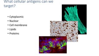
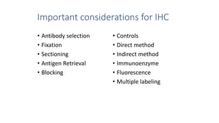
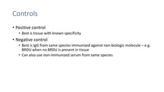
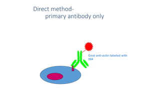
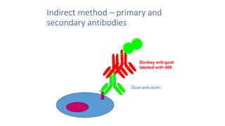
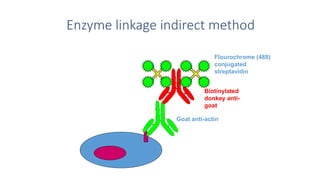
Immunohistochemistry (IHC) combines histological and immunological techniques to identify specific tissue components using antigen-antibody reactions tagged with visible labels. IHC allows visualization of the distribution and localization of cellular components. Antibodies bind specifically to antigens, providing spatial location of particular cells and proteins. Important considerations for IHC include antibody selection, fixation, antigen retrieval, controls, and detection methods such as direct, indirect, and enzyme-linked methods. IHC is useful for tumor diagnosis, classification, predictive markers, and distinguishing between benign and malignant lesions.
Immunohistochemistry (IHC) is a highly sensitive method that allows the localization of antigen within a cell or a tissue with high resolution. The method is based on the use of a primary antibody that specifically binds to its complementary antigen. The bound antibody may then be visualized by a variety of methods such as colorimetric end points.
This method is used to visualise the localisation and quantity of a protein of interest. The target protein is bound to by a specific primary antibody, which in turn is detected by a secondary antibody conjugated to a fluorophore. A fluorescent or confocal microscope is used to visualise the protein.
Immunocytochemistry (ICC) differs from immunohistochemistry (IHC) in that the former is performed on samples of intact cells that have had most, if not all, of their surrounding extracellular matrix removed. In contrast, immunohistochemical samples are sections of biological tissue, where each cell is surrounded by tissue architecture and other cells normally found in the intact tissue. These differences cause the samples to be prepared differently. For ICC, the sample requires permeabilisation so that the antibodies can reach the intracellular targets. Depending on the thickness of the sample, IHC samples do not require this.
Do you have a technical question? Get in touch: info@stjohnslabs.com
This is a powerpoint presentation of Immunohistochemistry of lesions of prostate. This presentation will be helpful for postgraduate pathology students and practitioners alike. We are also on youtube. Please visit our channel at https://www.youtube.com/channel/UCwjkzK-YnJ-ra4HMOqq3Fkw
TEGENE ALEMU CANCER BIOLOGY SURGERY DEPARTMENTTegeneAlemu
Cancer Biology by Tegene Alemu Jimma Ethiopia
Surgery Role on Oncology
On this seminar
Hallmark of cancer
Cell cycles
Apoptosis of cell
Diagnosis
Therapy
This PPT is about immune system and immune therapy, some basic knowledge about Chimeric Antigen Receptor or CAR technology and its application on tumor therapy.
This is needs to be updated as new Biomarkers and Dx antibodies come into existence in remarkable numbers every day! You have to construct an algorithm based on your need and availability of antibodies. Selection of Antibodies for the algorithm also depends on your ability to recognize the characterization of the specific antibody.
- Video recording of this lecture in English language: https://youtu.be/kqbnxVAZs-0
- Video recording of this lecture in Arabic language: https://youtu.be/SINlygW1Mpc
- Link to download the book free: https://nephrotube.blogspot.com/p/nephrotube-nephrology-books.html
- Link to NephroTube website: www.NephroTube.com
- Link to NephroTube social media accounts: https://nephrotube.blogspot.com/p/join-nephrotube-on-social-media.html
- Video recording of this lecture in English language: https://youtu.be/lK81BzxMqdo
- Video recording of this lecture in Arabic language: https://youtu.be/Ve4P0COk9OI
- Link to download the book free: https://nephrotube.blogspot.com/p/nephrotube-nephrology-books.html
- Link to NephroTube website: www.NephroTube.com
- Link to NephroTube social media accounts: https://nephrotube.blogspot.com/p/join-nephrotube-on-social-media.html
Knee anatomy and clinical tests 2024.pdfvimalpl1234
This includes all relevant anatomy and clinical tests compiled from standard textbooks, Campbell,netter etc..It is comprehensive and best suited for orthopaedicians and orthopaedic residents.
Title: Sense of Smell
Presenter: Dr. Faiza, Assistant Professor of Physiology
Qualifications:
MBBS (Best Graduate, AIMC Lahore)
FCPS Physiology
ICMT, CHPE, DHPE (STMU)
MPH (GC University, Faisalabad)
MBA (Virtual University of Pakistan)
Learning Objectives:
Describe the primary categories of smells and the concept of odor blindness.
Explain the structure and location of the olfactory membrane and mucosa, including the types and roles of cells involved in olfaction.
Describe the pathway and mechanisms of olfactory signal transmission from the olfactory receptors to the brain.
Illustrate the biochemical cascade triggered by odorant binding to olfactory receptors, including the role of G-proteins and second messengers in generating an action potential.
Identify different types of olfactory disorders such as anosmia, hyposmia, hyperosmia, and dysosmia, including their potential causes.
Key Topics:
Olfactory Genes:
3% of the human genome accounts for olfactory genes.
400 genes for odorant receptors.
Olfactory Membrane:
Located in the superior part of the nasal cavity.
Medially: Folds downward along the superior septum.
Laterally: Folds over the superior turbinate and upper surface of the middle turbinate.
Total surface area: 5-10 square centimeters.
Olfactory Mucosa:
Olfactory Cells: Bipolar nerve cells derived from the CNS (100 million), with 4-25 olfactory cilia per cell.
Sustentacular Cells: Produce mucus and maintain ionic and molecular environment.
Basal Cells: Replace worn-out olfactory cells with an average lifespan of 1-2 months.
Bowman’s Gland: Secretes mucus.
Stimulation of Olfactory Cells:
Odorant dissolves in mucus and attaches to receptors on olfactory cilia.
Involves a cascade effect through G-proteins and second messengers, leading to depolarization and action potential generation in the olfactory nerve.
Quality of a Good Odorant:
Small (3-20 Carbon atoms), volatile, water-soluble, and lipid-soluble.
Facilitated by odorant-binding proteins in mucus.
Membrane Potential and Action Potential:
Resting membrane potential: -55mV.
Action potential frequency in the olfactory nerve increases with odorant strength.
Adaptation Towards the Sense of Smell:
Rapid adaptation within the first second, with further slow adaptation.
Psychological adaptation greater than receptor adaptation, involving feedback inhibition from the central nervous system.
Primary Sensations of Smell:
Camphoraceous, Musky, Floral, Pepperminty, Ethereal, Pungent, Putrid.
Odor Detection Threshold:
Examples: Hydrogen sulfide (0.0005 ppm), Methyl-mercaptan (0.002 ppm).
Some toxic substances are odorless at lethal concentrations.
Characteristics of Smell:
Odor blindness for single substances due to lack of appropriate receptor protein.
Behavioral and emotional influences of smell.
Transmission of Olfactory Signals:
From olfactory cells to glomeruli in the olfactory bulb, involving lateral inhibition.
Primitive, less old, and new olfactory systems with different path
263778731218 Abortion Clinic /Pills In Harare ,sisternakatoto
263778731218 Abortion Clinic /Pills In Harare ,ABORTION WOMEN’S CLINIC +27730423979 IN women clinic we believe that every woman should be able to make choices in her pregnancy. Our job is to provide compassionate care, safety,affordable and confidential services. That’s why we have won the trust from all generations of women all over the world. we use non surgical method(Abortion pills) to terminate…Dr.LISA +27730423979women Clinic is committed to providing the highest quality of obstetrical and gynecological care to women of all ages. Our dedicated staff aim to treat each patient and her health concerns with compassion and respect.Our dedicated group ABORTION WOMEN’S CLINIC +27730423979 IN women clinic we believe that every woman should be able to make choices in her pregnancy. Our job is to provide compassionate care, safety,affordable and confidential services. That’s why we have won the trust from all generations of women all over the world. we use non surgical method(Abortion pills) to terminate…Dr.LISA +27730423979women Clinic is committed to providing the highest quality of obstetrical and gynecological care to women of all ages. Our dedicated staff aim to treat each patient and her health concerns with compassion and respect.Our dedicated group of receptionists, nurses, and physicians have worked together as a teamof receptionists, nurses, and physicians have worked together as a team wwww.lisywomensclinic.co.za/
2. Introduction
• Immunohistochemistry (IHC) combines histological, immunological
and biochemical techniques for the identification of specific tissue
components by means of a specific antigen/antibody reaction tagged
with a visible label
• IHC makes it possible to visualize the distribution and localization of
specific cellular components within a cell or tissue.
3. • Antibodies bind to antigen in specific manner
• Gives a spatial location
• Can be used to locate particular cells and proteins
• Can be used to identify cellular events – e.g.apoptosis
4. History
• The principle has existed since the 1930s.
• Started in 1941 when Coons identified pneumococci using
a direct fluorescent method.
• Indirect method
• Addition of horseradish peroxidase
• Peroxidase anti-peroxidase technique in 1979
• Use of Avidin & Biotin complex in early 1980’s
5. What cellular antigens can we
target?
• Cytoplasmic
• Nuclear
• Cell membrane
• Lipids
• Proteins
8. Monoclonal v. polyclonal
• Monoclonal
• Mouse or rabbit
hybridoma
• Tends to be ‘cleaner’
• Very consistent batch-to-
batch
• More likely to get false
negative results
• Polyclonal
• Many different species
• Tends to have more non-
specific reactivity
• Can have very different
avidity/affinity batch-to-
batch
• More likely to have
success in an unknown
application
9. Fixation
Aldehyde
10% NBF
4% formaldehyde with
PBS buffer
2% formaldehyde with
picric acid and PBS
The paraformaldehyde
paradox
24-72 hours
Many others
Best for good
architecture
Frozen
With or without sucrose
OCT
Fix with acetone or
methanol (fix by
coagulation, also
permeabilizes)
Best for cell membrane
antigens, cytokines
10. Sectioning
• Paraffin
• Must heat and process
through xylenes and
alcohols – ruins some
antigens
• Most commonly used
• BEST if not stored more
than two weeks – lose
antigenicity after that
time
• Frozen
• Better survival of many
antigens
• Poor morphology
• Poor resolution at higher
mag
• Special storage
• Cutting difficulty
11. Antigen retrieval
HIER
Use
MW/steamer/pressure
cooker ~ 20 minutes,
slow cool
Citrate 6.0
Tris-EDTA 9.0
EDTA 8.0
Must determine for each
new antibody/antigen
target
PIER
Proteinase K
Trypsin
Pepsin
Pronase,etc.
Destroys some epitopes
Bad for morphology
12. Improving antibody penetration
Need this for intracellular (cytoplasmic, nuclear) or membrane
components when epitope is inside cell membrane
Detergents most popular
Triton-X
Tween
Also decreases surface tension – better coverage
Can’t use for membrane proteins
Acetone/Methanol
Precipitate proteins outside cell membranes- more accessible
Saponin
Punches holes in cell membrane – holes close up when removed
13. Blocking
• Background staining
• Specific
• Polyclonal antibodies – impure antigen used
• Inadequate fixation – diffusion of antigen – often worse in center of large
block
• Non-specific
• Non-immunologic binding – usually uniform
• Endogenous peroxidases
• Endogenous biotin
14. Controls
• Positive control
• Best is tissue with known specificity
• Negative control
• Best is IgG from same species immunized against non-biologic molecule – e.g.
BRDU when no BRDU is present in tissue
• Can also use non-immunized serum from same species
18. Enzymatic detection methods
Brightfield microscope sufficient for analysis of specimens
Suitable for tissue analysis at low magnification
Resolution of subcellular structures not as good as with fluorescence methods,
but can be combined with electron microscopy
Unimited shelf life of labelled specimens
Substrate reagents often toxic/carcinogenic
19. Usefulness of IHC tests.
• Poorly differentiated tumors and mixed carcinomas
• Undifferentiated tumors of unknown origin
• Treatment based on sub-type of cancer: Personalized Medicine eg., Breast Ca
• Monitoring progress of cancer (Predictive and Prognosis)
• In Malignant Lymphoma,
• In identifying Carcinoid (Neuroendocrine) tumors
• In Cytologic specimens
20.
21. Histiogenic Dx of Neoplasm
A) Expression of cytokeratin AE1/AE3 in lung carcinosarcoma ; B) chromogranin
expression in gastric neuroendocrine carcinoma ; C) HMB 45 immunostainning in
melanoma .
Epithelium NeuroEndocrine Melanocyte
26. Urothelial Ca vs Prostate Ca
• EMA
• CK7
• P63
• CK5/6
• EpCam
• CD57
• PSA
• PAP
• NKX3.1
• Prostein
+ -
+ -
+/- 0
+/- 0
+/- 0
-/+ +/-
0 +
0 +/-
0 +
0 +
0 +
Urothelial ca Prostate ca
Hammerich KH.Archives of Pathology and Laboratory Medicine;132(3):432-440
Urothelial ca
Prostate ca
27. Risk Biomarkers (or screening biomarkers) Describe risk of cancer
occurrence or cancer progression because they
are implicated in neoplastic progression and include:
1. Genetic predisposition (e.g., BRCA1/2)
2. Over expression of genes (e.g., BCR-ABLTyrosin Kinase Inhibitor in CML, HER-2/neu in
Br Ca,PTEN, RAS,Colo Rectal Cancer AKTin Pancreatic cancer)
4.Environmental factors and lifestyle (e.g., HPV or HBV infection
http://www.esmo.org/content/download/8713/176680/file/
The-use-of-Biomarkers-for-Treatment-Sessa-Fasolo.pdf
28. Undifferentiated Tumors
Use of IHC to differentiate broad lineage
Pan CK
AE1/AE3
CD45
(LCA)
HMB45
or S100
VIM
Carcinoma Positive Negative Negative Negative
Melanoma Negative Negative Positive Positive
Sarcoma Negative Negative Negative Positive
Lymphoma Negative Positive Negative Negative
29. Carcinoma Use of CK7 and CK20
CK7 +
CK20 +
CK7 +
CK20 -
CK7 –
CK20 +
CK7 –
CK20 -
Urothelial Ca
Pancreatic Ad Ca
OvarianMucinous
Ca
Ad Ca of Bladder
Gastric Ad Ca
Cholangio Ca
subset
Breast Ca
EndoMetrial Ad Ca
EndoCervical Ad Ca
Ovarian Cerous Ca
Lung Ad Ca
Cholangio Ca
LungSmCC
Mesothelioma
Thyroid Ca
SCC of Cervix
SalivaryGland
tumors
Urothelial Ca subset
Pancreatic and
Gastric Ad Ca
subset
Colorectal Ad Ca
Markel Cell ca
Gastric Ad Ca
subset
Prostate Ad Ca
SCC
RCC
HCC
Mesothelioma
AdrenoCortical ca
NonSeminoma
LungSmCC
minorSubset
Gastric Ad Ca
subset
http://www.pathinformatics.com/department/documents/SusanE%20Lecture.pdf
30. Immunohistochemistry stains in
squamous cell carcinoma and
adenocarcinoma of lung. H&E:
hematoxylin and eosin; CK:
cytokeratin; TTF-1: thyroid
transcription factor 1. Squamous
carcinomas are typically positive
for CK5/6 and P63, and negative
for CK7 and TTF-1, with the reverse
profile for adenocarcinoma
although this case of squamous cell
carcinoma demonstrates focal
weak staining for CK7.
SqCC AdCa
“Ancillary Testing in Lung Cancer Diagnosis”
Dublinski et al.
Openi.nlm.nih.gov