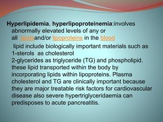
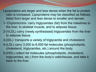
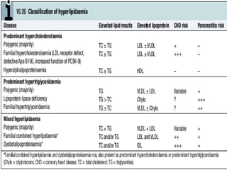
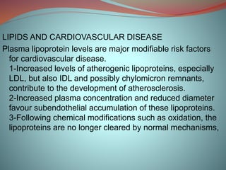
Hyperlipidemia involves abnormally elevated levels of lipids and lipoproteins in the blood. Lipids include cholesterol, triglycerides, and phospholipids, which are transported around the body within lipoproteins like chylomicrons, VLDL, LDL, IDL, and HDL. High cholesterol and triglycerides are major risk factors for cardiovascular disease, and very high triglycerides can cause pancreatitis. Hyperlipidemias can be primary or secondary, and are classified based on which lipids are predominantly elevated, such as cholesterol, triglycerides, or both. Primary causes include genetic defects or polygenic influences, while secondary causes include hypothyroidism, pregnancy, liver disease
the aim of sharing this material to help students and provide delayed information regarding topic.You all are most welcome for you suggestion to make i more easy, graspable and attractive.(easy to learn in creative way)
Hyperlipidemia , dyslipidemia , and drug therapy
also Fat transport and metabolisim and pathophysiology of lipoprotein
clincal importance of
1. Hypertriglycredemia
2. Hypercholesterolemia
3.Combined hyperlipidemia
4. Some other lipoprotein disorders
Including disorder of HDL_C
The high risks of lipids and its relevance towards the development of different cardiovascular diseases has been known to all where this present slide focuses on that only along with the different treatment procedures,.
"48 SLIDES???!!", my friends shouted.
A boring "48 slides" is depend on how you arrange it. And this is not the one for sure.
I always love to prepare a short and sweet presentation. Or maybe long but sweet presentation? Oh yeah! Enjoy!
#SLIDESKILLSvsSLIDEKILLS
Basics of hyperlipoproreinemia in an easy and understandable way.gives a brief picture of the disease , it's cauusitive agents and clinical sequelae following it.
Complications of abnormal lipid levels
Generally, a high total cholesterol level (which includes LDL, HDL, and VLDL cholesterol), particularly a high level of LDL (the "bad") cholesterol, increases the risk of atherosclerosis and thus the risk of heart attack or strok
The prostate is an exocrine gland of the male mammalian reproductive system
It is a walnut-sized gland that forms part of the male reproductive system and is located in front of the rectum and just below the urinary bladder
Function is to store and secrete a clear, slightly alkaline fluid that constitutes 10-30% of the volume of the seminal fluid that along with the spermatozoa, constitutes semen
A healthy human prostate measures (4cm-vertical, by 3cm-horizontal, 2cm ant-post ).
It surrounds the urethra just below the urinary bladder. It has anterior, median, posterior and two lateral lobes
It’s work is regulated by androgens which are responsible for male sex characteristics
Generalised disease of the prostate due to hormonal derangement which leads to non malignant enlargement of the gland (increase in the number of epithelial cells and stromal tissue)to cause compression of the urethra leading to symptoms (LUTS
Title: Sense of Taste
Presenter: Dr. Faiza, Assistant Professor of Physiology
Qualifications:
MBBS (Best Graduate, AIMC Lahore)
FCPS Physiology
ICMT, CHPE, DHPE (STMU)
MPH (GC University, Faisalabad)
MBA (Virtual University of Pakistan)
Learning Objectives:
Describe the structure and function of taste buds.
Describe the relationship between the taste threshold and taste index of common substances.
Explain the chemical basis and signal transduction of taste perception for each type of primary taste sensation.
Recognize different abnormalities of taste perception and their causes.
Key Topics:
Significance of Taste Sensation:
Differentiation between pleasant and harmful food
Influence on behavior
Selection of food based on metabolic needs
Receptors of Taste:
Taste buds on the tongue
Influence of sense of smell, texture of food, and pain stimulation (e.g., by pepper)
Primary and Secondary Taste Sensations:
Primary taste sensations: Sweet, Sour, Salty, Bitter, Umami
Chemical basis and signal transduction mechanisms for each taste
Taste Threshold and Index:
Taste threshold values for Sweet (sucrose), Salty (NaCl), Sour (HCl), and Bitter (Quinine)
Taste index relationship: Inversely proportional to taste threshold
Taste Blindness:
Inability to taste certain substances, particularly thiourea compounds
Example: Phenylthiocarbamide
Structure and Function of Taste Buds:
Composition: Epithelial cells, Sustentacular/Supporting cells, Taste cells, Basal cells
Features: Taste pores, Taste hairs/microvilli, and Taste nerve fibers
Location of Taste Buds:
Found in papillae of the tongue (Fungiform, Circumvallate, Foliate)
Also present on the palate, tonsillar pillars, epiglottis, and proximal esophagus
Mechanism of Taste Stimulation:
Interaction of taste substances with receptors on microvilli
Signal transduction pathways for Umami, Sweet, Bitter, Sour, and Salty tastes
Taste Sensitivity and Adaptation:
Decrease in sensitivity with age
Rapid adaptation of taste sensation
Role of Saliva in Taste:
Dissolution of tastants to reach receptors
Washing away the stimulus
Taste Preferences and Aversions:
Mechanisms behind taste preference and aversion
Influence of receptors and neural pathways
Impact of Sensory Nerve Damage:
Degeneration of taste buds if the sensory nerve fiber is cut
Abnormalities of Taste Detection:
Conditions: Ageusia, Hypogeusia, Dysgeusia (parageusia)
Causes: Nerve damage, neurological disorders, infections, poor oral hygiene, adverse drug effects, deficiencies, aging, tobacco use, altered neurotransmitter levels
Neurotransmitters and Taste Threshold:
Effects of serotonin (5-HT) and norepinephrine (NE) on taste sensitivity
Supertasters:
25% of the population with heightened sensitivity to taste, especially bitterness
Increased number of fungiform papillae
TEST BANK for Operations Management, 14th Edition by William J. Stevenson, Ve...kevinkariuki227
TEST BANK for Operations Management, 14th Edition by William J. Stevenson, Verified Chapters 1 - 19, Complete Newest Version.pdf
Flu Vaccine Alert in Bangalore Karnatakaaddon Scans
As flu season approaches, health officials in Bangalore, Karnataka, are urging residents to get their flu vaccinations. The seasonal flu, while common, can lead to severe health complications, particularly for vulnerable populations such as young children, the elderly, and those with underlying health conditions.
Dr. Vidisha Kumari, a leading epidemiologist in Bangalore, emphasizes the importance of getting vaccinated. "The flu vaccine is our best defense against the influenza virus. It not only protects individuals but also helps prevent the spread of the virus in our communities," he says.
This year, the flu season is expected to coincide with a potential increase in other respiratory illnesses. The Karnataka Health Department has launched an awareness campaign highlighting the significance of flu vaccinations. They have set up multiple vaccination centers across Bangalore, making it convenient for residents to receive their shots.
To encourage widespread vaccination, the government is also collaborating with local schools, workplaces, and community centers to facilitate vaccination drives. Special attention is being given to ensuring that the vaccine is accessible to all, including marginalized communities who may have limited access to healthcare.
Residents are reminded that the flu vaccine is safe and effective. Common side effects are mild and may include soreness at the injection site, mild fever, or muscle aches. These side effects are generally short-lived and far less severe than the flu itself.
Healthcare providers are also stressing the importance of continuing COVID-19 precautions. Wearing masks, practicing good hand hygiene, and maintaining social distancing are still crucial, especially in crowded places.
Protect yourself and your loved ones by getting vaccinated. Together, we can help keep Bangalore healthy and safe this flu season. For more information on vaccination centers and schedules, residents can visit the Karnataka Health Department’s official website or follow their social media pages.
Stay informed, stay safe, and get your flu shot today!
Couples presenting to the infertility clinic- Do they really have infertility...Sujoy Dasgupta
Dr Sujoy Dasgupta presented the study on "Couples presenting to the infertility clinic- Do they really have infertility? – The unexplored stories of non-consummation" in the 13th Congress of the Asia Pacific Initiative on Reproduction (ASPIRE 2024) at Manila on 24 May, 2024.
These lecture slides, by Dr Sidra Arshad, offer a quick overview of physiological basis of a normal electrocardiogram.
Learning objectives:
1. Define an electrocardiogram (ECG) and electrocardiography
2. Describe how dipoles generated by the heart produce the waveforms of the ECG
3. Describe the components of a normal electrocardiogram of a typical bipolar leads (limb II)
4. Differentiate between intervals and segments
5. Enlist some common indications for obtaining an ECG
Study Resources:
1. Chapter 11, Guyton and Hall Textbook of Medical Physiology, 14th edition
2. Chapter 9, Human Physiology - From Cells to Systems, Lauralee Sherwood, 9th edition
3. Chapter 29, Ganong’s Review of Medical Physiology, 26th edition
4. Electrocardiogram, StatPearls - https://www.ncbi.nlm.nih.gov/books/NBK549803/
5. ECG in Medical Practice by ABM Abdullah, 4th edition
6. ECG Basics, http://www.nataliescasebook.com/tag/e-c-g-basics
Ozempic: Preoperative Management of Patients on GLP-1 Receptor Agonists Saeid Safari
Preoperative Management of Patients on GLP-1 Receptor Agonists like Ozempic and Semiglutide
ASA GUIDELINE
NYSORA Guideline
2 Case Reports of Gastric Ultrasound
Explore natural remedies for syphilis treatment in Singapore. Discover alternative therapies, herbal remedies, and lifestyle changes that may complement conventional treatments. Learn about holistic approaches to managing syphilis symptoms and supporting overall health.
Title: Sense of Smell
Presenter: Dr. Faiza, Assistant Professor of Physiology
Qualifications:
MBBS (Best Graduate, AIMC Lahore)
FCPS Physiology
ICMT, CHPE, DHPE (STMU)
MPH (GC University, Faisalabad)
MBA (Virtual University of Pakistan)
Learning Objectives:
Describe the primary categories of smells and the concept of odor blindness.
Explain the structure and location of the olfactory membrane and mucosa, including the types and roles of cells involved in olfaction.
Describe the pathway and mechanisms of olfactory signal transmission from the olfactory receptors to the brain.
Illustrate the biochemical cascade triggered by odorant binding to olfactory receptors, including the role of G-proteins and second messengers in generating an action potential.
Identify different types of olfactory disorders such as anosmia, hyposmia, hyperosmia, and dysosmia, including their potential causes.
Key Topics:
Olfactory Genes:
3% of the human genome accounts for olfactory genes.
400 genes for odorant receptors.
Olfactory Membrane:
Located in the superior part of the nasal cavity.
Medially: Folds downward along the superior septum.
Laterally: Folds over the superior turbinate and upper surface of the middle turbinate.
Total surface area: 5-10 square centimeters.
Olfactory Mucosa:
Olfactory Cells: Bipolar nerve cells derived from the CNS (100 million), with 4-25 olfactory cilia per cell.
Sustentacular Cells: Produce mucus and maintain ionic and molecular environment.
Basal Cells: Replace worn-out olfactory cells with an average lifespan of 1-2 months.
Bowman’s Gland: Secretes mucus.
Stimulation of Olfactory Cells:
Odorant dissolves in mucus and attaches to receptors on olfactory cilia.
Involves a cascade effect through G-proteins and second messengers, leading to depolarization and action potential generation in the olfactory nerve.
Quality of a Good Odorant:
Small (3-20 Carbon atoms), volatile, water-soluble, and lipid-soluble.
Facilitated by odorant-binding proteins in mucus.
Membrane Potential and Action Potential:
Resting membrane potential: -55mV.
Action potential frequency in the olfactory nerve increases with odorant strength.
Adaptation Towards the Sense of Smell:
Rapid adaptation within the first second, with further slow adaptation.
Psychological adaptation greater than receptor adaptation, involving feedback inhibition from the central nervous system.
Primary Sensations of Smell:
Camphoraceous, Musky, Floral, Pepperminty, Ethereal, Pungent, Putrid.
Odor Detection Threshold:
Examples: Hydrogen sulfide (0.0005 ppm), Methyl-mercaptan (0.002 ppm).
Some toxic substances are odorless at lethal concentrations.
Characteristics of Smell:
Odor blindness for single substances due to lack of appropriate receptor protein.
Behavioral and emotional influences of smell.
Transmission of Olfactory Signals:
From olfactory cells to glomeruli in the olfactory bulb, involving lateral inhibition.
Primitive, less old, and new olfactory systems with different path
New Drug Discovery and Development .....NEHA GUPTA
The "New Drug Discovery and Development" process involves the identification, design, testing, and manufacturing of novel pharmaceutical compounds with the aim of introducing new and improved treatments for various medical conditions. This comprehensive endeavor encompasses various stages, including target identification, preclinical studies, clinical trials, regulatory approval, and post-market surveillance. It involves multidisciplinary collaboration among scientists, researchers, clinicians, regulatory experts, and pharmaceutical companies to bring innovative therapies to market and address unmet medical needs.
Pulmonary Thromboembolism - etilogy, types, medical- Surgical and nursing man...VarunMahajani
Disruption of blood supply to lung alveoli due to blockage of one or more pulmonary blood vessels is called as Pulmonary thromboembolism. In this presentation we will discuss its causes, types and its management in depth.
Lung Cancer: Artificial Intelligence, Synergetics, Complex System Analysis, S...Oleg Kshivets
RESULTS: Overall life span (LS) was 2252.1±1742.5 days and cumulative 5-year survival (5YS) reached 73.2%, 10 years – 64.8%, 20 years – 42.5%. 513 LCP lived more than 5 years (LS=3124.6±1525.6 days), 148 LCP – more than 10 years (LS=5054.4±1504.1 days).199 LCP died because of LC (LS=562.7±374.5 days). 5YS of LCP after bi/lobectomies was significantly superior in comparison with LCP after pneumonectomies (78.1% vs.63.7%, P=0.00001 by log-rank test). AT significantly improved 5YS (66.3% vs. 34.8%) (P=0.00000 by log-rank test) only for LCP with N1-2. Cox modeling displayed that 5YS of LCP significantly depended on: phase transition (PT) early-invasive LC in terms of synergetics, PT N0—N12, cell ratio factors (ratio between cancer cells- CC and blood cells subpopulations), G1-3, histology, glucose, AT, blood cell circuit, prothrombin index, heparin tolerance, recalcification time (P=0.000-0.038). Neural networks, genetic algorithm selection and bootstrap simulation revealed relationships between 5YS and PT early-invasive LC (rank=1), PT N0—N12 (rank=2), thrombocytes/CC (3), erythrocytes/CC (4), eosinophils/CC (5), healthy cells/CC (6), lymphocytes/CC (7), segmented neutrophils/CC (8), stick neutrophils/CC (9), monocytes/CC (10); leucocytes/CC (11). Correct prediction of 5YS was 100% by neural networks computing (area under ROC curve=1.0; error=0.0).
CONCLUSIONS: 5YS of LCP after radical procedures significantly depended on: 1) PT early-invasive cancer; 2) PT N0--N12; 3) cell ratio factors; 4) blood cell circuit; 5) biochemical factors; 6) hemostasis system; 7) AT; 8) LC characteristics; 9) LC cell dynamics; 10) surgery type: lobectomy/pneumonectomy; 11) anthropometric data. Optimal diagnosis and treatment strategies for LC are: 1) screening and early detection of LC; 2) availability of experienced thoracic surgeons because of complexity of radical procedures; 3) aggressive en block surgery and adequate lymph node dissection for completeness; 4) precise prediction; 5) adjuvant chemoimmunoradiotherapy for LCP with unfavorable prognosis.
Lung Cancer: Artificial Intelligence, Synergetics, Complex System Analysis, S...
Hyperlipidemia
1.
2. Hyperlipidemia, hyperlipoproteinemia:involves
abnormally elevated levels of any or
all lipidsand/or lipoproteins in the blood
lipid include biologically important materials such as
1-sterols as cholesterol
2-glycerides as triglyceride (TG) and phospholipid.
these lipid transported within the body by
incorporating lipids within lipoproteins. Plasma
cholesterol and TG are clinically important because
they are major treatable risk factors for cardiovascular
disease also severe hypertriglyceridaemia can
predisposes to acute pancreatitis.
3. Lipoproteins are larger and less dense when the fat to protein
ratio is increased. Lipoproteins may be classified as follows,
listed from larger and less dense to smaller and denser.
1- Chylomicrons :carry triglycerides (fat) from the intestines to
the liver, to skeletal muscle, and to adipose tissue.
2-(VLDL) carry (newly synthesised) triglycerides from the liver
to adipose tissue.
3-(IDL): transports a variety of triglyceride and cholesterol
4-(LDL) carry 3,000 to 6,000 fat molecules (phospholipids,
cholesterol, triglycerides, etc.) around the body.
5-(HDL) collect fat molecules (phospholipids, cholesterol,
triglycerides, etc.) from the body's cells/tissues, and take it
back to the liver.
4.
5. It is also possible to classify lipoproteins as "alpha" and
"beta", according to the classification of proteins
in serum protein electrophoresis
Etiology of hyperlipidemias:
Primary: Following the exclusion of secondary causes
primary lipid abnormalities may be diagnosed.the
Fredrickson classification (types I to V) is no longer in
use because it adds little to clinical decision-making.
Instead, primary lipid abnormalities are classified
according to the predominant lipid problem:
hypercholesterolaemia , hypertriglyceridaemia or mixed
hyperlipidemia.
6. A- Predominant hypercholesterolaemia:
Classified into
1-Polygenic hypercholesterolaemia is the most
common cause of mild to moderate increase in LDL-C
This condition is caused by a susceptible genotype
aggravated by 1 or more factors, including atherogenic
diet (excessive intake of saturated fat, trans fat, and, to
a lesser extent, cholesterol), obesity, and sedentary
lifestyle.
Physical signs such as corneal arcus and xanthelasma
may be found.
Risk of cardiovascular disease is proportional to the
degree of LDL-C elevation, together with other major
risk factors, particularly low HDL-C.
7. 2-Familial hypercholesterolaemia (FH) causes
moderate to severe hypercholesterolaemia with a
prevalence of at least 0.2%. in most It is usually due
to an autosomal dominantly inherited mutation of the
LDL receptor gene
3-defects in the apolipoprotein B100 or increased
function of sterol-sensitive protease known as pcsk9
(Proprotein convertase subtilisin/kexin type 9)
LDL-C is removed from the blood when it binds to an
LDLR on the surface of liver cells, and is taken inside
the cells. When PCSK9 binds to an LDLR, the
receptor is destroyed along with the LDL particle. But
if PCSK9 does not bind, the receptor can return to
the surface of the cell and remove more cholesterol.
8. Number of monoclonal antibody bind pcsk9 as evolocumab
& alirocumab.
Most patients with these abnormalities exhibit LDL levels that
are approximately twice as high as in unaffected subjects of
the same age and gender.
Family history reveals that approximately 50% of each
generation suffers hypercholesterolaemia, often with very
premature cardiovascular disease.
FH may be accompanied by xanthomas of the Achilles or
extensor digitorum tendons which are strongly suggestive
(but not pathognomonic) for FH. The onset of corneal arcus
before age 40 is also suggestive of this condition.
9. In populations in which there is a 'founder gene' effect or
consanguineous marriage, homozygous FH
occasionally occurs, resulting in more extensive
xanthomas and precocious cardiovascular disease in
childhood.
4-Hyperalphalipoproteinaemia refers to increased levels
of HDL-C. In the absence of an increase in LDL-C, this
condition does not cause CVS disease, so it should not
be regarded as pathological.
Familial combined hyperlipidaemia, and
dysbetalipoproteinaemia, may present with the pattern
of predominant hypercholesterolaemia
10. B-Predominant hypertriglyceridaemia:
Classified into
1-Polygenic hypertriglyceridaemia is the most common
primary cause of TG elevation. It also commonly occurs
secondary to
1-excess alcohol 2-medications
3-type 2 diabetes 4-impaired glucose tolerance
5-central obesity
It is often accompanied by post-prandial hyperlipidaemia
and reduced HDL-C, both of which may contribute to
cardiovascular risk.
Excessive dietary fat intake or other exacerbating factors
may precipitate a massive increase in TG levels, which, if
they exceed 10 mmol/l (880 mg/dl), may pose a risk of
acute pancreatitis.
11. 2-Lipoprotein lipase deficiency: Lipoprotein lipase
deficiency is an infrequent autosomal recessive
disorder due to hereditary deficiency of lipoprotein
lipase (LPL) resulting in elevated chylomicrons,
3-Familial apoprotein CII deficiency a condition
caused by a lack of lipoprotein lipase activator. Or
due to circulating inhibitor of lipoprotein lipase.
It causes massive hypertriglyceridaemia that is
resistant to drug treatment.
usually presents in childhood with eruptive
xanthomata and abdominal colic.
12. Complications:
1-retinal vein occlusion 2-acute pancreatitis
3-steatosis(abnormal retention of lipids within a cell)
4-hepatosplenomegaly 5-lipaemia
retinalis( milky appearance of the veins and arteries of t
he retina
4-Familial hypertriglyceridemia: is an autosomal
dominant condition occurring in approximately 1% of
the population.
Triglyceride levels, but not cholesterol, are elevated as
a result of excess hepatic production of VLDL or
heterozygous LPL deficiency.
Unlike familial hypercholesterolemia, there is no
association with premature coronary disease..
13. However, affected individuals are at risk for
chylomicronemia syndrome, characterized by elevated
chylomicrons in the blood. It has been suggested that it
may represent a secondary response to impaired bile
acid resorption
On the other hand, it predisposes to levels of
hypertriglyceridaemia that are sufficient to pose a risk of
pancreatitis .Familial combined hyperlipidaemia, and
dysbetalipoproteinaemia, may present with the pattern
of predominant hypertriglyceridaemia.
14. C-Mixed hyperlipidaemia :The term 'mixed' usually
implies the presence of hypertriglyceridaemia as well as
an increase in LDL-c or IDL-c. classified into
1- polygenic :Primary mixed hyperlipidaemia is usually
polygenic and, like predominant hypertriglyceridaemia,
often occurs in association with type 2 diabetes,
impaired glucose tolerance, central obesity HT.
15. 2-Familial combined hyperlipidaemia is a dominantly
inherited disorder caused by overproduction of
atherogenic apolipoprotein B-containing lipoproteins.
It results in elevation of cholesterol, TG or both in
different family members at different times.
It is associated with an increased risk of cardiovascular
disease but it does not produce any pathognomonic
physical signs. In practice, this relatively common
condition is substantially modified by factors such as
age and weight.
16. 3-Dysbetalipoproteinaemia
It is caused by homozygous inheritance of the
apolipoprotein E2 allele, which is the isoform least
avidly recognised by the LDL receptor. In conjunction
with other exacerbating factors such as obesity and
diabetes, it leads to accumulation of atherogenic IDL
and chylomicron remnants.
Premature cardiovascular disease is common and it
may also result in the formation of palmar xanthomas,
tuberous xanthomas or tendon xanthomas.
17.
18.
19. hyperlipopr
oteinemia
defect Increased
lipoprotei
n
Main
symptoms
serum
Appeara
nce
Treatment
Type Ia or
Buerger-
Gruetz
syndrome or
familial
hyperchylom
icronemia
Decreased lip
oprotein
lipase (LPL)
Chylomicro
ns
Acute
pancreatitis, lipe
mia retinalis,
eruptive
skin xanthomas,
hepatosplenom
egaly
Creamy
top layer
Diet control
Type Ib
Familial
apoprotein
CII
deficiency
Altered ApoC2 Chylomicro
ns
Same as a Creamy
top layer
Diet control
Type Ic LPL inhibitor in
blood
Chylomicro
ns
Same as a
Type IIa or
F.Hypercho.
LDL
receptordeficie
ncy
LDL Xanthelasma, ar
cus senilis,
tendon
xanthomas
Clear Bile acid
sequestrants
,statins, niaci
n
Type IIb or
Familial
Decreased LD
L receptor and
=======
=
20. hyperlipopr
oteinemia
defect Increased
lipoprotein
Main
symptoms
serum
Appearance
Treatment
Type III OR
Familial
dysbetalipop
roteinemia
Defect
in Apo E
2synthesis
IDL Tuboeruptive
xanthomas
and palmar
xanthomas
Turbid
Fibrate,
statins
Type IV or
Familial
hypertriglyce
ridemia
Increased
VLDL
production &
decreased
elimination
VLDL Can
cause pancr
eatitis at
high
triglyceride
levels
same Fibrate,
niacin,
statins
Type V Increased
VLDL
production
and
decreased L
PL
VLDL and
chylomicrons
Creamy top
layer and
turbid bottom
Niacin,
fibrate
21. Rare dyslipidaemias: Several rare disturbances of lipid
metabolism have been described .They provide important
insights into lipid metabolism and its impact on risk of
cardiovascular disease. Fish eye disease and Apo A1
Milano demonstrate that very low HDL levels do not
necessarily cause cardiovascular disease, but Apo A1
deficiency, Tangier disease and LCAT deficiency
demonstrate that low HDL-C can be atherogenic under
some circumstances. Sitosterolaemia and cerebrotendinous
xanthomatosis demonstrate that sterols other than
cholesterol can cause xanthomas and cardiovascular
disease, while abetalipoproteinaemia and
hypobetalipoproteinaemia suggest that low levels of
apolipoprotein B-containing lipoproteins reduce the risk of
cardiovascular disease at the expense of fat-soluble vitamin
deficiency, leading to retinal lesions and peripheral
neuropathy.
24. LIPIDS AND CARDIOVASCULAR DISEASE
Plasma lipoprotein levels are major modifiable risk factors
for cardiovascular disease.
1-Increased levels of atherogenic lipoproteins, especially
LDL, but also IDL and possibly chylomicron remnants,
contribute to the development of atherosclerosis.
2-Increased plasma concentration and reduced diameter
favour subendothelial accumulation of these lipoproteins.
3-Following chemical modifications such as oxidation, the
lipoproteins are no longer cleared by normal mechanisms,
25. They trigger a self-perpetuating inflammatory response
during which they are taken up by macrophages to
form foam cells(lipid laden macrophage) a hallmark
of the atherosclerotic process.
cholesterol-laden cells release cholesterol to HDL for
reverse cholesterol transport to the liver for
excretion.
HDL may also counteract some components of the
inflammatory response, such as the expression of
vascular adhesion molecules by the endothelium.
Consequently, low HDL cholesterol levels also
predispose to atherosclerosis.
26. Diagnosis
LIPID MEASUREMENT
Measurement of plasma cholesterol alone is not sufficient for
comprehensive assessment.
Levels of total cholesterol (TC), triglyceride (TG) and HDL
cholesterol (HDL-C) should be obtained after a 12-hour fast
to permit the calculation of LDL cholesterol (LDL-C)
according to the Friedewald formula
LDL-C = TC - HDL-C - (TG/2.2) mmol/l
Before the formula is applied, lipid levels in mg/dl can be
converted to mmol/l by dividing by 38 for cholesterol and 88
for triglycerides.
The formula becomes unreliable when TG levels exceed 4
mmol/l (350 mg/dl). Non-fasting samples are unaffected in
terms of TC and measured LDL-C, but they differ in terms of
TG and HDL-C, and hence the calculation of LDL-C is
invalidated.
27. Consideration must be given to influencing factors such
as recent illness, after which cholesterol levels
temporarily decrease in proportion to severity.
Measurements that will affect major decisions, such as
initiation of drug therapy, should be confirmed with a
repeat measurement. Elevated TG, which is common
in obesity, diabetes and insulin resistance is frequently
associated with low HDL and increased 'small, dense'
LDL. Under these circumstances, LDL-C may
underestimate risk. This is one situation in which
measurement of apolipoprotein B may provide
additional useful information.
28. MANAGEMENT OF DYSLIPIDAEMIA
Lipid-lowering therapies have a key role in the secondary
and primary prevention of cardiovascular diseases.
1-Assessment of absolute risk
2-treatment of all modifiable risk factors
3-optimisation of lifestyle factors, especially diet and
exercise, are central to management in all cases.
Public health organisations recommend thresholds for
the introduction of lipid-lowering therapy based on the
identification of patients in very high-risk categories or
those calculated to be at high absolute risk according to
algorithms or tables such as the Joint British Societies
Coronary Risk Prediction Chart.
29. 1-patients who already have cardiovascular disease
2-diabetes mellitus
3-an absolute risk of cardiovascular disease of greater
than 20% in the ensuing 10 years, regarded as having
sufficient risk to justify drug treatment.
Public health organisations also recommend target
levels for patients receiving drug treatment. High-risk
patients should aim for
1-HDL-C > 1 mmol/l (38 mg/dl)
2-fasting TG < 2 mmol/l (∼180 mg/dl)
3-LDL-C have been reduced from 2.5 to 2.0 mmol/l (76
mg/dl) or less.In general, total cholesterol should be < 5
mmol/l (190 mg/dl) during treatment, and < 4 mmol/l
(∼150 mg/dl) in high-risk patients and in secondary
prevention of cardiovascular disease.
30. Non-pharmacological treatment
Patients with lipid abnormalities should receive medical
advice and, if necessary, dietary counselling to:
1-reduce intake of saturated and trans-unsaturated fat
to less than 7-10% of total energy
2-reduce the intake of cholesterol to less than 250
mg/day
3-replace sources of saturated fat and cholesterol with
alternative foods such as lean meat, low-fat dairy
products, polyunsaturated spreads and low glycaemic
index carbohydrates
4-reduce energy-dense foods such as fats and soft
drinks, whilst increasing activity and exercise to achieve
stable or negative energy balance (i.e. weight
maintenance or weight loss)
31. 5-increase consumption of cardioprotective and nutrient-
dense foods such as vegetables, unrefined
carbohydrates, fish, pulses, legumes, fruit etc.
6-adjust alcohol consumption, reducing intake if excessive
or if associated with hypertension, hypertriglyceridaemia
or central obesity
7-achieve additional benefits with supplementary intake of
foods containing lipid-lowering nutrients such as n-3 fatty
acids, dietary fibre and plant sterols.
Response to diet is usually apparent within 3-4 weeks but
dietary adjustment may need to be introduced gradually.
32. Hyperlipidaemia in general, and hypertriglyceridaemia in
particular, can be very responsive to these measures.
Explanation, encouragement and other measures
should be undertaken to reinforce patient compliance.
Even minor weight loss can substantially reduce
cardiovascular risk, especially in centrally obese
patients .
All other modifiable cardiovascular risk factors should
be assessed and treated.
Where possible, intercurrent drug treatments that
adversely affect the lipid profile should be replaced.
33. Pharmacological management: The main diagnostic
categories provide a useful framework for management
and the selection of first-line pharmacological treatment
1-HMG( 3-hydroxy-3-methyl-glutaryl)CoA reductase
inhibitors (statins) Statins inhibit cholesterol synthesis,
thereby up-regulating activity of the LDL receptor. This
increases clearance of LDL and its precursor, IDL,
thereby causing a secondary reduction in LDL
synthesis. As a result, statins
1-reduce LDL-C by up to 60%
2-reduce TG by up to 40%
3-increase HDL-C by up to 10%. They also reduce the
concentration of intermediate metabolites such as
isoprenes, which may lead to other effects such as
suppression of the inflammatory response.
34. There is clear evidence of protection against
1-stroke 2-total and coronary mortality
3-reduction in CVS events in high-risk patients..
Simvastatin (20-80 mg/day) Atorvastatin (10-80 mg/day)
Pravastatin (20-80 mg/day) Fluvastatin (20-80 mg/day)
Lovastatin (20-80 mg/day) Rosuvastatin (10-40 mg/day
SE:Statins are generally well tolerated and serious side-
effects are rare (well below 2%).
1-Myalgia
2-asymptomatic increase in creatine kinase (CK)
3-myositis
4-infrequently rhabdomyolysis along with liver function test
abnormalities are the most important category,.
Side-effects are more likely in patients who are elderly,
debilitated or receiving other drugs that interfere with statin
degradation, which usually involves cytochrome P450 3A4.
35. 2-Cholesterol absorption inhibitors, such as ezetimibe
These inhibit the intestinal mucosal transporter NPC1L1
that absorbs dietary and biliary cholesterol.
Depletion of hepatic cholesterol up-regulates hepatic
LDL receptor activity.
This mechanism of action is synergistic with the effect of
statins.
Monotherapy with the standard 10 mg/day dose reduces
LDL-C by 15-20%. Slightly greater (17-25%)
incremental LDL-C reduction occurs when ezetimibe is
added to statins.
36. SE Hepatitis, abdominal pain, back pain, arthralgias
3-Bile acid sequestering resins, such as colestyramine
and colestipol
These prevent the reabsorption of bile acids, thereby
increasing de novo bile acid synthesis from hepatic
cholesterol. As with ezetimibe, the resultant depletion of
hepatic cholesterol up-regulates LDL receptor activity
and reduces LDL-C in a manner that is synergistic with
the action of statins.
37. High doses (24 g/day colestyramine) can achieve
substantial reductions in LDL-C and modest increases
in HDL-C, but TG may rise.
Resins are safe, but they are poorly tolerated because of
their gastrointestinal effects and they may interfere with
bio-availability of other drugs.
4-Nicotinic acid (vitamin B3)
In pharmacological doses
1-reduces peripheral fatty acid release
2- decrease cholesterol and TG
3-HDL-C increases
38. side-effects
1-Flushing occurs universally
2- gastric irritation
3-liver function disturbances
4- exacerbation of gout and hyperglycaemia Slow-
release formulations and low-dose aspirin may reduce
flushing. Trials suggest a beneficial effect on
atherosclerosis and cardiovascular events.
Routine treatment of predominant hypercholesterolaemia
generally requires continuation of diet + statin in
sufficient doses to achieve target LDL-C levels.
39. Therapy may have to be interrupted or ceased if there
are
1-clear-cut muscle side-effects
2- CK elevation beyond 10 times the upper limit of
normal
3-sustained ALT elevation beyond 2-3 times the upper
limit of normal (and not accounted for by fatty liver).
Patients who do not reach LDL targets on the highest
tolerated statin dose may receive ezetimibe, plant
sterols, nicotinic acid or resins, and these agents may
also be added when patients are intolerant of statins.
40. Nicotinic acid may also be used as an alternative in
statin intolerance. It is also very effective in
combination with a statin, but caution is required
because the risk of side-effects is increased.
Post-menopausal oestrogen replacement therapy,
which may reduce LDL-C and increase HDL-C and
TG, is no longer recommended for cardiovascular
disease prevention
41. Predominant hypertriglyceridaemia
1-Fibrates: Bezafibrate, Ciprofibrate,Clofibrate, Gemfibrozil
Fenofibrate
These stimulate peroxisome proliferator activated receptor
(PPAR)-alpha, which controls the expression of gene
products that mediate the metabolism of triglyceride and
HDL.
As a result, synthesis of fatty acids, triglyceride and VLDL is
reduced whilst that of lipoprotein lipase, which catabolises
TG, is enhanced. leading to increased reverse cholesterol
transport via HDL.
Consequently fibrates
1-reduce TG by up to 50%
2-increase HDL-C by up to 20%
3-LDL-C changes are variable.
42. reduced rates of cardiovascular disease have been
reported in studies amongst patients with low HDL-C
levels and in subgroups of patients with the clinical
picture of insulin resistance
The FIELD trial suggests that fibrates represent
selective adjuvant therapy rather than first-line lipid-
lowering therapy in most patients with type 2 diabetes.
Fibrates are generally well tolerated side-effects:
1-myalgia 2-myopathy
3-abnormal liver function tests
4-they may increase the risk of cholelithiasis
5-prolong the action of anticoagulants.
43. 2-highly polyunsaturated long-chain n-3 fatty acids
Eicosapentaenoic acid (EPA) and docosahexaenoic acid
(DHA) comprise approximately 30% of the fatty acids in fish
oil.
1-EPA and DHA are potent inhibitors of VLDL TG formation.
Intakes of greater than 2 g n-3 fatty acid (equivalent to 6 g of
most forms of fish oil) per day lower TG in a dose-dependent
fashion.
Up to 50% reduction in TG may be achieved with 15 g fish
oil per day. Changes in LDL-C and HDL-C are variable.
2- Fish oil fatty acids have also inhibit platelet aggregation
3- improve models of ventricular arrhythmia.
4-Dietary and pharmacological trials indicate that n-3 fatty
acids reduce mortality from coronary heart disease..
44. Mixed hyperlipidaemia
Mixed hyperlipidaemia can be difficult to treat. Statins
alone are less effective first-line therapy once fasting
TG exceeds approximately 4 mmol/l (∼350 mg/dl).
Fibrates alone are first-line therapy for
dysbetalipoproteinaemia, but they may not control the
cholesterol component in other forms of mixed
hyperlipidaemia. Combination therapy is often required.
Statin plus fish oil is relatively safe and effective when
TG is not too high and in future fibrate plus ezetimibe
may be effective.
Statin plus nicotinic acid or statin plus fibrate is effective,
but the risk of myopathy is greater.
45.
46. Monitoring of therapy
The effect of drug therapy can be assessed after 6 weeks
(12 weeks for fibrates), and it is prudent to review
1-side-effects 2-lipid response
3-CK 4-LFT
5- cardiovascular symptoms or signs
6- measurement of weight
7- blood pressure
Follow-up should encourage continued compliance
(especially diet and exercise)