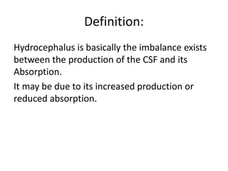
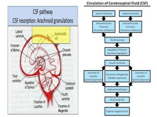
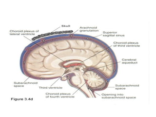
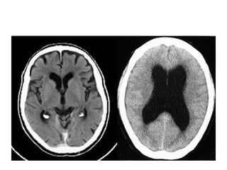
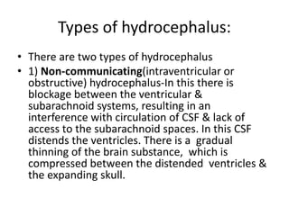
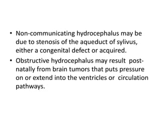
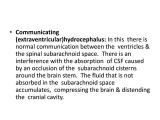
Hydrocephalus
Dr. Shaheer Anwar
Neurosurgery Resident.
Objectives:
• To define hydrocephalus.
• To enlist the causes of hydrocephalus.
• Physiology of formation of CSF production &
flow.
• Types of hydrocephalus & their pathophysiology.
• Clinical manifestation in infants, childhood and
adults.
• Diagnostic evaluation for hydrocephalus.
• Management of hydrocephalus- Medical,
Surgical.
Definition:
Hydrocephalus is basically the imbalance exists
between the production of the CSF and its
Absorption.
It may be due to its increased production or
reduced absorption.
Causes of hydrocephalus:
• CONGENITAL HYDROCEPHALUS:
• Intrauterine infections: Rubella,
Cytomegalovirus, Toxoplasmosis.
• Trauma: Subarachnoid, Intracranial,
Intraventricular haemorrhages.
• Congenital malformations:
• Dandy-walker syndrome: Atresia of foramina
of megendie and luschka.
• Aqueduct stenosis: Stenosis of aqueduct of
sylvius causes dilatation of lateral and 3rd
ventricles.
• Arnold-Chiari syndrome: Portions of
cerebellum & brainstem herniating into
cervical spinal canal, blocking the flow of CSF
into the posterior fossa.
• ACQUIRED HYDROCEPHALUS: -
• Tuberculosis, chronic & pyogenic meningitis.
• Post-intraventricular hemorrhage.
• Posterior-fossa tumors.
• Arteriovenousmalformations, intracranial
hemorrhage, ruptured aneurysm.
Types of hydrocephalus:
• There are two types of hydrocephalus
• 1) Non-communicating(intraventricular or
obstructive) hydrocephalus-In this there is
blockage between the ventricular &
subarachnoid systems, resulting in an
interference with circulation of CSF & lack of
access to the subarachnoid spaces. In this CSF
distends the ventricles. There is a gradual
thinning of the brain substance, which is
compressed between the distended ventricles &
the expanding skull.
• Non-communicating hydrocephalus may be
due to stenosis of the aqueduct of sylivus,
either a congenital defect or acquired.
• Obstructive hydrocephalus may result post-
natally from brain tumors that puts pressure
on or extend into the ventricles or circulation
pathways.
• Communicating
(extraventricular)hydrocephalus: In this there is
normal communication between the ventricles &
the spinal subarachnoid space. There is an
interference with the absorption of CSF caused
by an occlusion of the subarachnoid cisterns
around the brain stem. The fluid that is not
absorbed in the subarachnoid space
accumulates, compressing the brain & distending
the cranial cavity.
• Communicating hydrocephalus may be due to
subarachnoid hemorrhage or meningitis,
toxoplasmosis or cytomegalovirus infection,
in which there is an obliteration of the
subarachnoid spaces by fibrous tissue
reaction, or to diseases of connective tissue.
Clinical Manifestations:
Infants:
• Head grows at abnormal rate.
• Anterior fontanel is tense, often bulging, & non pulsatile.
• Scalp veins are dilated & markedly so when infant cries.
• Macewen’s sign- with increase in intracranial volume, the
bones of the skull become thin & the sutures become
palpably separated to produce the cracked pot sound on
the percussion of the skull.
• Frontal bossing with depressed eyes.
• Sun-set sign- eyes rotated downward, in which sclera may
be visible above iris.
Young Children:
Acute onset:
1. Irritability.
2. Impaired conscious level.
3. Vomiting.
Gradual onset:
1. Mental retardation.
2. Failure to thrive.
Adults:
Acute Onset:
• Headache.
• Vomiting.
• Deterioration of conscious level.
• Impaired upward gaze(Perinaud’s phenomenon).
Gradual onset:
• Dementia.
• Gait ataxia.
• Incontinence.
Diagnostic evaluation:
• Xray Skull.
• Routine daily head (occipitofrontal)
circumference measurements in infants.
• A head CT scan is one of the best tests for
identifying hydrocephalus.
• Lumbar puncture and examination of the
cerebrospinal fluid (rarely done).
Management of Hydrocephalus:
• Medical management.
• Surgical management.
Medical Management:
• Acetazolamide 50mg/kg/day diminishes CSF
production.
• Oral Glycerol.
Surgical management:
• The removal of the obstruction (tumor, hemorrhage or cyst) to the
flow of CSF.
• Reduction in the amount of CSF produced through destruction of a
portion of the choroid plexus or a third or fourth ventriculostomy.
• Shunting of CSF from the ventricle to another site in the normal
circulatory passageway of this fluid.
• Shunting of CSF from the ventricle to an area outside the CNS, an
extracranial body compartment.
• Shunting is the most common procedure to be done in the surgical
management of hydrocephalus.
• Most shunt systems consist of a ventricular catheter, a flush pump,
a unidirectional flow valve & a distal catheter. All are radiopaque
for easy visualization after placement & all are tested before
insertion.
• Endoscopic Third ventriculostomy(ETV).
Types of shunts:
• Ventriculoperitonial(VP) shunt.
• Ventriculoatrial(VA) shunt.
• Ventriculopleural shunt.
• Lumboperitoneal Shunt.
• Ventriculoperitonial(VP) shunt: This is the
preferred procedure especially in neonates &
young infants. There is greater allowance for
excess tubing, which minimizes the number
of revisions needed as the child grows. In this
ventricular catheter is inserted into the
anterior portion of a lateral ventricle through
a burr hole in the skull.
• Ventriculoatrial(VA) shunt: It is reserved for
older children who have attained most of their
somatic growth & children with abdominal
pathology. It requires repeated lengthening as
child grows. A silicon catheter is inserted in
lateral ventricle & down through the internal
jugular vein into left atrium of the heart.
The CSF drains into circulating blood. This type of
shunt may become easily obstructed or infected. If
an infection occurs, bacterial endocarditis,
ventriculitis & bacteremia may result.
• Endoscopic third ventriculostomy: It is a
procedure that has potential for greater
independence from VP or VA shunting in
children with non-communicating
hydrocephalus. In this procedure a small
opening is made in the floor of the 3rd
ventricle, allowing CSF to flow freely through
previously blocked ventricle, thus bypassing
the aqueduct of sylvius.
Compications of Shunting:
1. Infection.
2. Malfunction.
3. Subdural hematoma caused by rapid
reduction of ICP & size.
Hydrocephalus

Hydrocephalus

  • 1.
  • 2.
    Objectives: • To definehydrocephalus. • To enlist the causes of hydrocephalus. • Physiology of formation of CSF production & flow. • Types of hydrocephalus & their pathophysiology. • Clinical manifestation in infants, childhood and adults. • Diagnostic evaluation for hydrocephalus. • Management of hydrocephalus- Medical, Surgical.
  • 3.
    Definition: Hydrocephalus is basicallythe imbalance exists between the production of the CSF and its Absorption. It may be due to its increased production or reduced absorption.
  • 6.
    Causes of hydrocephalus: •CONGENITAL HYDROCEPHALUS: • Intrauterine infections: Rubella, Cytomegalovirus, Toxoplasmosis. • Trauma: Subarachnoid, Intracranial, Intraventricular haemorrhages. • Congenital malformations: • Dandy-walker syndrome: Atresia of foramina of megendie and luschka.
  • 7.
    • Aqueduct stenosis:Stenosis of aqueduct of sylvius causes dilatation of lateral and 3rd ventricles. • Arnold-Chiari syndrome: Portions of cerebellum & brainstem herniating into cervical spinal canal, blocking the flow of CSF into the posterior fossa.
  • 8.
    • ACQUIRED HYDROCEPHALUS:- • Tuberculosis, chronic & pyogenic meningitis. • Post-intraventricular hemorrhage. • Posterior-fossa tumors. • Arteriovenousmalformations, intracranial hemorrhage, ruptured aneurysm.
  • 10.
    Types of hydrocephalus: •There are two types of hydrocephalus • 1) Non-communicating(intraventricular or obstructive) hydrocephalus-In this there is blockage between the ventricular & subarachnoid systems, resulting in an interference with circulation of CSF & lack of access to the subarachnoid spaces. In this CSF distends the ventricles. There is a gradual thinning of the brain substance, which is compressed between the distended ventricles & the expanding skull.
  • 11.
    • Non-communicating hydrocephalusmay be due to stenosis of the aqueduct of sylivus, either a congenital defect or acquired. • Obstructive hydrocephalus may result post- natally from brain tumors that puts pressure on or extend into the ventricles or circulation pathways.
  • 12.
    • Communicating (extraventricular)hydrocephalus: Inthis there is normal communication between the ventricles & the spinal subarachnoid space. There is an interference with the absorption of CSF caused by an occlusion of the subarachnoid cisterns around the brain stem. The fluid that is not absorbed in the subarachnoid space accumulates, compressing the brain & distending the cranial cavity.
  • 13.
    • Communicating hydrocephalusmay be due to subarachnoid hemorrhage or meningitis, toxoplasmosis or cytomegalovirus infection, in which there is an obliteration of the subarachnoid spaces by fibrous tissue reaction, or to diseases of connective tissue.
  • 14.
    Clinical Manifestations: Infants: • Headgrows at abnormal rate. • Anterior fontanel is tense, often bulging, & non pulsatile. • Scalp veins are dilated & markedly so when infant cries. • Macewen’s sign- with increase in intracranial volume, the bones of the skull become thin & the sutures become palpably separated to produce the cracked pot sound on the percussion of the skull. • Frontal bossing with depressed eyes. • Sun-set sign- eyes rotated downward, in which sclera may be visible above iris.
  • 15.
    Young Children: Acute onset: 1.Irritability. 2. Impaired conscious level. 3. Vomiting. Gradual onset: 1. Mental retardation. 2. Failure to thrive.
  • 16.
    Adults: Acute Onset: • Headache. •Vomiting. • Deterioration of conscious level. • Impaired upward gaze(Perinaud’s phenomenon). Gradual onset: • Dementia. • Gait ataxia. • Incontinence.
  • 17.
    Diagnostic evaluation: • XraySkull. • Routine daily head (occipitofrontal) circumference measurements in infants. • A head CT scan is one of the best tests for identifying hydrocephalus. • Lumbar puncture and examination of the cerebrospinal fluid (rarely done).
  • 18.
    Management of Hydrocephalus: •Medical management. • Surgical management.
  • 19.
    Medical Management: • Acetazolamide50mg/kg/day diminishes CSF production. • Oral Glycerol.
  • 20.
    Surgical management: • Theremoval of the obstruction (tumor, hemorrhage or cyst) to the flow of CSF. • Reduction in the amount of CSF produced through destruction of a portion of the choroid plexus or a third or fourth ventriculostomy. • Shunting of CSF from the ventricle to another site in the normal circulatory passageway of this fluid. • Shunting of CSF from the ventricle to an area outside the CNS, an extracranial body compartment. • Shunting is the most common procedure to be done in the surgical management of hydrocephalus. • Most shunt systems consist of a ventricular catheter, a flush pump, a unidirectional flow valve & a distal catheter. All are radiopaque for easy visualization after placement & all are tested before insertion. • Endoscopic Third ventriculostomy(ETV).
  • 21.
    Types of shunts: •Ventriculoperitonial(VP) shunt. • Ventriculoatrial(VA) shunt. • Ventriculopleural shunt. • Lumboperitoneal Shunt.
  • 22.
    • Ventriculoperitonial(VP) shunt:This is the preferred procedure especially in neonates & young infants. There is greater allowance for excess tubing, which minimizes the number of revisions needed as the child grows. In this ventricular catheter is inserted into the anterior portion of a lateral ventricle through a burr hole in the skull.
  • 23.
    • Ventriculoatrial(VA) shunt:It is reserved for older children who have attained most of their somatic growth & children with abdominal pathology. It requires repeated lengthening as child grows. A silicon catheter is inserted in lateral ventricle & down through the internal jugular vein into left atrium of the heart. The CSF drains into circulating blood. This type of shunt may become easily obstructed or infected. If an infection occurs, bacterial endocarditis, ventriculitis & bacteremia may result.
  • 24.
    • Endoscopic thirdventriculostomy: It is a procedure that has potential for greater independence from VP or VA shunting in children with non-communicating hydrocephalus. In this procedure a small opening is made in the floor of the 3rd ventricle, allowing CSF to flow freely through previously blocked ventricle, thus bypassing the aqueduct of sylvius.
  • 25.
    Compications of Shunting: 1.Infection. 2. Malfunction. 3. Subdural hematoma caused by rapid reduction of ICP & size.