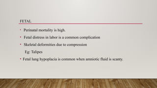
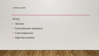
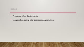
HYDRAMNIOS
POLYHYDRAMNIOS
RISK FACTIORS
MATERNAL
• Diabetes mellitus, About 50% of diabetic pregnancies have hydramnios.
• Cardiac or renal disease
FETAL
• Fetal malformation (eg. Spina bifida, hydrocephaly)
• Hydrops fetalis in Rh- incompatibility
• Multiple pregnancy
• Esophageal or duodenal atresia
• aneuploidy
SYMPTOMS
• The maternal symptoms depend on the stage of gestation .
• Mild symptoms include
Abdominal discomfort
Slight dyspnea
• IN SEVERE CASES (OVER 4000ML OF FLUID)
 Abdominal pain
 Dyspnea
 Orthopnea
Oedema of the abdomen, legs, vulva
 Nausea and Vomiting
TYPES OF POLYHYDRAMNIOS
 Acute polyhydramnios
 Chronic polyhydramnios
ACUTE POLYHYDRAMNIOS
This condition is extremely rare. It usually occurs before 20 weeks of
pregnancy.
SYMPTOMS
• Abdominal pain
• Nausea
• Vomiting
TREATMENT
 Most often spontaneous abortion occurs
 In case with severe TTT’s (Twin - Twin Transfusion synderome) repetitive
amnioreduction until the AFI is normal, may improve the perinatal
outcome.
 Laser ablation may cure the cause of TTT’s.
CHRONIC POLYHYDRAMNIOS
This occurs later in pregnancy usually between 32 and 40 weeks. The
prognosis depends on the underlying causes.
SYMPTOMS
• Respiratory – Dyspnea
• Palpitation
• Edema of the legs , varicositis in the legs or vulva and hemorrhoids.
SIGNS
• The patient may be in a dyspneic state in the lying down position
• Evidence of pre- eclampsia (edema, hypertension and proteinuria) may be
present.
MANAGEMENT
• Supportive therapy like bed rest
• Drainage of excess amniotic fluid.
DIAGNOSIS
Abdominal Examination
• The uterus is larger than expected
• The fetal parts are difficult to outline.
• The fetal heart rate cannot be heard clerly.
Ultra sonography:
• Ultrasonography helps to establish the diagnosis and detect the presence of many fetal
anamolies
• To detect abnormally large echo-free space between the fetus and uterine wall.
• AFI (is more than 25cm is polyhydramnios)
• Detect fetal congenital malformations such as anencephly spina bifida meningocele
• Multiple pregnancy
• Fetal malformation
MANAGEMENT
• In mild cases rest and mild sedation is helpful.
• In severe cases the hospitalization is required and removal of fluid it may feel
comfortable to the mother temporarily.
• The fetus is matured, induction of labour and delivery may be recommended.
• Major congenital abnormalities are diagnosed termination of pregnancy is
advisable.
COMPLICATION
• Fetal malpresentation
• Premature rupture of membranes
• Prolonged labour
• Amniotic fluid embolism
PROGNOSIS
• Major fetal congenital malformations occur in 20%
• Incidence of prematurity is more than twice the general rate.
• Placental abruption due to rapid decomposition of the uterus when the
membranes rupture and result in uncreased morbidity and mortality.
POLY HYDRAMNIOS
ETIOLOGY
FETAL
• Fetal chromosomal abnormalities
• Renal agencies
• Obstructed uropathy
• Spontaneous rupture of membrane
• IUGR
MATERNAL
• Hypertensive disorder
• Uteroplacental insufficiency
• Dehydration
EFFECTS OF OLIGOHYDRAMNIOS
EARLY PREGNANCY
• Pressure deformities, such as club feet
• Pulmonary hypoplacia has been reported
• The skin becomes dry and wrinkled
LATE PREGNANCY
• Close adaption between the fetus and the uterine wall can lead to pressure
on the umblical cord and obstruction cord and obstruction to the flow of
blood and from the fetus
• Fetal hypoxia may result
• Meconium passed into an amniotic sac. Aspiration of thick meconium by
the fetus will lead to aspiration pneumonia after birth.
DIAGNOSIS
• The uterine size appears smaller than gestational period.
• Fetal malpresentation
• On abdominal palpation
• ULTRSOUND
No pocket of amniotic fluid is greater than 1 cm.
HAZARDS
MTERNAL
• There is high incidence of operative because of fetal distress and mal
presentation.
FETAL
• Perinatal mortality is high.
• Fetal distress in labor is a common complication
• Skeletal deformities due to compression
Eg: Talipes
• Fetal lung hypoplacia is common when amniotic fluid is scanty.
COMPLICATION
FETAL
• Abortion
• Fetal pulmonary hypoplacia
• Cord compression
• High fetal mortality
MATERNAL
• Prolonged labor due to inertia.
• Increased operative interference malpresentation
THANK YOU

HYDRAMNIOS poly and oligohydramnios.pptx

  • 1.
  • 2.
    POLYHYDRAMNIOS RISK FACTIORS MATERNAL • Diabetesmellitus, About 50% of diabetic pregnancies have hydramnios. • Cardiac or renal disease
  • 3.
    FETAL • Fetal malformation(eg. Spina bifida, hydrocephaly) • Hydrops fetalis in Rh- incompatibility • Multiple pregnancy • Esophageal or duodenal atresia • aneuploidy
  • 4.
    SYMPTOMS • The maternalsymptoms depend on the stage of gestation . • Mild symptoms include Abdominal discomfort Slight dyspnea
  • 5.
    • IN SEVERECASES (OVER 4000ML OF FLUID)  Abdominal pain  Dyspnea  Orthopnea Oedema of the abdomen, legs, vulva  Nausea and Vomiting
  • 6.
    TYPES OF POLYHYDRAMNIOS Acute polyhydramnios  Chronic polyhydramnios
  • 7.
    ACUTE POLYHYDRAMNIOS This conditionis extremely rare. It usually occurs before 20 weeks of pregnancy. SYMPTOMS • Abdominal pain • Nausea • Vomiting
  • 8.
    TREATMENT  Most oftenspontaneous abortion occurs  In case with severe TTT’s (Twin - Twin Transfusion synderome) repetitive amnioreduction until the AFI is normal, may improve the perinatal outcome.  Laser ablation may cure the cause of TTT’s.
  • 9.
    CHRONIC POLYHYDRAMNIOS This occurslater in pregnancy usually between 32 and 40 weeks. The prognosis depends on the underlying causes. SYMPTOMS • Respiratory – Dyspnea • Palpitation • Edema of the legs , varicositis in the legs or vulva and hemorrhoids.
  • 10.
    SIGNS • The patientmay be in a dyspneic state in the lying down position • Evidence of pre- eclampsia (edema, hypertension and proteinuria) may be present. MANAGEMENT • Supportive therapy like bed rest • Drainage of excess amniotic fluid.
  • 11.
    DIAGNOSIS Abdominal Examination • Theuterus is larger than expected • The fetal parts are difficult to outline. • The fetal heart rate cannot be heard clerly.
  • 12.
    Ultra sonography: • Ultrasonographyhelps to establish the diagnosis and detect the presence of many fetal anamolies • To detect abnormally large echo-free space between the fetus and uterine wall. • AFI (is more than 25cm is polyhydramnios) • Detect fetal congenital malformations such as anencephly spina bifida meningocele • Multiple pregnancy • Fetal malformation
  • 13.
    MANAGEMENT • In mildcases rest and mild sedation is helpful. • In severe cases the hospitalization is required and removal of fluid it may feel comfortable to the mother temporarily. • The fetus is matured, induction of labour and delivery may be recommended. • Major congenital abnormalities are diagnosed termination of pregnancy is advisable.
  • 14.
    COMPLICATION • Fetal malpresentation •Premature rupture of membranes • Prolonged labour • Amniotic fluid embolism
  • 15.
    PROGNOSIS • Major fetalcongenital malformations occur in 20% • Incidence of prematurity is more than twice the general rate. • Placental abruption due to rapid decomposition of the uterus when the membranes rupture and result in uncreased morbidity and mortality.
  • 16.
    POLY HYDRAMNIOS ETIOLOGY FETAL • Fetalchromosomal abnormalities • Renal agencies • Obstructed uropathy • Spontaneous rupture of membrane • IUGR
  • 17.
    MATERNAL • Hypertensive disorder •Uteroplacental insufficiency • Dehydration
  • 18.
    EFFECTS OF OLIGOHYDRAMNIOS EARLYPREGNANCY • Pressure deformities, such as club feet • Pulmonary hypoplacia has been reported • The skin becomes dry and wrinkled
  • 19.
    LATE PREGNANCY • Closeadaption between the fetus and the uterine wall can lead to pressure on the umblical cord and obstruction cord and obstruction to the flow of blood and from the fetus • Fetal hypoxia may result • Meconium passed into an amniotic sac. Aspiration of thick meconium by the fetus will lead to aspiration pneumonia after birth.
  • 20.
    DIAGNOSIS • The uterinesize appears smaller than gestational period. • Fetal malpresentation • On abdominal palpation • ULTRSOUND No pocket of amniotic fluid is greater than 1 cm.
  • 21.
    HAZARDS MTERNAL • There ishigh incidence of operative because of fetal distress and mal presentation.
  • 22.
    FETAL • Perinatal mortalityis high. • Fetal distress in labor is a common complication • Skeletal deformities due to compression Eg: Talipes • Fetal lung hypoplacia is common when amniotic fluid is scanty.
  • 23.
    COMPLICATION FETAL • Abortion • Fetalpulmonary hypoplacia • Cord compression • High fetal mortality
  • 24.
    MATERNAL • Prolonged labordue to inertia. • Increased operative interference malpresentation
  • 25.