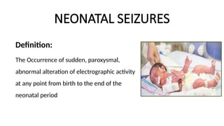
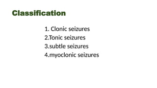
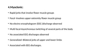
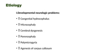
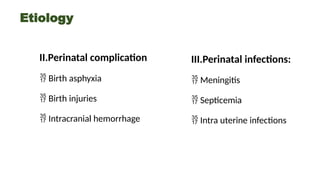
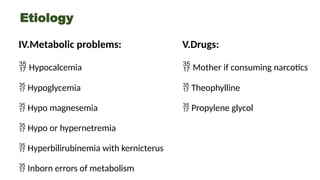
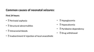
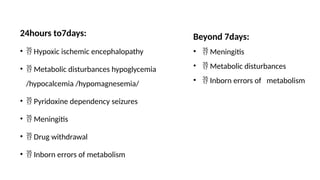
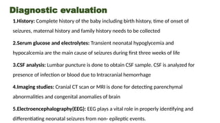
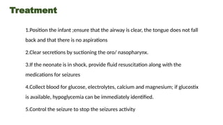
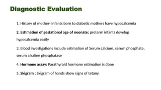
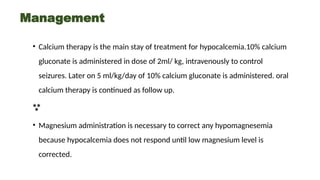
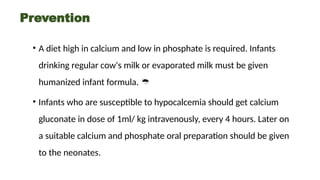
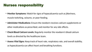
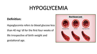
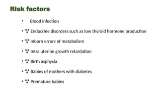
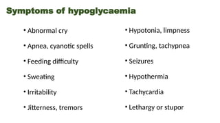
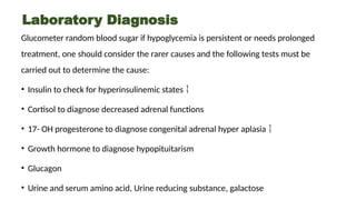
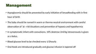
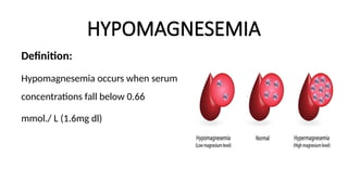
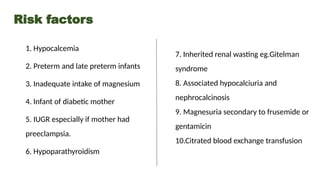
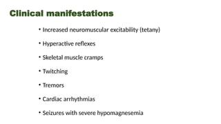
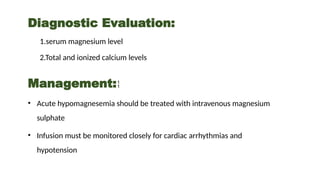
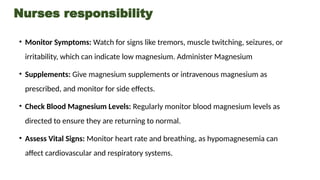
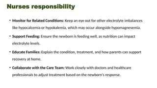
NEONATAL SEIZURES
Definition:
The Occurrence of sudden, paroxysmal,
abnormal alteration of electrographic activity
at any point from birth to the end of the
neonatal period
Classification
1. Clonic seizures
2.Tonic seizures
3.subtle seizures
4.myoclonic seizures
1.Clonic seizures
Slow rhythmic jerking movements approximately 1-3 per second. Focal- Involves
face, upper or lower extremities on one side of body. May involve neck or trunk
infant is conscious during events. Multi focal- may migrate randomly from one part
of the body to another movements may start at different times.
2.Tonic seizures:
Extension, stiffening moment Generalized -Extension of the all four limbs. Upper
limbs maintained in a stiffly flexed position. Focal- sustained posturing of a limb
Asymmetric posturing of trunk or neck
3.Subtle seizures:
May develop in either full term or preterm infants but is more common in preterm.
Often overlooked by inexperienced observers.
Signs:
• Horizontal eye deviation
• Repititive blinking or fluttering of the eyelids
• Sucking or other oral buccal lingual movements.
• Arm movements that resemble rowing or swimming
• Leg movements described as pedaling or bicycling
4.Myoclonic:
• Rapid jerks that involve flexor muscle groups
• Focal -Involves upper extremity flexor muscle group
• No electro encephalogram (EEG )discharge observed
• Multi focal-Asynchronous twitching of several parts of the body
• No associated EEG discharges observed
• Generalized -Bilateral jerks of upper and lower limbs
• Associated with EEG discharges.
Etiology
I.Developmental neurologic problems:
•  Congenital hydrocephalus
•  Microcephaly
•  Cerebral dysgenesis
•  Porencephaly
•  Polymicrogyria
•  Agenesis of corpus callosum
Etiology
II.Perinatal complication
 Birth asphyxia
 Birth injuries
 Intracranial hemorrhage
III.Perinatal infections:
 Meningitis
 Septicemia
 Intra uterine infections
Etiology
IV.Metabolic problems:
 Hypocalcemia
 Hypoglycemia
 Hypo magnesemia
 Hypo or hypernetremia
 Hyperbilirubinemia with kernicterus
 Inborn errors of metabolism
V.Drugs:
 Mother if consuming narcotics
 Theophylline
 Propylene glycol
Common causes of neonatal seizures:
First 24 hours:
•  Perinatal asphyxia
•  Structural abnormalities
•  Intracranial bleeds
•  Inadvertment IV injection of local anaesthetic
•  Hypoglycemia
•  Hypocalcemia
•  Pyridoxine dependency
•  Drug withdrawal
24hours to7days:
•  Hypoxic ischemic encephalopathy
•  Metabolic disturbances hypoglycemia
/hypocalcemia /hypomagnesemia/
•  Pyridoxine dependency seizures
•  Meningitis
•  Drug withdrawal
•  Inborn errors of metabolism
Beyond 7days:
•  Meningitis
•  Metabolic disturbances
•  Inborn errors of metabolism
Diagnostic evaluation
1.History: Complete history of the baby including birth history, time of onset of
seizures, maternal history and family history needs to be collected
2.Serum glucose and electrolytes: Transient neonatal hypoglycemia and
hypocalcemia are the main cause of seizures during first three weeks of life
3.CSF analysis: Lumbar puncture is done to obtain CSF sample. CSF is analyzed for
presence of infection or blood due to Intracranial hemorrhage
4.Imaging studies: Cranial CT scan or MRI is done for detecting parenchymal
abnormalities and congenital anomalies of brain
5.Electroencephalography(EEG): EEG plays a vital role in properly identifying and
differentiating neonatal seizures from non- epileptic events.
Treatment
1.Position the infant ;ensure that the airway is clear, the tongue does not fall
back and that there is no aspirations
2.Clear secretions by suctioning the oro/ nasopharynx.
3.If the neonate is in shock, provide fluid resuscitation along with the
medications for seizures
4.Collect blood for glucose, electrolytes, calcium and magnesium; if glucostix
is available, hypoglycemia can be immediately identified.
5.Control the seizure to stop the seizures activity
Follow up:
Continuation of anticonvulsants:
• Once seizures are controlled in the acute phase,it is not necessary to
continue anticonvulsant. 
• Occasionally, for seizures that are difficult to control, a maintenance dose of
phenobarbitone(5mg/kg/day) is continued. 
• Discontinue all anticonvulsants before the neonate is discharged from the
hospital if the infant is neurologically normal on clinical examination.
Evaluation:
The infant must be evaluated for hydrocephalus by serial measurement of
the head circumference, neurosonography and MRI. This is especially relevant if
the seizures were due to meningitis, intracranial bleeds or structural anomalies of
the brain. Hearing and visual assessment should also be carried out to facilitate
appropriate therapy if required
Prognosis:
The combination of a 5 minutes apgar score of ˂3and fits and signs of
encephalopathy is associated with 33%mortality and 55% handicap.
Nurses responsibility
1. Monitor Seizures: Watch for signs of seizures and document details.
2. Administer Medications: Give seizure medications and monitor effects.
3. Check Vital Signs: Regularly monitor heart rate, breathing, and oxygen levels.
4. Assist with Tests: Help with EEGs and other diagnostic tests.
5. Work with the Team: Communicate with doctors and healthcare staff about the
baby’s condition.
6. Educate Families: Explain seizures and treatments to the baby’s family.
7. Ensure Safety: Take steps to protect the baby during and after seizures.
HYPOCALCEMIA
Definition:
Hypocalcemia is defined as total serum calcium
concentration of less than 8.5mg/ dl in children, less
than 8mg /dl in term neonates and less than7mg /dl
in preterm neonates.
Incidence:
Hypocalcemia is seen in approximately 1 of every
4000 newborns.
Etiology
I.Vitamin D deficiency.It occurs due to:
•  Malabsorption
•  Malnutrition
•  Prolonged phenytoin therapy
•  Abnormal phenytoin therapy
•  Abnormal metabolism of vitamin D
•  Congenital rickets
II. Metabolic causes
•  Hypoparathyroidism
•  Pseudo hypoparathyroidism
•  Hypomagnesemia
•  Hyperphosphatemia
Etiology
III. Increased calcium loss from the body
•  Hypercalciuria
•  Prolong corticosteroid therapy
•  Prolong frusemide therapy
•  Renal tubular necrosis
IV.Other causes:
•  Metabolic alkalosis
•  Hypoproteinuria
•  Infants of diabetic mother
•  Prematurity
•  Acute pancreatitis
•  IV lipid infusion
Types
1. Early onset:
Appears in first 48 hours; appears in preterm infants who experienced
perinatal hypoxia or sometimes in infant of diabetic mother.
2.Late onset:
Cow's milk- induced hypocalcemia (neonatal tetany); apparent after
first 3-4 days (high phosphorus/calcium ratio of cow’s milk depresses
parathyroid activity, reducing serum calcium levels); infants with
intestinal malabsorption, hypoparathyroidism or hypomagnesemia.
Clinical features
a) Early neonatal hypocalcemia oçcurs with in 48-72 hours of birth
and manifestation are
i) High pitch cry
ii) Intermittent cyanosis
iii)Oedema
iv) Vomiting
v) Abdominal distention
vi) Periods of apnea
Clinical features
b) Late neonatal hypocalcemia occurs 3-7days after birth
or even as 6 weeks of age .manifestations include:
i) Irritability and jitterness
ii) Muscular twitching
Diagnostic Evaluation
1. History of mother- Infants born to diabetic mothers have hypocalcemia
2. Estimation of gestational age of neonate: preterm infants develop
hypocalcemia easily
3. Blood investigations include estimation of Serum calcium, serum phosphate,
serum alkaline phosphatase
4. Hormone assay: Parathyroid hormone estimation is done
5. Skigram : Skigram of hands show signs of tetany.
Management
• Calcium therapy is the main stay of treatment for hypocalcemia.10% calcium
gluconate is administered in dose of 2ml/ kg, intravenously to control
seizures. Later on 5 ml/kg/day of 10% calcium gluconate is administered. oral
calcium therapy is continued as follow up.

• Magnesium administration is necessary to correct any hypomagnesemia
because hypocalcemia does not respond until low magnesium level is
corrected.
Prevention
• A diet high in calcium and low in phosphate is required. Infants
drinking regular cow's milk or evaporated milk must be given
humanized infant formula. 
• Infants who are susceptible to hypocalcemia should get calcium
gluconate in dose of 1ml/ kg intravenously, every 4 hours. Later on
a suitable calcium and phosphate oral preparation should be given
to the neonates.
Nurses responsibility
• Monitor Symptoms: Watch for signs of hypocalcaemia such as jitteriness,
muscle twitching, seizures, or poor feeding.
• Administer Medications: Ensure the newborn receives calcium supplements or
other medications as prescribed, and monitor for any side effects.
• Check Blood Calcium Levels: Regularly monitor the newborn’s blood calcium
levels as directed by the healthcare team.
• Assess Vital Signs: Keep track of heart rate, respiratory rate, and overall stability,
as hypocalcaemia can affect heart and breathing functions.
• Support Feeding: Encourage proper feeding, as hypocalcaemia can
sometimes be related to feeding issues in newborns.
• Educate Families: Explain the condition, its treatment, and how parents can
help with feeding and monitoring at home.
• Collaborate with the Care Team: Communicate regularly with the doctors and
healthcare team about the baby’s condition and any changes in symptoms or
test results
Nurses responsibility
HYPOGLYCEMIA
Definition:
Hypoglycemia refers to blood glucose less
than 40 mg/ dl for the first four weeks of
life irrespective of birth weight and
gestational age.
Types
1.Increased or impaired glucose utilization:
Large or normal- size infants who appear to suffer from hyperinsulinism
infants born to women with diabetes; Infants with increased metabolic
demands such as those with cold stress, sepsis or after resuscitation;
infants with enzymatic or metabolic endocrine defects
2.Decreased glucose stores:
Small or growth restricted infants, preterm infants.
Causes
1.Increased utilisation of
glucose- hyperinsulinism:
• Infants of diabetic mother
(IDM)
• Large for gestational age
infants
• Erythroblastosis
• Islet cell hyperplasia
• Insulin producing tumors
2.Decreased production
stress:
• Prematurity
• Intrauterine growth
restriction
• Inadequate calorie intake
• Delayed onset of feeding
3.Increased Utilisation
and/ or decreased
production:
Perinatal stress:
• Shock
• Sepsis
• Asphyxia
• Hypothermia
Risk factors
• Blood infection
•  Endocrine disorders such as low thyroid hormone production
•  Inborn errors of metabolism
•  Intra uterine growth retardation
•  Birth asphyxia
•  Babies of mothers with diabetes
•  Premature babies
Symptoms of hypoglycaemia
• Abnormal cry
• Apnea, cyanotic spells
• Feeding difficulty
• Sweating
• Irritability
• Jitterness, tremors
• Hypotonia, limpness
• Grunting, tachypnea
• Seizures
• Hypothermia
• Tachycardia
• Lethargy or stupor
Laboratory Diagnosis
Glucometer random blood sugar if hypoglycemia is persistent or needs prolonged
treatment, one should consider the rarer causes and the following tests must be
carried out to determine the cause:
• Insulin to check for hyperinsulinemic states 
• Cortisol to diagnose decreased adrenal functions
• 17- OH progesterone to diagnose congenital adrenal hyper aplasia 
• Growth hormone to diagnose hypopituitarism
• Glucagon
• Urine and serum amino acid, Urine reducing substance, galactose
Management
• Hypoglycemia should be prevented by early initiation of breastfeeding with in first
hour of birth
• The baby should be nursed in warm or thermo neutral environment with careful
observation of ‘at –risk’situations and prevention of hypoxia and hypothermia
• In symptomatic infant with convulsions, 10% dextrose 2ml/kg intravenously is given
as a bolus.
• Blood glucose level to be checked every 1/2hourly
• Oral feeds are introduced gradually and glucose infusion is tapered off
Nursing management:
•  Identify infants at risk or with hypoglycemia
•  Administer IV glucose as prescribed
•  Initiate early breastfeeding or formula feedings in healthy infant
•  Ensure adequate intake of carbohydrates (breast milk or formula)
Complications: 
• Developmental delay
• Seizures
• Heart failure
Nurses responsibility
• Monitor Blood Glucose Levels: Regularly check the newborn’s blood sugar levels
using a glucometer, as directed by the healthcare team.
• Observe for Symptoms: Watch for signs of hypoglycemia such as jitteriness,
lethargy, poor feeding, rapid breathing, or seizures.
• Administer Treatment: Provide glucose supplementation, either orally or
intravenously, as prescribed to raise blood sugar levels.
• Ensure Proper Feeding: Encourage frequent feedings, whether breastfeeding or
formula feeding, to help stabilize blood sugar levels.
Nurses responsibility
• Monitor Vital Signs: Keep track of the baby’s heart rate, breathing, and
temperature, which can be affected by low blood sugar.
• Prevent Recurrences: Work with the care team to ensure regular feeding
schedules and monitor glucose levels to prevent further hypoglycemic
episodes.
• Educate Families: Teach parents about the importance of feeding and
monitoring the baby for signs of hypoglycemia at home.
HYPOMAGNESEMIA
Definition:
Hypomagnesemia occurs when serum
concentrations fall below 0.66
mmol./ L (1.6mg dl)
Risk factors
1. Hypocalcemia
2. Preterm and late preterm infants
3. Inadequate intake of magnesium
4. Infant of diabetic mother
5. IUGR especially if mother had
preeclampsia.
6. Hypoparathyroidism
7. Inherited renal wasting eg.Gitelman
syndrome
8. Associated hypocalciuria and
nephrocalcinosis
9. Magnesuria secondary to frusemide or
gentamicin
10.Citrated blood exchange transfusion
Clinical manifestations
• Increased neuromuscular excitability (tetany)
• Hyperactive reflexes
• Skeletal muscle cramps
• Twitching
• Tremors
• Cardiac arrhythmias
• Seizures with severe hypomagnesemia
Diagnostic Evaluation:
1.serum magnesium level
2.Total and ionized calcium levels
Management:
• Acute hypomagnesemia should be treated with intravenous magnesium
sulphate
• Infusion must be monitored closely for cardiac arrhythmias and
hypotension
Nurses responsibility
• Monitor Symptoms: Watch for signs like tremors, muscle twitching, seizures, or
irritability, which can indicate low magnesium. Administer Magnesium
• Supplements: Give magnesium supplements or intravenous magnesium as
prescribed, and monitor for side effects.
• Check Blood Magnesium Levels: Regularly monitor blood magnesium levels as
directed to ensure they are returning to normal.
• Assess Vital Signs: Monitor heart rate and breathing, as hypomagnesemia can
affect cardiovascular and respiratory systems.
Nurses responsibility
• Monitor for Related Conditions: Keep an eye out for other electrolyte imbalances
like hypocalcemia or hypokalemia, which may occur alongside hypomagnesemia.
• Support Feeding: Ensure the newborn is feeding well, as nutrition can impact
electrolyte levels.
• Educate Families: Explain the condition, treatment, and how parents can support
recovery at home.
• Collaborate with the Care Team: Work closely with doctors and healthcare
professionals to adjust treatment based on the newborn's response.
THANK YOU

NEONATAL SEIZURES.pptx obstetrical condition

  • 1.
    NEONATAL SEIZURES Definition: The Occurrenceof sudden, paroxysmal, abnormal alteration of electrographic activity at any point from birth to the end of the neonatal period
  • 2.
    Classification 1. Clonic seizures 2.Tonicseizures 3.subtle seizures 4.myoclonic seizures
  • 3.
    1.Clonic seizures Slow rhythmicjerking movements approximately 1-3 per second. Focal- Involves face, upper or lower extremities on one side of body. May involve neck or trunk infant is conscious during events. Multi focal- may migrate randomly from one part of the body to another movements may start at different times. 2.Tonic seizures: Extension, stiffening moment Generalized -Extension of the all four limbs. Upper limbs maintained in a stiffly flexed position. Focal- sustained posturing of a limb Asymmetric posturing of trunk or neck
  • 4.
    3.Subtle seizures: May developin either full term or preterm infants but is more common in preterm. Often overlooked by inexperienced observers. Signs: • Horizontal eye deviation • Repititive blinking or fluttering of the eyelids • Sucking or other oral buccal lingual movements. • Arm movements that resemble rowing or swimming • Leg movements described as pedaling or bicycling
  • 5.
    4.Myoclonic: • Rapid jerksthat involve flexor muscle groups • Focal -Involves upper extremity flexor muscle group • No electro encephalogram (EEG )discharge observed • Multi focal-Asynchronous twitching of several parts of the body • No associated EEG discharges observed • Generalized -Bilateral jerks of upper and lower limbs • Associated with EEG discharges.
  • 6.
    Etiology I.Developmental neurologic problems: • Congenital hydrocephalus •  Microcephaly •  Cerebral dysgenesis •  Porencephaly •  Polymicrogyria •  Agenesis of corpus callosum
  • 7.
    Etiology II.Perinatal complication  Birthasphyxia  Birth injuries  Intracranial hemorrhage III.Perinatal infections:  Meningitis  Septicemia  Intra uterine infections
  • 8.
    Etiology IV.Metabolic problems:  Hypocalcemia Hypoglycemia  Hypo magnesemia  Hypo or hypernetremia  Hyperbilirubinemia with kernicterus  Inborn errors of metabolism V.Drugs:  Mother if consuming narcotics  Theophylline  Propylene glycol
  • 9.
    Common causes ofneonatal seizures: First 24 hours: •  Perinatal asphyxia •  Structural abnormalities •  Intracranial bleeds •  Inadvertment IV injection of local anaesthetic •  Hypoglycemia •  Hypocalcemia •  Pyridoxine dependency •  Drug withdrawal
  • 10.
    24hours to7days: • Hypoxic ischemic encephalopathy •  Metabolic disturbances hypoglycemia /hypocalcemia /hypomagnesemia/ •  Pyridoxine dependency seizures •  Meningitis •  Drug withdrawal •  Inborn errors of metabolism Beyond 7days: •  Meningitis •  Metabolic disturbances •  Inborn errors of metabolism
  • 11.
    Diagnostic evaluation 1.History: Completehistory of the baby including birth history, time of onset of seizures, maternal history and family history needs to be collected 2.Serum glucose and electrolytes: Transient neonatal hypoglycemia and hypocalcemia are the main cause of seizures during first three weeks of life 3.CSF analysis: Lumbar puncture is done to obtain CSF sample. CSF is analyzed for presence of infection or blood due to Intracranial hemorrhage 4.Imaging studies: Cranial CT scan or MRI is done for detecting parenchymal abnormalities and congenital anomalies of brain 5.Electroencephalography(EEG): EEG plays a vital role in properly identifying and differentiating neonatal seizures from non- epileptic events.
  • 12.
    Treatment 1.Position the infant;ensure that the airway is clear, the tongue does not fall back and that there is no aspirations 2.Clear secretions by suctioning the oro/ nasopharynx. 3.If the neonate is in shock, provide fluid resuscitation along with the medications for seizures 4.Collect blood for glucose, electrolytes, calcium and magnesium; if glucostix is available, hypoglycemia can be immediately identified. 5.Control the seizure to stop the seizures activity
  • 13.
    Follow up: Continuation ofanticonvulsants: • Once seizures are controlled in the acute phase,it is not necessary to continue anticonvulsant.  • Occasionally, for seizures that are difficult to control, a maintenance dose of phenobarbitone(5mg/kg/day) is continued.  • Discontinue all anticonvulsants before the neonate is discharged from the hospital if the infant is neurologically normal on clinical examination.
  • 14.
    Evaluation: The infant mustbe evaluated for hydrocephalus by serial measurement of the head circumference, neurosonography and MRI. This is especially relevant if the seizures were due to meningitis, intracranial bleeds or structural anomalies of the brain. Hearing and visual assessment should also be carried out to facilitate appropriate therapy if required Prognosis: The combination of a 5 minutes apgar score of ˂3and fits and signs of encephalopathy is associated with 33%mortality and 55% handicap.
  • 15.
    Nurses responsibility 1. MonitorSeizures: Watch for signs of seizures and document details. 2. Administer Medications: Give seizure medications and monitor effects. 3. Check Vital Signs: Regularly monitor heart rate, breathing, and oxygen levels. 4. Assist with Tests: Help with EEGs and other diagnostic tests. 5. Work with the Team: Communicate with doctors and healthcare staff about the baby’s condition. 6. Educate Families: Explain seizures and treatments to the baby’s family. 7. Ensure Safety: Take steps to protect the baby during and after seizures.
  • 16.
    HYPOCALCEMIA Definition: Hypocalcemia is definedas total serum calcium concentration of less than 8.5mg/ dl in children, less than 8mg /dl in term neonates and less than7mg /dl in preterm neonates. Incidence: Hypocalcemia is seen in approximately 1 of every 4000 newborns.
  • 17.
    Etiology I.Vitamin D deficiency.Itoccurs due to: •  Malabsorption •  Malnutrition •  Prolonged phenytoin therapy •  Abnormal phenytoin therapy •  Abnormal metabolism of vitamin D •  Congenital rickets II. Metabolic causes •  Hypoparathyroidism •  Pseudo hypoparathyroidism •  Hypomagnesemia •  Hyperphosphatemia
  • 18.
    Etiology III. Increased calciumloss from the body •  Hypercalciuria •  Prolong corticosteroid therapy •  Prolong frusemide therapy •  Renal tubular necrosis IV.Other causes: •  Metabolic alkalosis •  Hypoproteinuria •  Infants of diabetic mother •  Prematurity •  Acute pancreatitis •  IV lipid infusion
  • 19.
    Types 1. Early onset: Appearsin first 48 hours; appears in preterm infants who experienced perinatal hypoxia or sometimes in infant of diabetic mother. 2.Late onset: Cow's milk- induced hypocalcemia (neonatal tetany); apparent after first 3-4 days (high phosphorus/calcium ratio of cow’s milk depresses parathyroid activity, reducing serum calcium levels); infants with intestinal malabsorption, hypoparathyroidism or hypomagnesemia.
  • 20.
    Clinical features a) Earlyneonatal hypocalcemia oçcurs with in 48-72 hours of birth and manifestation are i) High pitch cry ii) Intermittent cyanosis iii)Oedema iv) Vomiting v) Abdominal distention vi) Periods of apnea
  • 21.
    Clinical features b) Lateneonatal hypocalcemia occurs 3-7days after birth or even as 6 weeks of age .manifestations include: i) Irritability and jitterness ii) Muscular twitching
  • 22.
    Diagnostic Evaluation 1. Historyof mother- Infants born to diabetic mothers have hypocalcemia 2. Estimation of gestational age of neonate: preterm infants develop hypocalcemia easily 3. Blood investigations include estimation of Serum calcium, serum phosphate, serum alkaline phosphatase 4. Hormone assay: Parathyroid hormone estimation is done 5. Skigram : Skigram of hands show signs of tetany.
  • 23.
    Management • Calcium therapyis the main stay of treatment for hypocalcemia.10% calcium gluconate is administered in dose of 2ml/ kg, intravenously to control seizures. Later on 5 ml/kg/day of 10% calcium gluconate is administered. oral calcium therapy is continued as follow up.  • Magnesium administration is necessary to correct any hypomagnesemia because hypocalcemia does not respond until low magnesium level is corrected.
  • 24.
    Prevention • A diethigh in calcium and low in phosphate is required. Infants drinking regular cow's milk or evaporated milk must be given humanized infant formula.  • Infants who are susceptible to hypocalcemia should get calcium gluconate in dose of 1ml/ kg intravenously, every 4 hours. Later on a suitable calcium and phosphate oral preparation should be given to the neonates.
  • 25.
    Nurses responsibility • MonitorSymptoms: Watch for signs of hypocalcaemia such as jitteriness, muscle twitching, seizures, or poor feeding. • Administer Medications: Ensure the newborn receives calcium supplements or other medications as prescribed, and monitor for any side effects. • Check Blood Calcium Levels: Regularly monitor the newborn’s blood calcium levels as directed by the healthcare team. • Assess Vital Signs: Keep track of heart rate, respiratory rate, and overall stability, as hypocalcaemia can affect heart and breathing functions.
  • 26.
    • Support Feeding:Encourage proper feeding, as hypocalcaemia can sometimes be related to feeding issues in newborns. • Educate Families: Explain the condition, its treatment, and how parents can help with feeding and monitoring at home. • Collaborate with the Care Team: Communicate regularly with the doctors and healthcare team about the baby’s condition and any changes in symptoms or test results Nurses responsibility
  • 27.
    HYPOGLYCEMIA Definition: Hypoglycemia refers toblood glucose less than 40 mg/ dl for the first four weeks of life irrespective of birth weight and gestational age.
  • 28.
    Types 1.Increased or impairedglucose utilization: Large or normal- size infants who appear to suffer from hyperinsulinism infants born to women with diabetes; Infants with increased metabolic demands such as those with cold stress, sepsis or after resuscitation; infants with enzymatic or metabolic endocrine defects 2.Decreased glucose stores: Small or growth restricted infants, preterm infants.
  • 29.
    Causes 1.Increased utilisation of glucose-hyperinsulinism: • Infants of diabetic mother (IDM) • Large for gestational age infants • Erythroblastosis • Islet cell hyperplasia • Insulin producing tumors 2.Decreased production stress: • Prematurity • Intrauterine growth restriction • Inadequate calorie intake • Delayed onset of feeding 3.Increased Utilisation and/ or decreased production: Perinatal stress: • Shock • Sepsis • Asphyxia • Hypothermia
  • 30.
    Risk factors • Bloodinfection •  Endocrine disorders such as low thyroid hormone production •  Inborn errors of metabolism •  Intra uterine growth retardation •  Birth asphyxia •  Babies of mothers with diabetes •  Premature babies
  • 31.
    Symptoms of hypoglycaemia •Abnormal cry • Apnea, cyanotic spells • Feeding difficulty • Sweating • Irritability • Jitterness, tremors • Hypotonia, limpness • Grunting, tachypnea • Seizures • Hypothermia • Tachycardia • Lethargy or stupor
  • 32.
    Laboratory Diagnosis Glucometer randomblood sugar if hypoglycemia is persistent or needs prolonged treatment, one should consider the rarer causes and the following tests must be carried out to determine the cause: • Insulin to check for hyperinsulinemic states  • Cortisol to diagnose decreased adrenal functions • 17- OH progesterone to diagnose congenital adrenal hyper aplasia  • Growth hormone to diagnose hypopituitarism • Glucagon • Urine and serum amino acid, Urine reducing substance, galactose
  • 33.
    Management • Hypoglycemia shouldbe prevented by early initiation of breastfeeding with in first hour of birth • The baby should be nursed in warm or thermo neutral environment with careful observation of ‘at –risk’situations and prevention of hypoxia and hypothermia • In symptomatic infant with convulsions, 10% dextrose 2ml/kg intravenously is given as a bolus. • Blood glucose level to be checked every 1/2hourly • Oral feeds are introduced gradually and glucose infusion is tapered off
  • 34.
    Nursing management: • Identify infants at risk or with hypoglycemia •  Administer IV glucose as prescribed •  Initiate early breastfeeding or formula feedings in healthy infant •  Ensure adequate intake of carbohydrates (breast milk or formula) Complications:  • Developmental delay • Seizures • Heart failure
  • 35.
    Nurses responsibility • MonitorBlood Glucose Levels: Regularly check the newborn’s blood sugar levels using a glucometer, as directed by the healthcare team. • Observe for Symptoms: Watch for signs of hypoglycemia such as jitteriness, lethargy, poor feeding, rapid breathing, or seizures. • Administer Treatment: Provide glucose supplementation, either orally or intravenously, as prescribed to raise blood sugar levels. • Ensure Proper Feeding: Encourage frequent feedings, whether breastfeeding or formula feeding, to help stabilize blood sugar levels.
  • 36.
    Nurses responsibility • MonitorVital Signs: Keep track of the baby’s heart rate, breathing, and temperature, which can be affected by low blood sugar. • Prevent Recurrences: Work with the care team to ensure regular feeding schedules and monitor glucose levels to prevent further hypoglycemic episodes. • Educate Families: Teach parents about the importance of feeding and monitoring the baby for signs of hypoglycemia at home.
  • 37.
    HYPOMAGNESEMIA Definition: Hypomagnesemia occurs whenserum concentrations fall below 0.66 mmol./ L (1.6mg dl)
  • 38.
    Risk factors 1. Hypocalcemia 2.Preterm and late preterm infants 3. Inadequate intake of magnesium 4. Infant of diabetic mother 5. IUGR especially if mother had preeclampsia. 6. Hypoparathyroidism 7. Inherited renal wasting eg.Gitelman syndrome 8. Associated hypocalciuria and nephrocalcinosis 9. Magnesuria secondary to frusemide or gentamicin 10.Citrated blood exchange transfusion
  • 39.
    Clinical manifestations • Increasedneuromuscular excitability (tetany) • Hyperactive reflexes • Skeletal muscle cramps • Twitching • Tremors • Cardiac arrhythmias • Seizures with severe hypomagnesemia
  • 40.
    Diagnostic Evaluation: 1.serum magnesiumlevel 2.Total and ionized calcium levels Management: • Acute hypomagnesemia should be treated with intravenous magnesium sulphate • Infusion must be monitored closely for cardiac arrhythmias and hypotension
  • 41.
    Nurses responsibility • MonitorSymptoms: Watch for signs like tremors, muscle twitching, seizures, or irritability, which can indicate low magnesium. Administer Magnesium • Supplements: Give magnesium supplements or intravenous magnesium as prescribed, and monitor for side effects. • Check Blood Magnesium Levels: Regularly monitor blood magnesium levels as directed to ensure they are returning to normal. • Assess Vital Signs: Monitor heart rate and breathing, as hypomagnesemia can affect cardiovascular and respiratory systems.
  • 42.
    Nurses responsibility • Monitorfor Related Conditions: Keep an eye out for other electrolyte imbalances like hypocalcemia or hypokalemia, which may occur alongside hypomagnesemia. • Support Feeding: Ensure the newborn is feeding well, as nutrition can impact electrolyte levels. • Educate Families: Explain the condition, treatment, and how parents can support recovery at home. • Collaborate with the Care Team: Work closely with doctors and healthcare professionals to adjust treatment based on the newborn's response.
  • 43.