Dr. abdelhakam Body effusion examination
•Download as PPTX, PDF•
0 likes•33 views

For medical & Health science

Recommended
More Related Content
What's hot
What's hot (20)
Similar to Dr. abdelhakam Body effusion examination
Similar to Dr. abdelhakam Body effusion examination (20)
Recently uploaded
Recently uploaded (20)
Dr. abdelhakam Body effusion examination
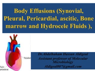
- 1. Body Effusions (Synovial, Pleural, Pericardial, ascitic, Bone marrow and Hydrocele Fluids ), Dr.Abdelhakam Hassan Aldigeal Assistant professor of Molecular Microbiology Aldigeal007@gmail.com
- 2. OriginFluid JointFromSynovial From the pleural cavity (space between the lungs and the inner chest wall) Pleural From the pericardial sac ( Membranous surrounding the heart) pericardial cavityFrom the peritoneal ( abdominal) Ascitic ( peritoneal) Usually from the sacHydrocele
- 3. Introduction • The human body is divided into five main body cavities: cranial, spinal, thoracic, abdominal, and pelvic. • Each cavity is lined with membranes, and within the body wall and these membranes, or between the membranes and organs, are small spaces filled with minute amounts of fluid.
- 4. Introduction • The purpose of this fluid is to bathe the organs and membranes, reducing the friction between organs. • Bacteria, fungi, virus, or parasite can invade any body tissue or sterile body fluid site. • All these fluids are considered normally sterile. Therefore, even one colony of a potentially pathogenic microorganism may be significant.
- 5. SPECIMENS FROM STERILE BODY SITES • FLUIDS • In response to infection, fluid may accumulate in any body cavity. • Infected solid tissue often presents as cellulitis or with abscess formation.
- 6. Pleural Fluid • Lining the entire thoracic cavity of the body is a serous membrane called the parietal pleura. • Covering the outer surface of the lung is another membrane called the visceral pleura. • Within the pleural space between the lung and chest wall is a small amount of fluid called pleural fluid. • that lubricates the surfaces of the pleura (the membranes surrounding the lungs and lining the chest cavity).
- 8. Fluid accumulates in the pleural space by three mechanisms: increased drainage of fluid into the space increased production of fluid by cells in the space decreased drainage of fluid from the space
- 9. CLINICAL FEATURES • History: Small pleural effusion: asymptomatic Large pleural effusion: pleuritic chest pain, abdominal pain, pain during inspiration or coughing The child may prefer to lie on the affected side (to decrease respiratory excursions) Cough Fever Respiratory distress, dyspnea.
- 10. TREATMENT Thoracentesis Diagnostic thoracentesis A needle is inserted into the chest wall to remove the collection of fluid 50-100ml of fluid is sent for analysis; Determines the type of fluid (transudate or exudate) temporarily relieve symptoms
- 13. Lab investigations • Normal pleural fluid contains few or no cells and has a consistency similar to serum, but with a lower protein count. • Pleural fluid containing numerous white blood cells is indicative of infections. • The fluid, or effusion, can then be analyzed for cell count, total protein, glucose, lactate dehydrogenase, amylase, cytology, and culture.
- 14. EMPYEMA • When effusions are extremely purulent or full of pus, the effusion is referred to as an empyema. • Empyema often arises as a complication of: • Pneumonia. • other infections near the lung (e.g. subdiaphragmatic infection) may seed microorganisms into the pleural cavity. • It has been estimated that 50% to 60% of patients develop empyema as a complication of pneumonia.
- 15. Peritoneal Fluid • The peritoneum is a large, moist, continuous sheet of membrane lining the walls of the abdominal pelvic cavity and the outer coat of the organs contained within the cavity. • Within the healthy human peritoneal cavity is a small amount of fluid that maintains the surface moisture of the peritoneum.
- 16. Peritoneal Fluid • In the abdomen, these two membrane linings are separated by a space called the peritoneal cavity, which contains or abuts the • liver, • pancreas, • spleen, • stomach and intestinal tract, • bladder, • and fallopian tubes and ovaries. • The kidneys occupy a retroperitoneal (behind the peritoneum) position.
- 17. ASCITES • During an infectious or inflammatory process, increased • amounts of fluid accumulate in the peritoneal cavity, a condition called ascites. • Most cases of ascites are due to liver disease, and in severe cases, the abdomen is often distended.
- 18. Lab Diagnosis • The fluid can be collected for testing by paracentesis • (the insertion of a needle into the abdomen and removal of fluid). • The peritoneal or ascites fluid can then be analyzed for amylase, protein, albumin, cell count, culture, and cytology. • Often ascitic fluid contains an increased number of inflammatory cells and an elevated protein level.
- 19. Pathogenicity • Agents of infection gain access to the peritoneum • through a perforation of the bowel, • through infection within abdominal viscera, by way of the bloodstream, or by external inoculation (as in surgery or trauma). • On occasion, as in pelvic inflammatory disease (PID), • organisms travel through the natural channels of the fallopian tubes into the peritoneal cavity.
- 20. Clinical features • 1- Primary peritonitis • is rare and results when infection spreads from the blood and lymph nodes with no apparent evidence of infection. • Causative organisms: • In children Streptococcus pneumoniae and group A streptococci, Enterobacteriaceae, other gram-negative bacilli, and staphylococci. • In adults, Escherichia coli , S. pneumoniae and group A streptococci. • Among sexually active young women, Neisseria gonorrhoeae and Chlamydia trachomatis • Fungal as Candida spp. may be recovered from immunosuppressed patients
- 21. Clinical features • 2- Secondary peritonitis is a complication • of a perforated viscus (organ), • surgery • traumatic injury • loss of bowel wall integrity following a destructive disease (e.g., ulcerative colitis, ruptured appendix, carcinoma), • obstruction, or a preceding infection (liver abscess, salpingitis, septicemia).
- 22. Secondary peritonitis • Causative organisms: • gonococci, anaerobes, or chlamydiae are isolated. • Enterobacteriaceae and enterococci • streptococci, Staphylococcus aureus • The organisms likely to be recovered include • E.coli, • the Bacteroides fragilis group, • Bilophila spp., • other anaerobic gram negative bacilli, • anaerobic gram-positive cocci, and clostridia.
- 23. Most severe cases associated with cirrhosis of the liver intra-abdominal malignancy
- 25. Pericardial Fluid • The area between the epicardium, which is the membrane surrounding the heart muscle, and the pericardium is called the pericardial space. • normally contains 15 to 20 mL of clear fluid. • During infection pericardium may become distended • and tight, and eventually tamponade (interference with • cardiac function and circulation) can ensue. • Up to 500 mL of fluid can accumulate during infection. • Myocarditis (inflammation of the heart muscle itself) • may accompany or follow pericarditis.
- 27. Causative organisms • Agents of pericarditis (inflammation of the pericardium) are usually viruses, especially Coxsackie virus. • Patients who develop pericarditis resulting from agents other than viruses (Parasites, bacteria, certain fungi) • are often immunocompromised or suffering from a chronic disease. • An example is infective endocarditis.
- 28. Common Etiologic Agents of Pericarditis and Myocarditis • Viruses • Enteroviruses (primary Coxsackie A and B and, less • frequently, echoviruses) • Adenoviruses • Influenza viruses • Bacteria (relatively uncommon) • Mycoplasma pneumoniae • Chlamydia trachomatis • Mycobacterium tuberculosis • Staphylococcus aureus • Streptococcus pneumoniae • Enterobacteriaceae and other gram-negative bacilli
- 29. Common Etiologic Agents of Pericarditis and Myocarditis • Fungi (relatively uncommon) • Coccidioides immitis • Aspergillus spp. • Candida spp. • Cryptococcus neoformans • Histoplasma capsulatum • Parasites (relatively uncommon) • Entamoeba histolytica • Toxoplasma gondii
- 30. Joint Fluid • Arthritis is an inflammation in a joint space. • Infection usually occurs secondary to hematogenous spread of bacteria • or, less often, fungi. • It may also occur after injection of material, especially corticosteroids, into joints • or after insertion of prosthetic material (e.g., total hip replacement).
- 31. Joint Fluid • Usually occurs at a single site (monoarticular) • but a preexisting bacteremia or fungemia may seed more than one joint to establish polyarticular infection. • In bacterial arthritis, the knees and hips are the most commonly affected joints.
- 32. Joint Fluid • self-limited arthritis caused by antigen-antibody interactions may follow an episode of infection, such as meningococcal meningitis. • When an etiologic agent cannot be isolated either the absence of viable agents or inadequate transport or culturing procedures may be the cause. • Borrelia burgdorferi is isolated from the joints of fewer than 20% of patients with Lyme disease.
- 33. Most Frequently Encountered Etiologic Agents of Infectious Arthritis • Staphylococcus aureus • Beta-hemolytic streptococci • Streptococci (other) • Haemophilus influenzae • Haemophilus spp. (other) • Bacteroides spp. • Fusobacterium spp. • Neisseria gonorrhoeae • Pseudomonas spp. • Salmonella spp. • Pasteurella multocida • Moraxella osloensis • Kingella kingae • Moraxella catarrhalis • Capnocytophaga spp. • Corynebacterium spp. • Clostridium spp. • Peptostreptococcus spp. • Eikenella corrodens • Actinomyces spp. • Mycobacterium spp. • Mycoplasma spp. • Ureaplasma urealyticum • Borrelia burgdorferi Bacterial
- 34. Most Frequently Encountered Etiologic Agents of Infectious Arthritis • Fungal • Candida spp. • Cryptococcus neoformans • Coccidioides immitis • Sporothrix schenckii • Viral • Hepatitis B • Mumps • Rubella • Other viruses (rarely)
- 35. Diagnosis • Diagnosis of joint infections requires an aspiration of • joint fluid for culture and microscopic examination. • Inoculating the fluid directly into blood culture bottles • may prevent the fluid from clotting. • Some of the fluid may be Gram stained • and inoculated onto blood as well as chocolate and anaerobic media. • The use of AFB (acid fast bacteria) and fungal media must also be considered.
- 37. Bone marrow • Bone marrow is typically aspirated from the interstitium of the iliac crest. • Usually, this material is not processed for routine bacteria, because blood cultures are equally useful, • and false-positive cultures for skin bacteria (Staphylococcus epidermidis) are frequent. • Bone removed at surgery or by percutaneous biopsy is sent to the laboratory in a • sterile container.
- 38. Occasionaly Wuchereria bancrofti microfilariae and rarely Brugia spp can be found. Hydrocele Fluid
- 39. Hydrocele Fluid examination • The fluid is placed on a slide • And air-dried to prevent distortion of the parasite. • The specimen should be stained with Giemsa, Wright’s, or hematoxylin stain and examined microscopically. • Polymerase chain reaction (PCR) amplification is available • for the rapid diagnosis of blood microfilariae including • W. bancrofti • and Brugia spp. • Serologic assays that measure antibody response have • limited utility in the diagnosis of infections with microfilariae.
- 40. Microfilaria of Wuchereria bancrofti in thick blood film.