Download to read offline

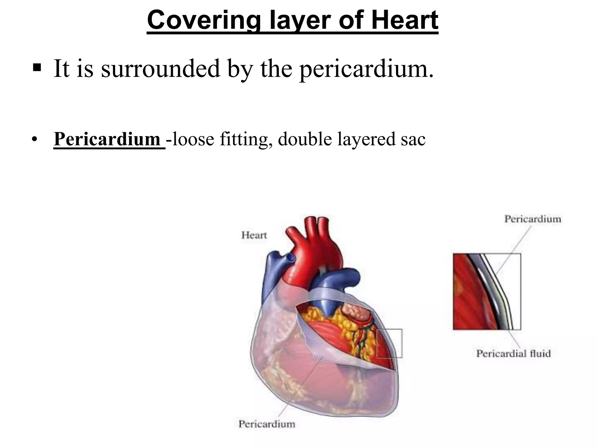

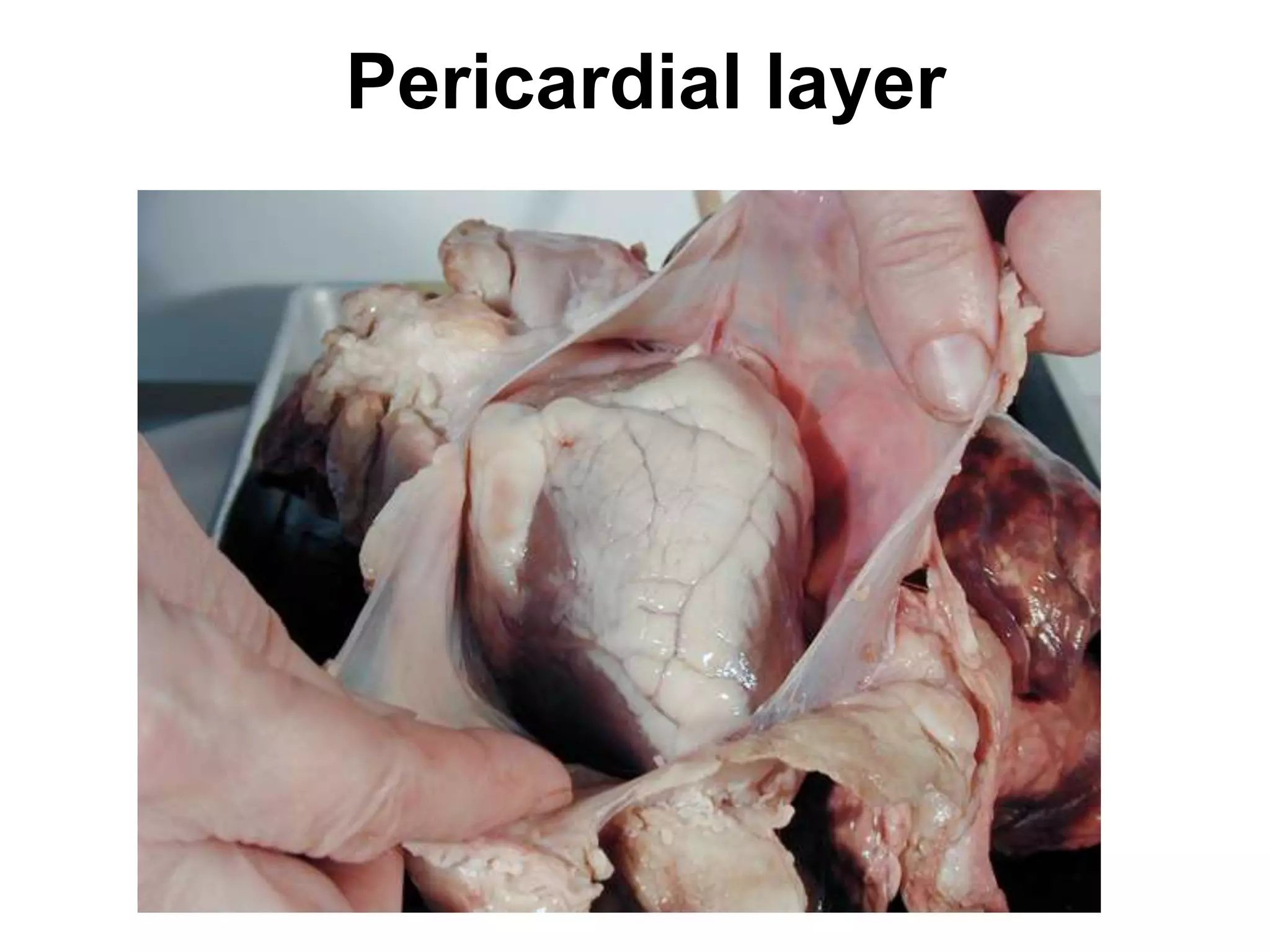
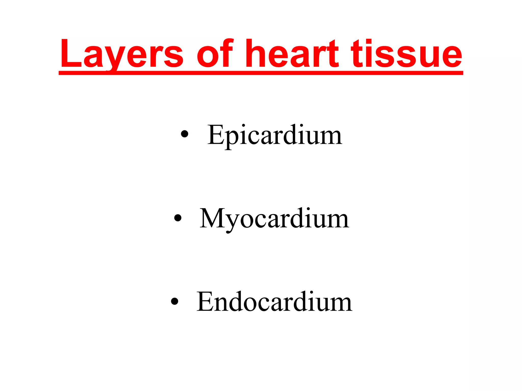


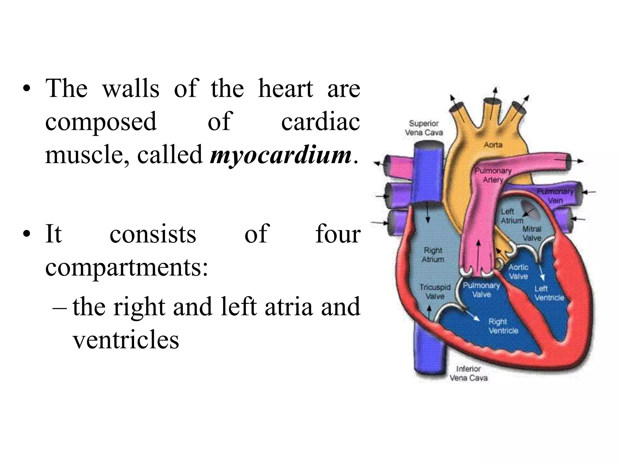
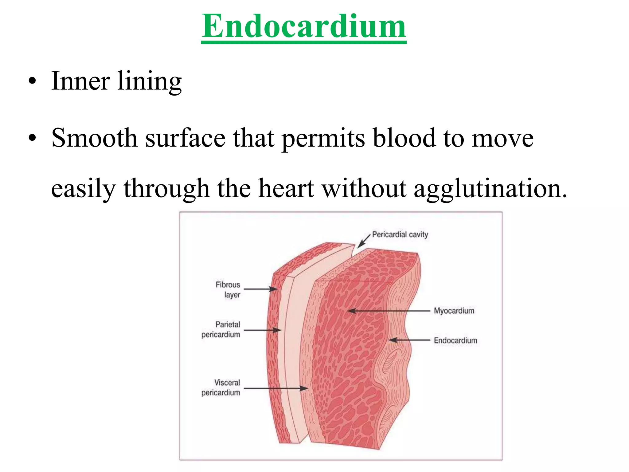
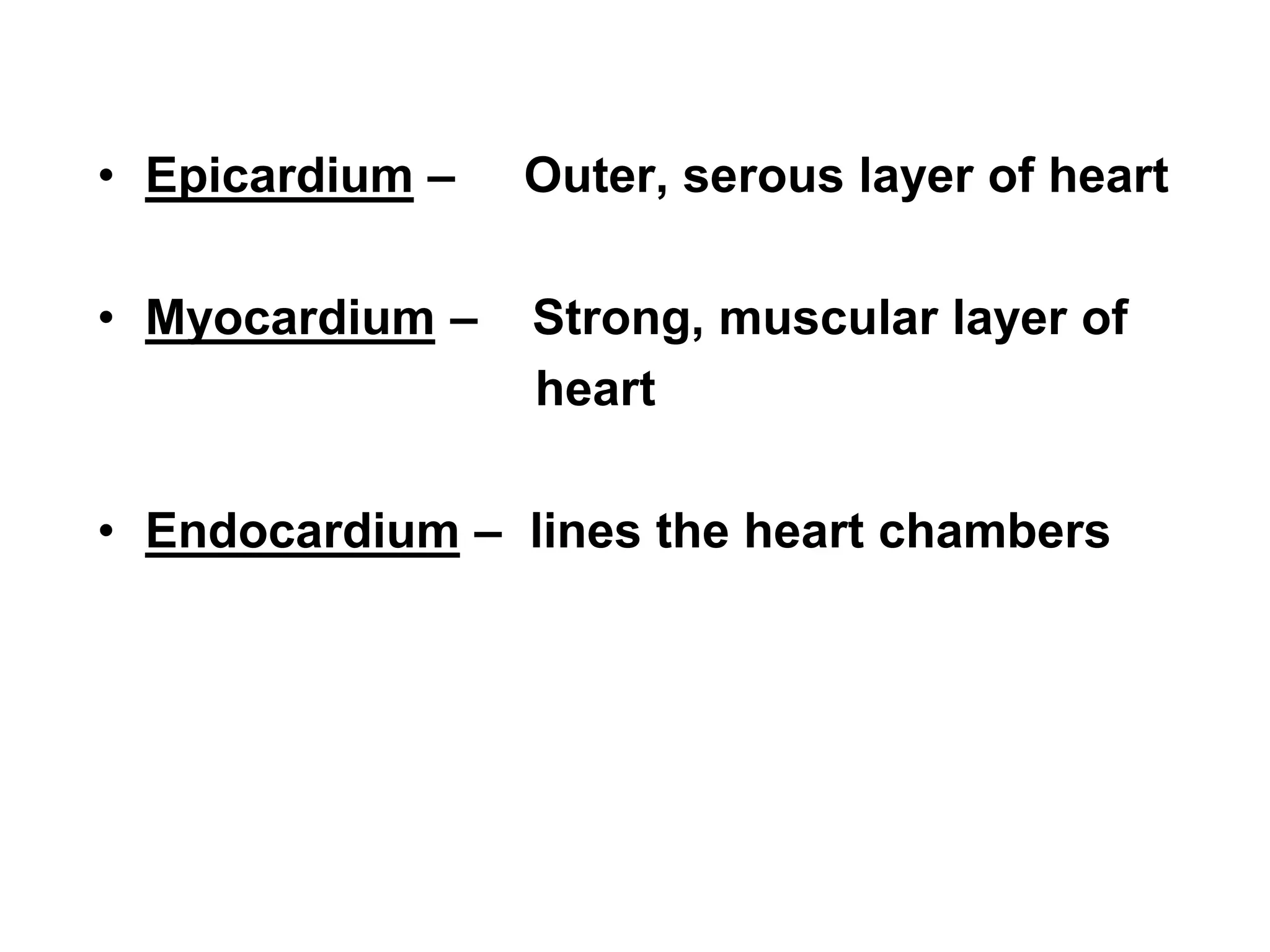


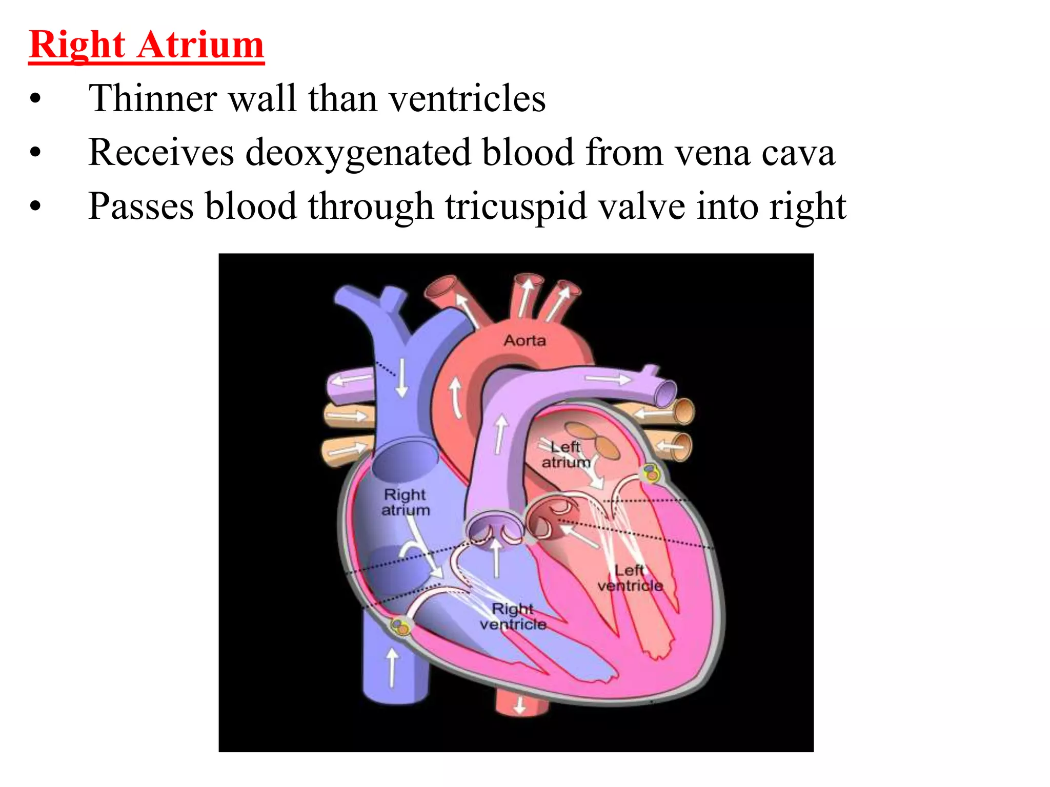
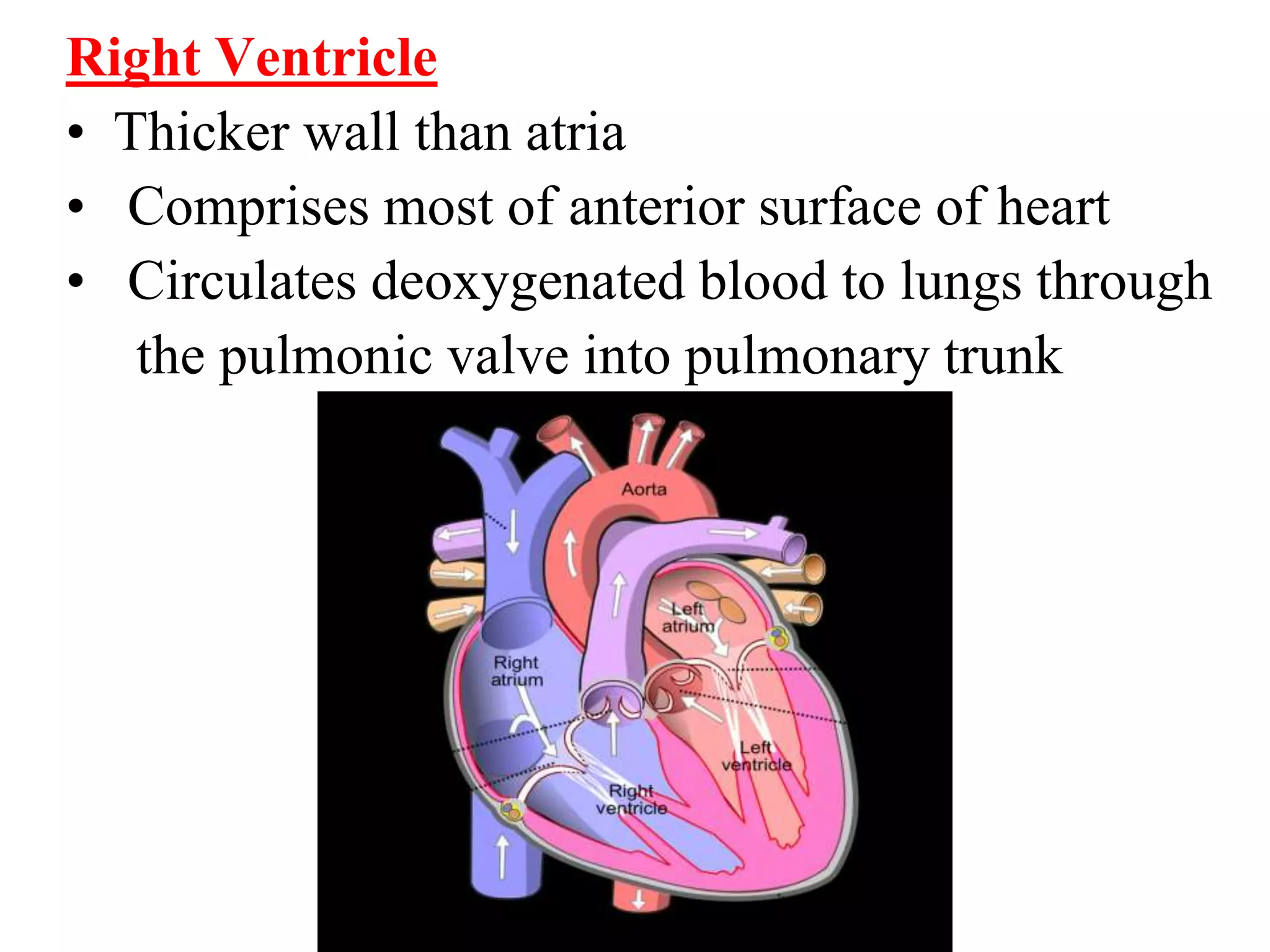


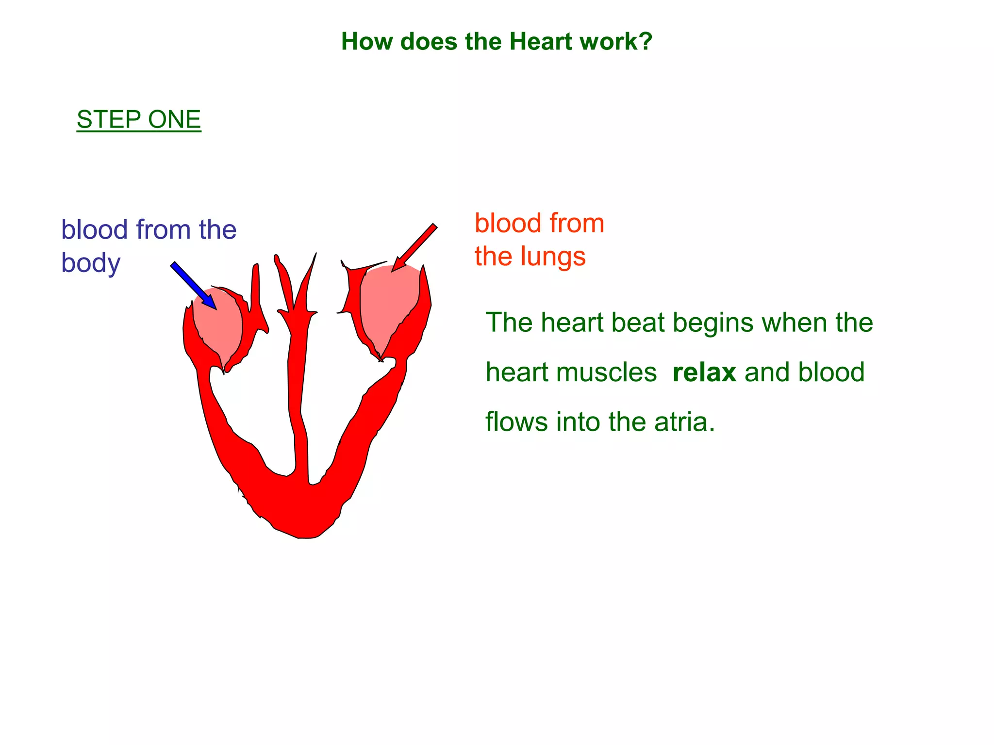
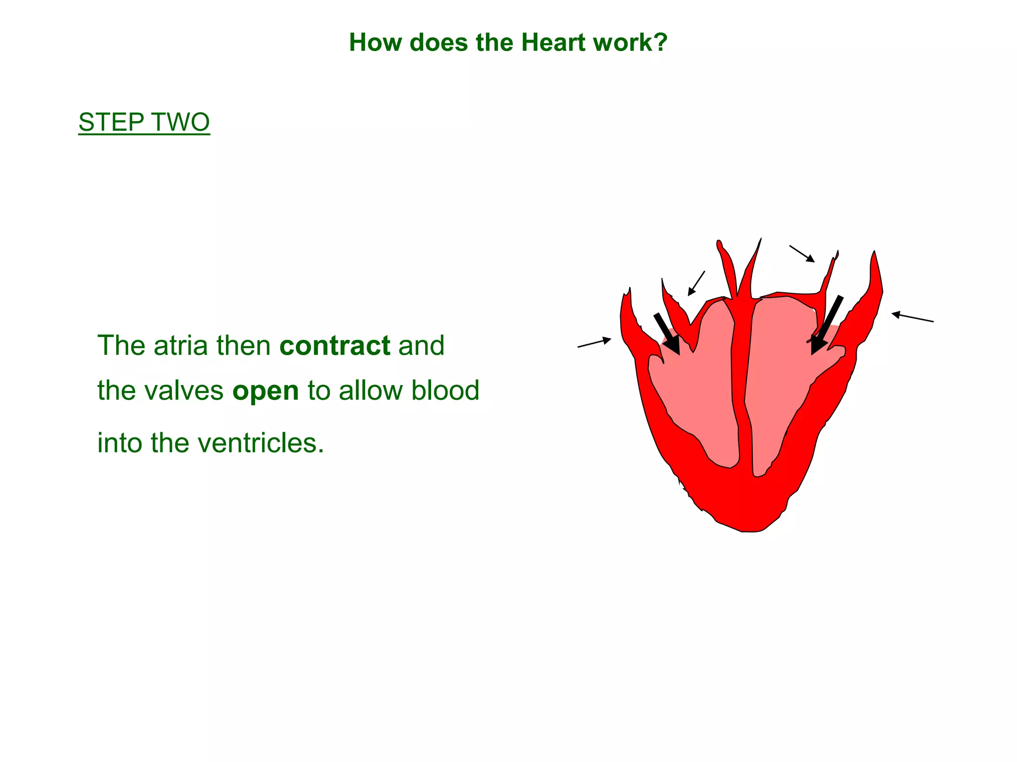


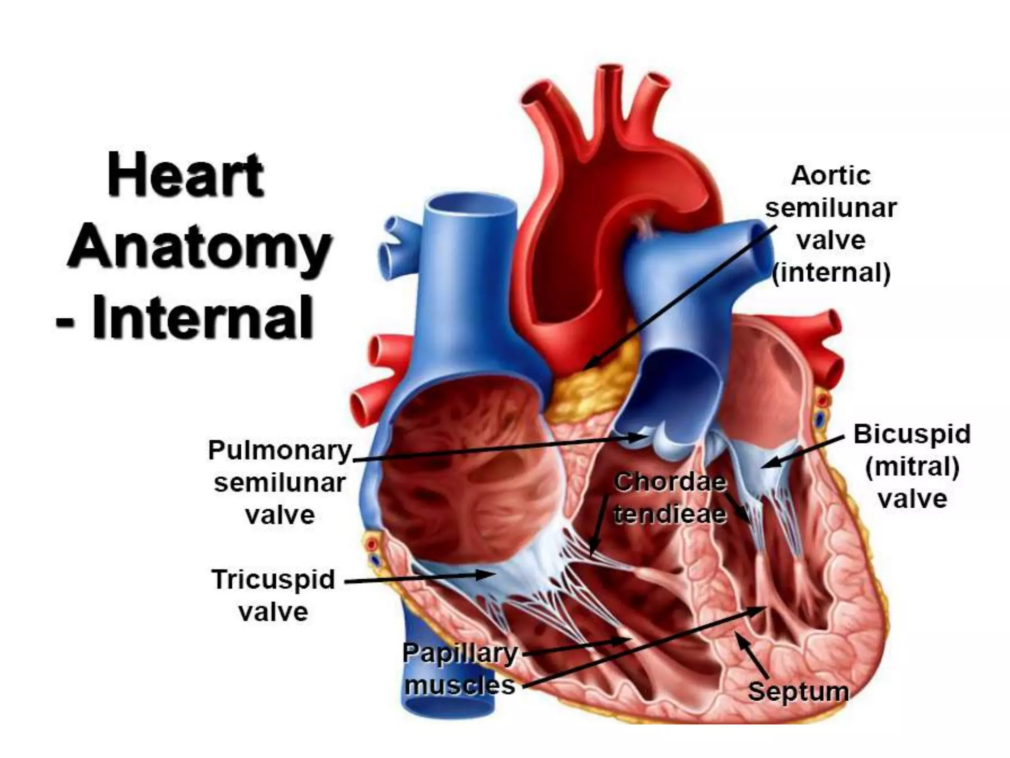


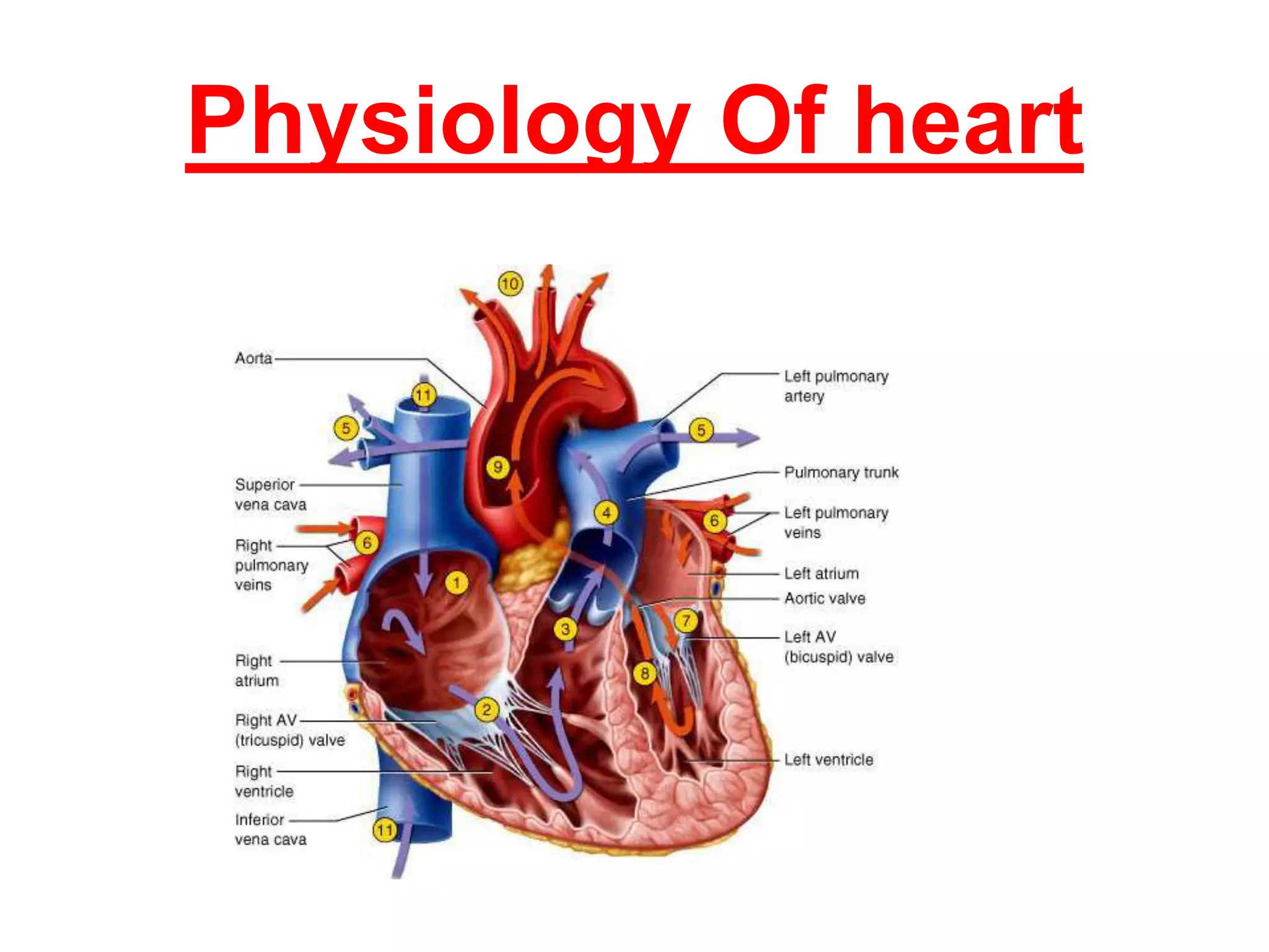


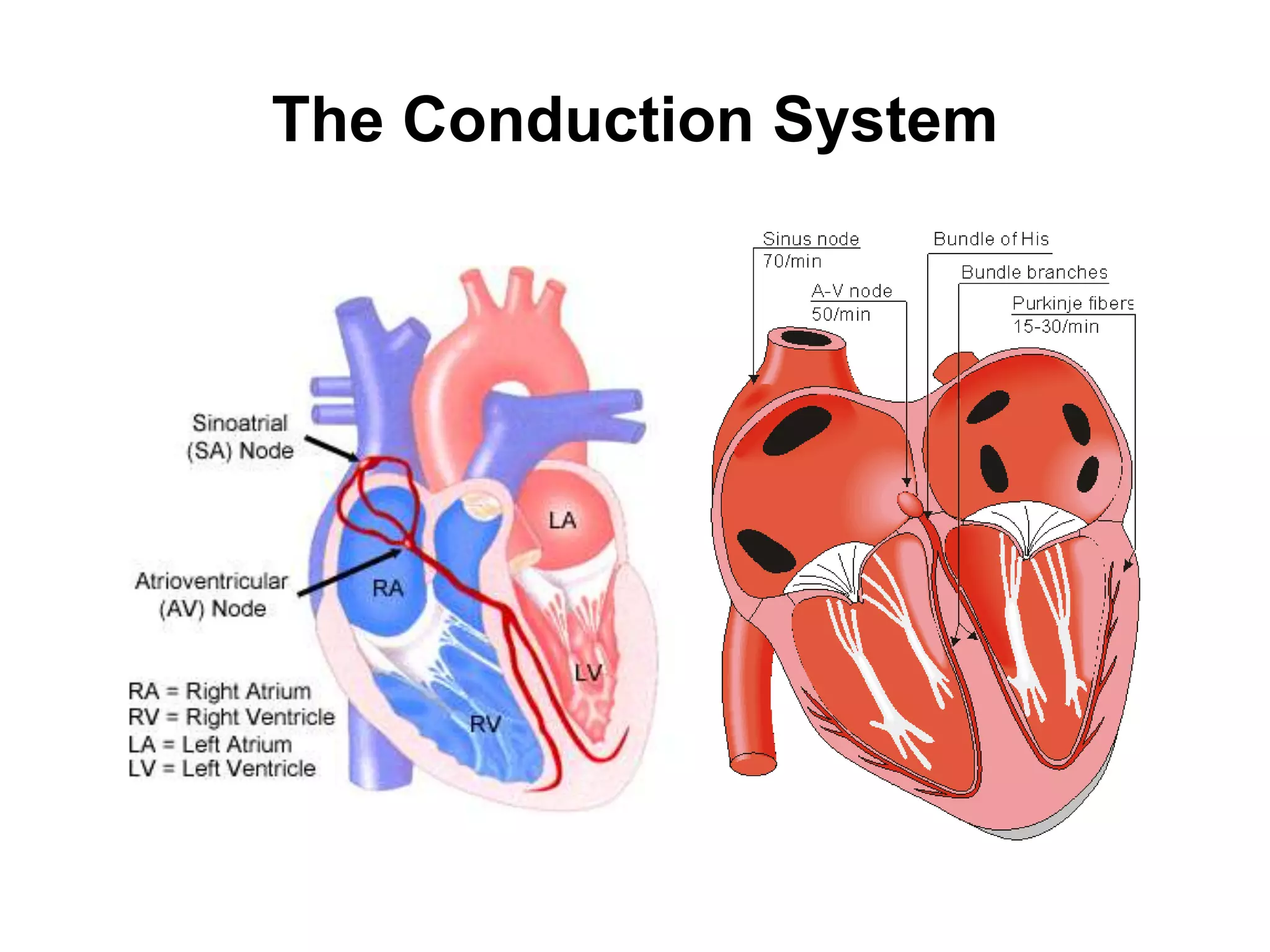


The document summarizes the layers of the heart and its internal structure. It discusses that the heart has two main layers - an outer covering layer called the pericardium and inner layers of heart tissue. The inner layers include the epicardium, myocardium and endocardium. It also describes the four chambers of the heart - the left and right atria which act as collecting reservoirs, and the left and right ventricles which act as pumps. Finally, it briefly discusses the conduction system which coordinates the heart's pumping action.


