Exoskeleton
Support important body
organ,enabling them to work
efficiently.
Protects internal structures
from damage and allows the
animal to move from place to
place.
5.
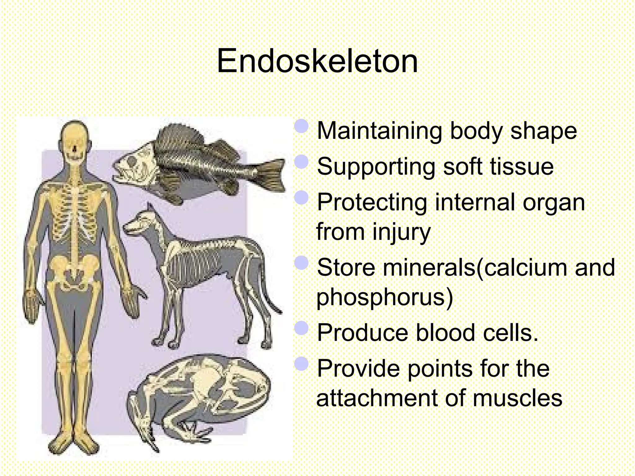
Endoskeleton
Maintaining body shape
Supportingsoft tissue
Protecting internal organ
from injury
Store minerals(calcium and
phosphorus)
Produce blood cells.
Provide points for the
attachment of muscles
6.
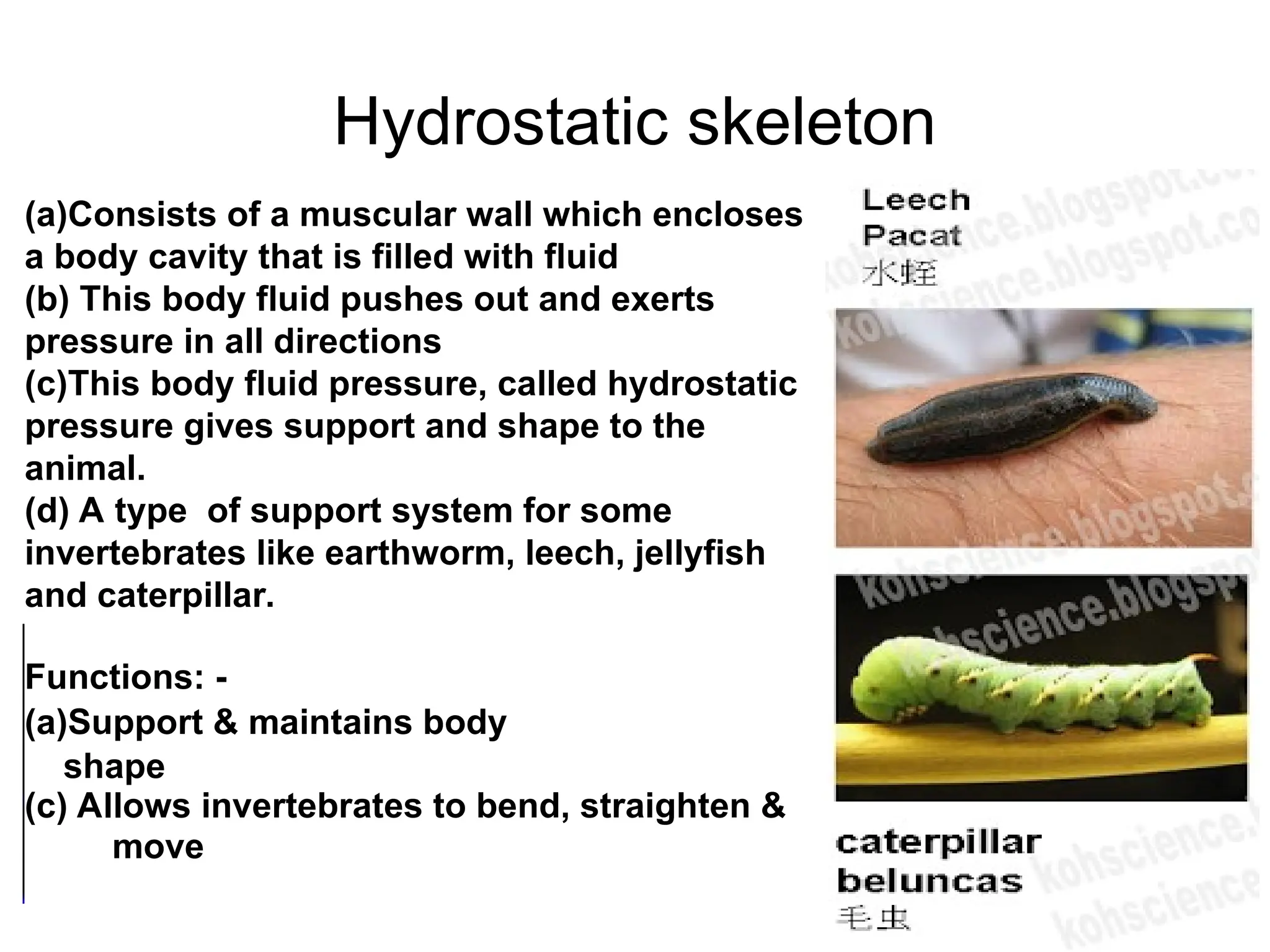
(a)Consists of amuscular wall which encloses
a body cavity that is filled with fluid
(b) This body fluid pushes out and exerts
pressure in all directions
(c)This body fluid pressure, called hydrostatic
pressure gives support and shape to the
animal.
(d) A type of support system for some
invertebrates like earthworm, leech, jellyfish
and caterpillar.
Functions: -
(a)Support & maintains body
shape
(c) Allows invertebrates to bend, straighten &
move
Hydrostatic skeleton
7.
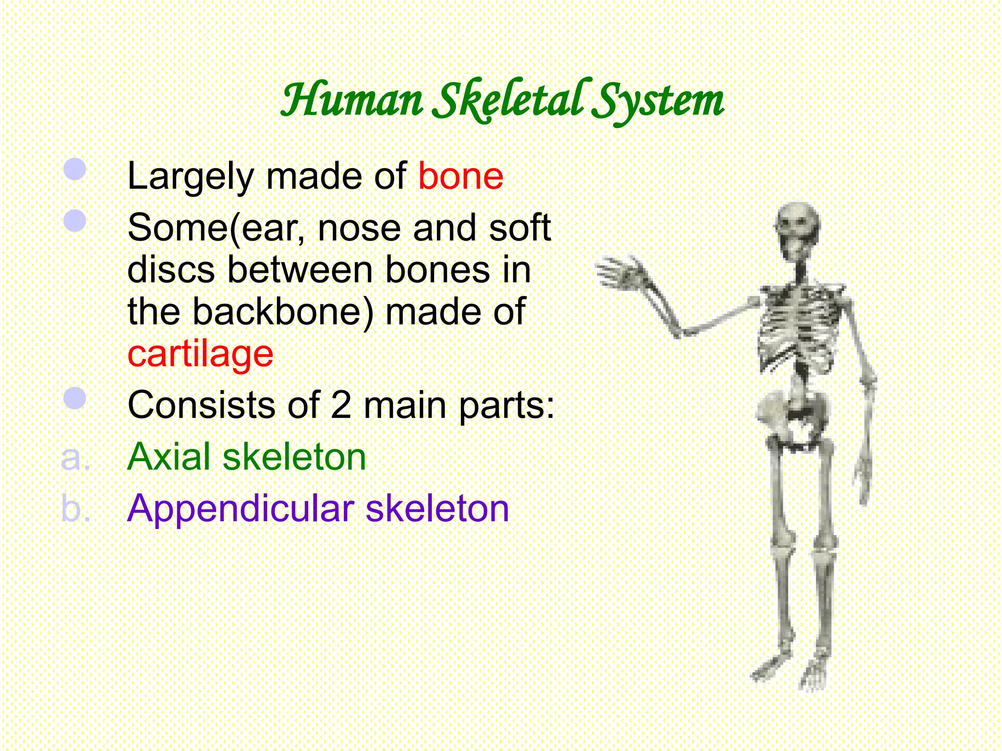
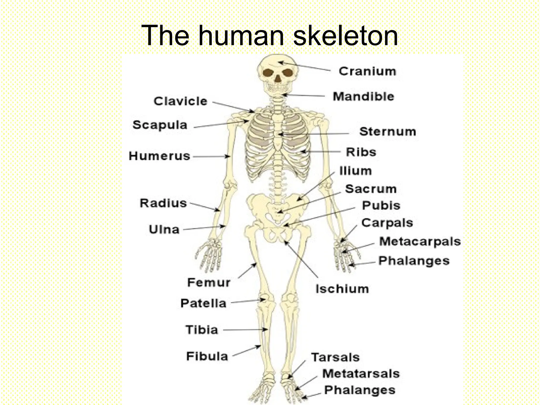
Human Skeletal System
Largely made of bone
Some(ear, nose and soft
discs between bones in
the backbone) made of
cartilage
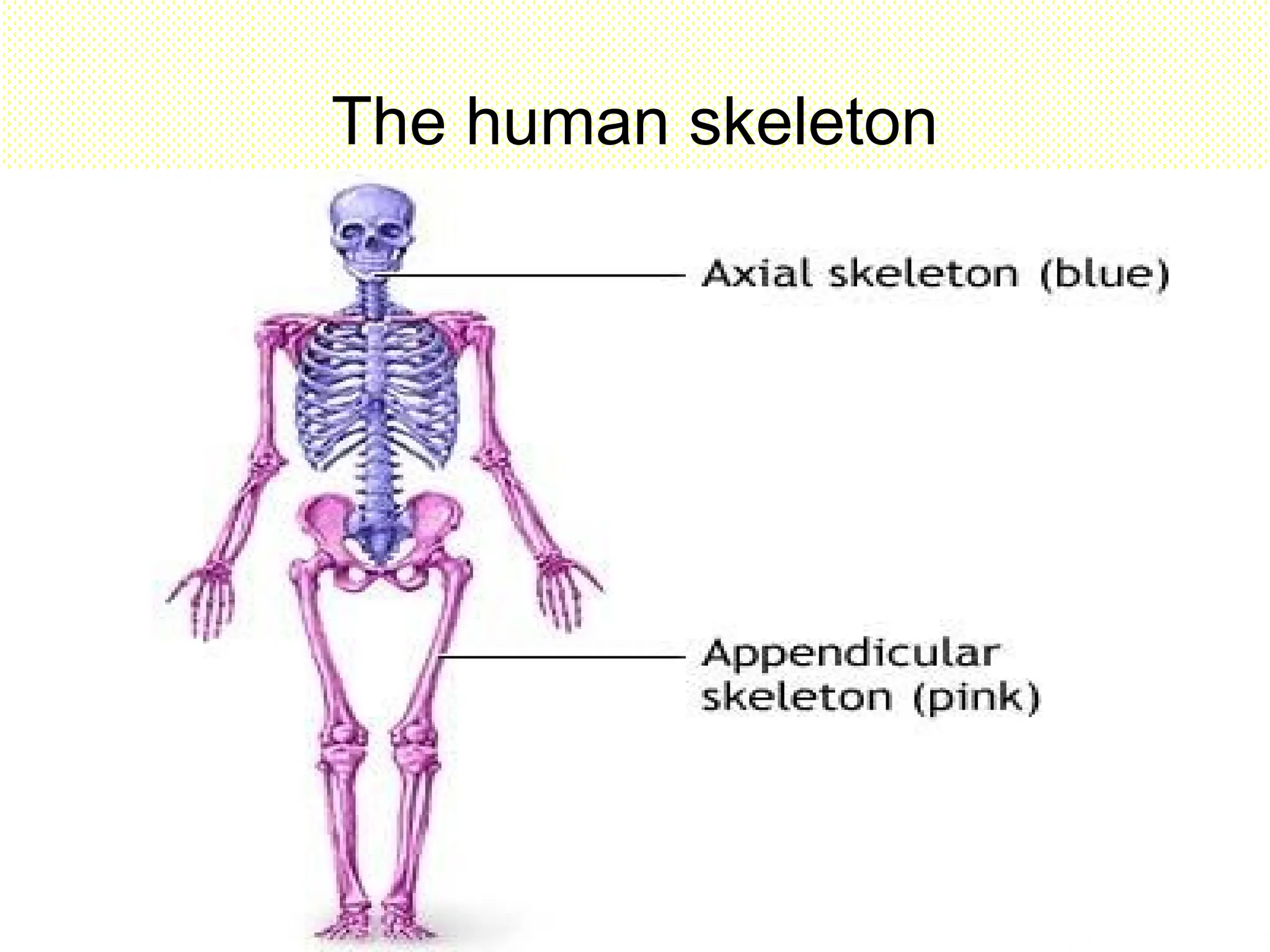
Consists of 2 main parts:
a. Axial skeleton
b. Appendicular skeleton
a. Skull
Madeup of:-
a. Cranium
- consists of 8
pieces of bones
which are fused
together to form
immovable
joints(suture).
– encloses & protect
the brain
Sutures
between the
bones of the
cranium
13.
a. Skull
Madeup of:-
b) Facial bones & jaw
- facial bones protect
the eyes & the ears
- upper jaw is fused
to the cranium & is
immovable
- joint between the
lower jaw & cranium
is movable.
14.
The bones atthe front of your skull hold
your eyes in place and form your facial
features.
Your mandible, or jawbone, is the largest,
strongest bone in your face.
It holds your lower teeth in place and you
move it to chew your food.
Skull is joined to the vertebral column at
the base of the cranium.
15.
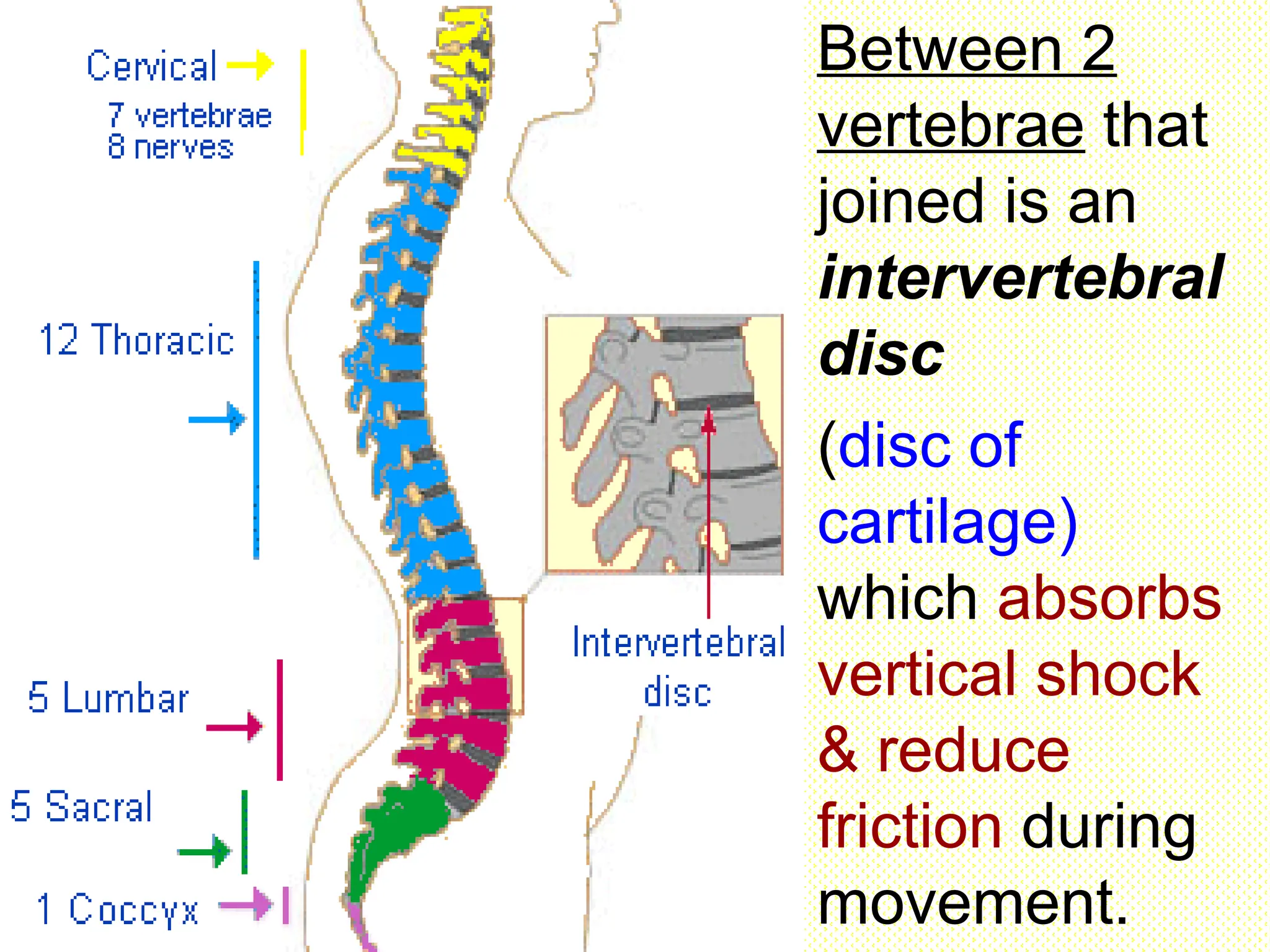
b. Vertebral column
Consists of 33 vertebrae joined to one another
but separated by cartilage.
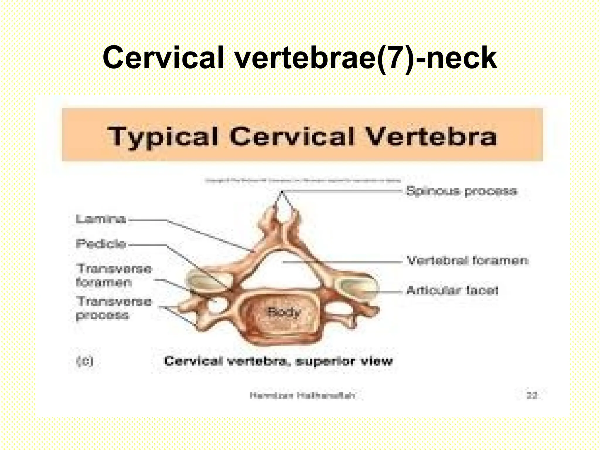
Five types :-
a. Cervical vertebrae (7) – neck
b. Thoracic vertebrae (12) – thorax
c. Lumbar vertebrae (5) – waist
d. Sacral vertebrae (5) fused together to form
sacrum at the hip
e. Caudal vertebrae (4) fused together to form
coccyx at the end of the vertebrae column.
16.
Between 2
vertebrae that
joinedis an
intervertebral
disc
(disc of
cartilage)
which absorbs
vertical shock
& reduce
friction during
movement.
Centrum Gives support& able to
withstand compression
force
Spinous process &
Transverse
process
For muscle & ligament
attachment
Vertebral foramen Contain the spinal cord
Transverse
foramen
Contains blood vessels
& nerves
Body / CentrumGives support & able to withstand
compression force
Spinous process / neural
spine
Transverse process
For muscle attachment
Neural arch / lamina Forms the neural canal which
protects the spinal cord
Facet / zygapophysis Join with another vertebra
Neural canal/vertebral
foramen
Contain the spinal cord
Transverse foramen Contains blood vessels & nerves
22.
Lumbar vertebrae(5)-waist
Largest andstrongest
vertebrae
Well developed
transverse processes,
and a short & flat
neural spine for
muscle attachment
Large & thick centrum
23.
Sacrum(5 sacral vertebrae)
Sacrumconsists of 5
sacral vertebrae hat
fused together.
Has 4 pairs of holes
through which nerves
leave the spinal cord.
Facets on both sides
of the first transverse
processes join with
the pelvic girdle.
Rib cage
Rib cageconsists of 12 pairs of
ribs which join with the thoracic
vertebrae & the sternum by
cartilages.
- The first 7 pairs joins directly to
the sternum are called true ribs.
- The 8th
, 9th
& 10th
pair of ribs join
to the sternum via the 7th
pair of
the ribs are called false ribs.
- The last 2 pairs are floating ribs &
not joined to the sternum.
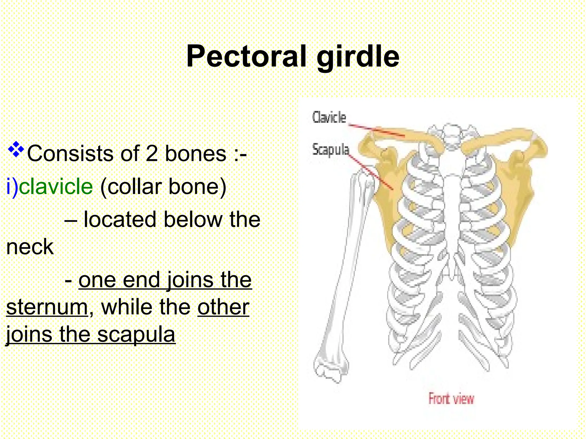
Pectoral girdle
Consists of2 bones :-
i)clavicle (collar bone)
– located below the
neck
- one end joins the
sternum, while the other
joins the scapula
29.
ii) Scapula (shoulderbone)
- Flat triangular bone
- On its dorsal surface, there is a ridge called
scapular spine that branches into 2 at its end.
- This is for the forelimb muscle attachment.
- At the end is a hollow cavity called glenoid cavity
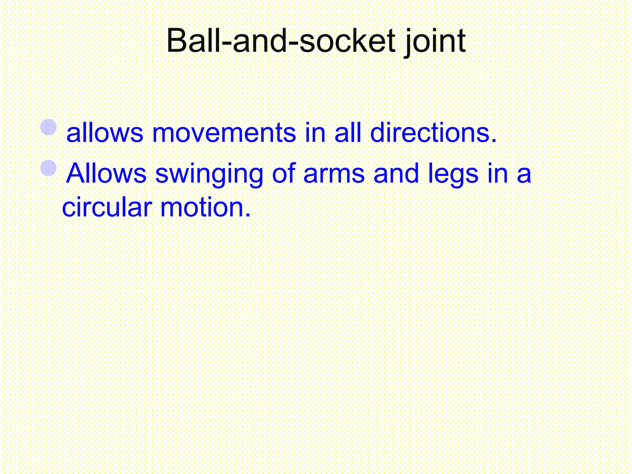
which articulates with the head of the humerus.
- This forms a ball-and-socket joint which allows
movements in all directions
30.
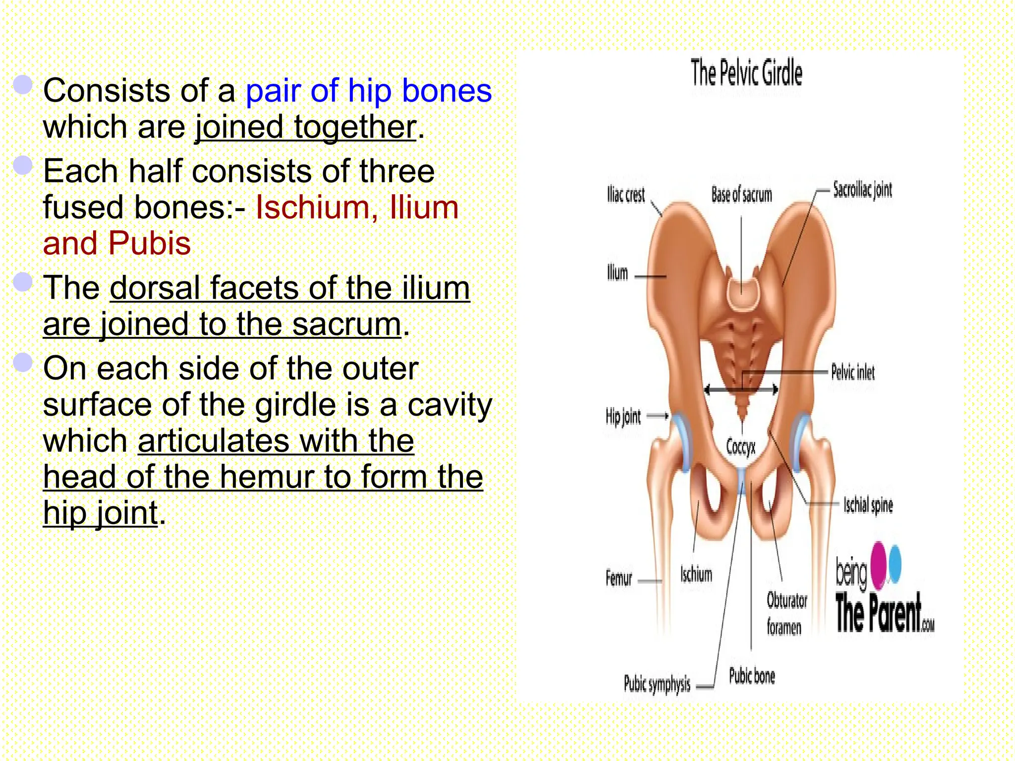
Pelvic girdle
Consists ofa pair of hip bones
which are joined together.
Each half consists of three
fused bones:- Ischium, Ilium
and Pubis
The dorsal facets of the ilium
are joined to the sacrum.
On each side of the outer
surface of the girdle is a cavity
which articulates with the
head of the hemur to form the
hip joint.
31.
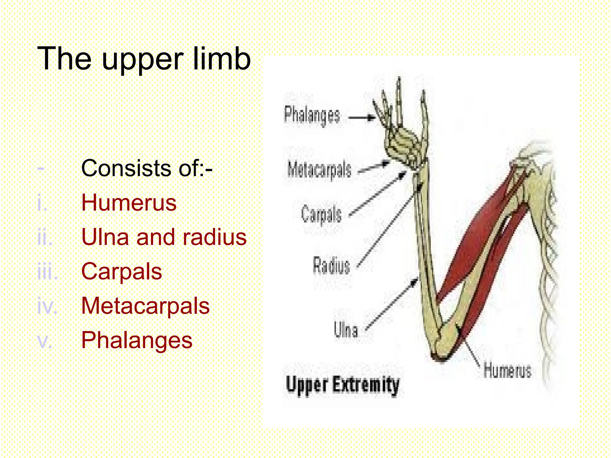
The upper limb
-Consists of:-
i. Humerus
ii. Ulna and radius
iii. Carpals
iv. Metacarpals
v. Phalanges
32.
i. Humerus
Long boneof the upper arm
Has a rounded head which joins
with the glenoid cavity to form
the ball-and-socket joint.
Lower end articulates with the
radius & ulna to form a hinge joint
at the elbow.
Hinge joint allows movement in only 1
direction.
33.
ii. Ulna andradius
2 long bones at the lower arm.
Both are parallel & held together at
both ends.
Radius is rod-shaped.
Ulna is longer/larger than the radius & has
projection at the upper end.
Upper end articulates with the humerus,
while lower end articulates with the
carpals at the wrist.
iv. Metacarpals &Phalanges
Are slender bones at the palm
There are 5 metacarpals
The distal ends of the metacarpals
articulate with the bones in the finger
called phalanges.
Each hand has 14 phalanges.
Thumb has 2 phalanges, while all the
other fingers have 3 phalanges each.
36.
The lower limb
Consists of femur, tibia,
fibula, tarsals,
metatarsals &
phalanges.
i) Femur
- Long bone in the thigh
- Upper end is a rounded
head which articulates
with the cavity in the
pelvic girdle.
37.
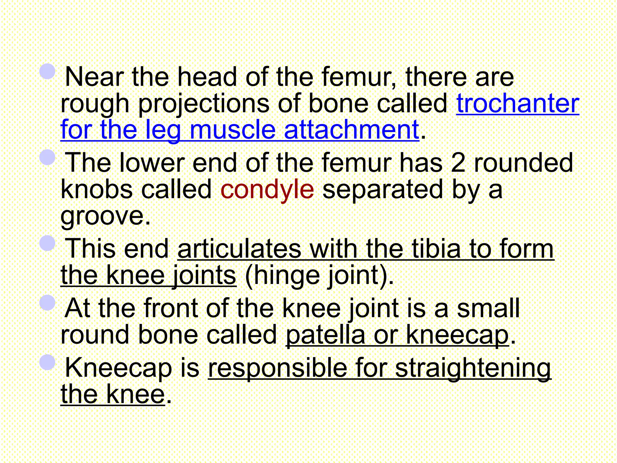
Near the headof the femur, there are
rough projections of bone called trochanter
for the leg muscle attachment.
The lower end of the femur has 2 rounded
knobs called condyle separated by a
groove.
This end articulates with the tibia to form
the knee joints (hinge joint).
At the front of the knee joint is a small
round bone called patella or kneecap.
Kneecap is responsible for straightening
the knee.
38.
ii. Tibia andfibula
- The leg bones
- Fibula is a smaller bone which lies on the
outside of the larger tibia.
- The lower end of it articulates with the
tarsals at the ankle.
39.
iii. Tarsals &metatarsals
Tarsals are ankle bones.
There are 7 tarsals but only one
articulates with the tibia and fibula.
The largest tarsal is the ankle bone forms
the heel.
Metatarsals consist of 5 bones that
articulate with the tarsals at the ankle &
phalanges at the toes.
40.
iv. Phalanges
Are bonesin the toes
Each toe has 3 phalanges except the big
toe which has only 2 phalanges.
42.
The Structure ofa Joint
Joint-the place where two
or more bones meet.
At joint, the bones are
held together by
ligaments.(tough sheets
of elastic fibres)
Allow bones to move
against one another and
prevent the dislocation of
the joint during movement
43.
Synovial joint
-freely moveable-
StructureFunction
Joint capsule Enclosure reinforced by
and strengthened with
ligaments
Synovial
membrane
Secrete synovial fluid
Synovial joint Cavity contains synovial
fluid(lubricant which
reduces the friction
between the ends of the
bones.
cartilage Cushions the joints,
absorbs shocks ,reduces
friction between the ends of
bones and protects the
Hinge joints
allowsmovement in only 1 direction
Lower end of humerus articulates with the
radius & ulna to form a hinge joint
at the elbow.
The lower end of the femur articulates with
the tibia to form the knee joints (hinge joint).
Tendons
Are fibrous connectivetissue.
Are flexible but they are not
elastic.
Connect skeletal muscles to
bones.
Ensure that the pulling force
exerted by the muscular
contraction is transmitted to
the bone, to pull the bone and
cause movement.
49.
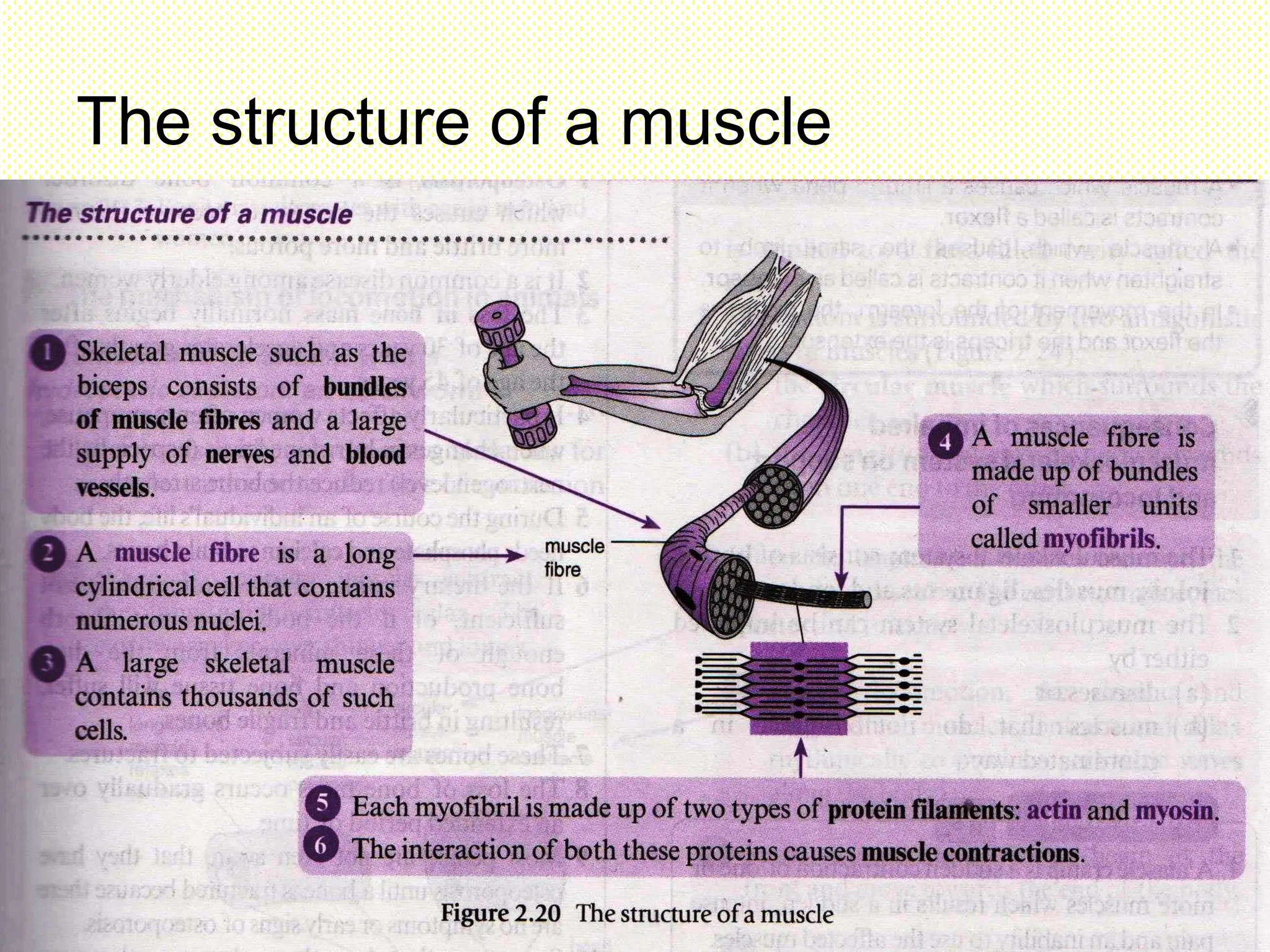
Muscles
made ofmuscle fibres (contract when
stimulated by a nerve to cause
movement)
Has 3 types :-
a. Skeletal muscle : attached to the bones
b. Smooth muscle : found in the wall of
blood vessels, stomach, digestive tract &
other internal organs.
c. Cardiac muscle : found in the walls of the
heart
50.
Skeletal muscles
Skeletal musclesproduce movements by exerting the
force to pull on the tendons which are attached to
bones.
Since a muscle cannot push, but only pull, it has to be
extended back to its original length by the opposing
action of another muscles.
Skeletal muscles exist as paired antagonistic muscles.
This means when one muscle contracts, the other
relaxes.
As the muscle contracts, it becomes shorter & thicker
exerting a pulling force to move the bones attached to it
and, thus causing movement.
51.
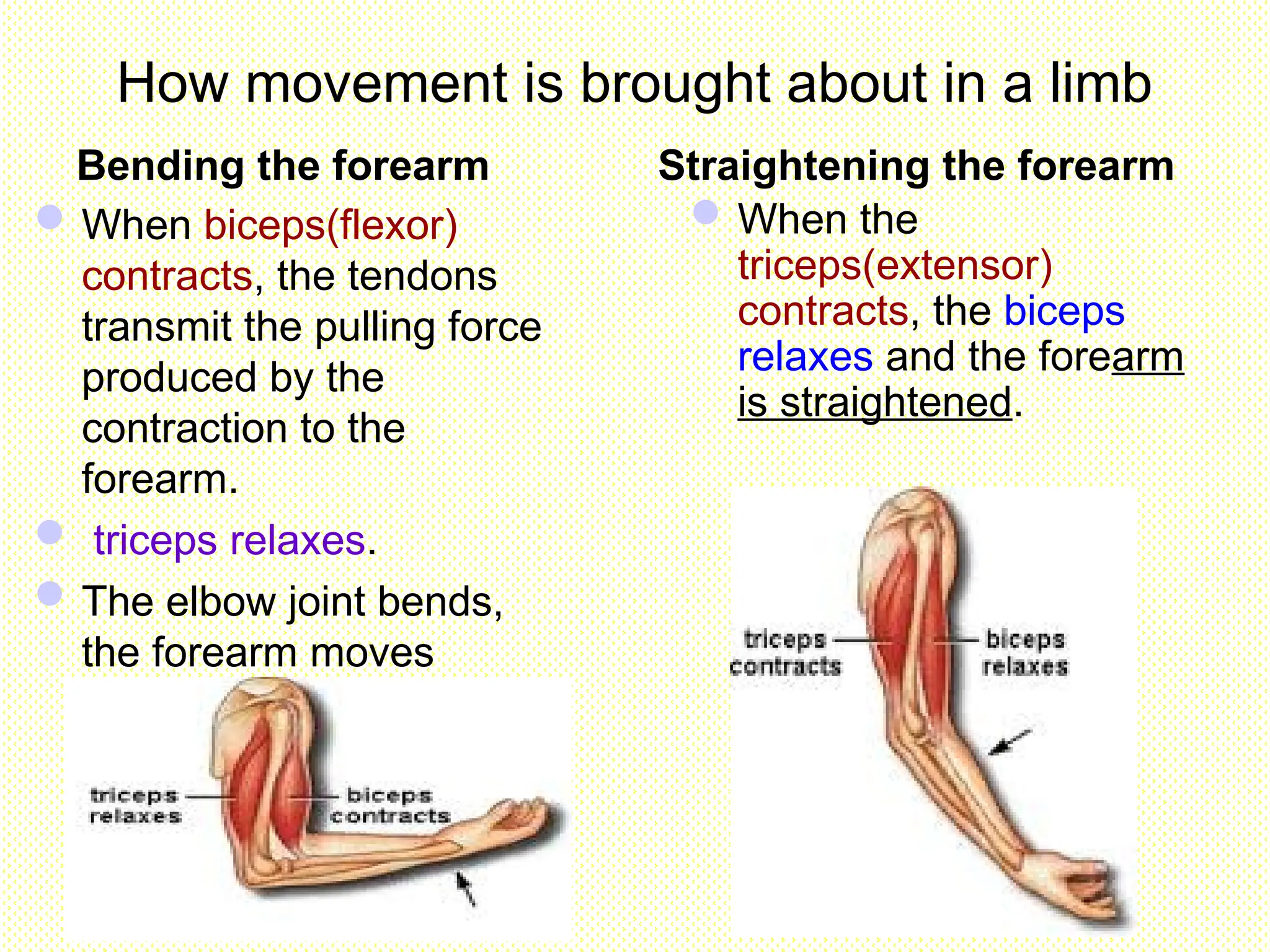
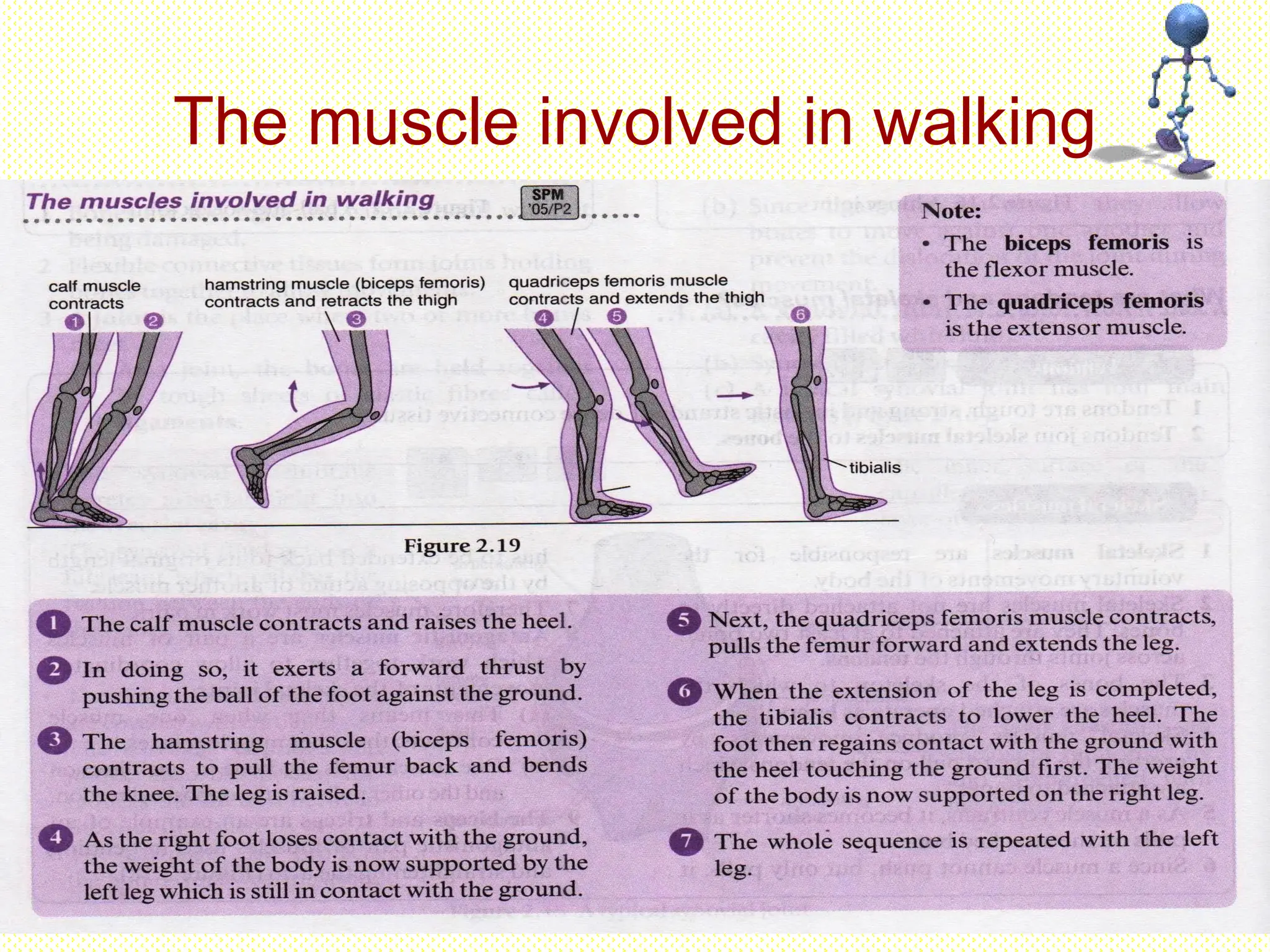
How movement isbrought about in a limb
Bending the forearm Straightening the forearm
When biceps(flexor)
contracts, the tendons
transmit the pulling force
produced by the
contraction to the
forearm.
triceps relaxes.
The elbow joint bends,
the forearm moves
upwards.
When the
triceps(extensor)
contracts, the biceps
relaxes and the forearm
is straightened.
52.
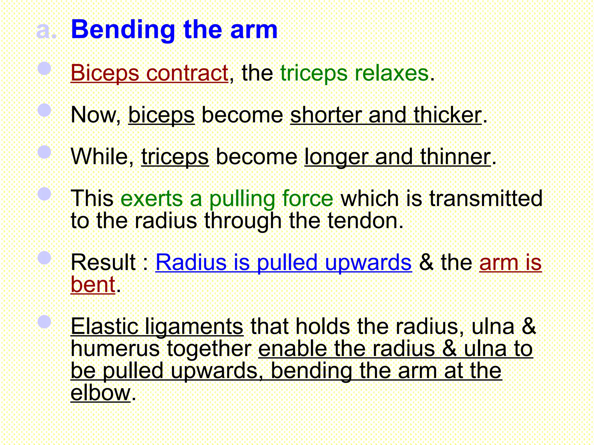
a. Bending thearm
Biceps contract, the triceps relaxes.
Now, biceps become shorter and thicker.
While, triceps become longer and thinner.
This exerts a pulling force which is transmitted
to the radius through the tendon.
Result : Radius is pulled upwards & the arm is
bent.
Elastic ligaments that holds the radius, ulna &
humerus together enable the radius & ulna to
be pulled upwards, bending the arm at the
elbow.
53.
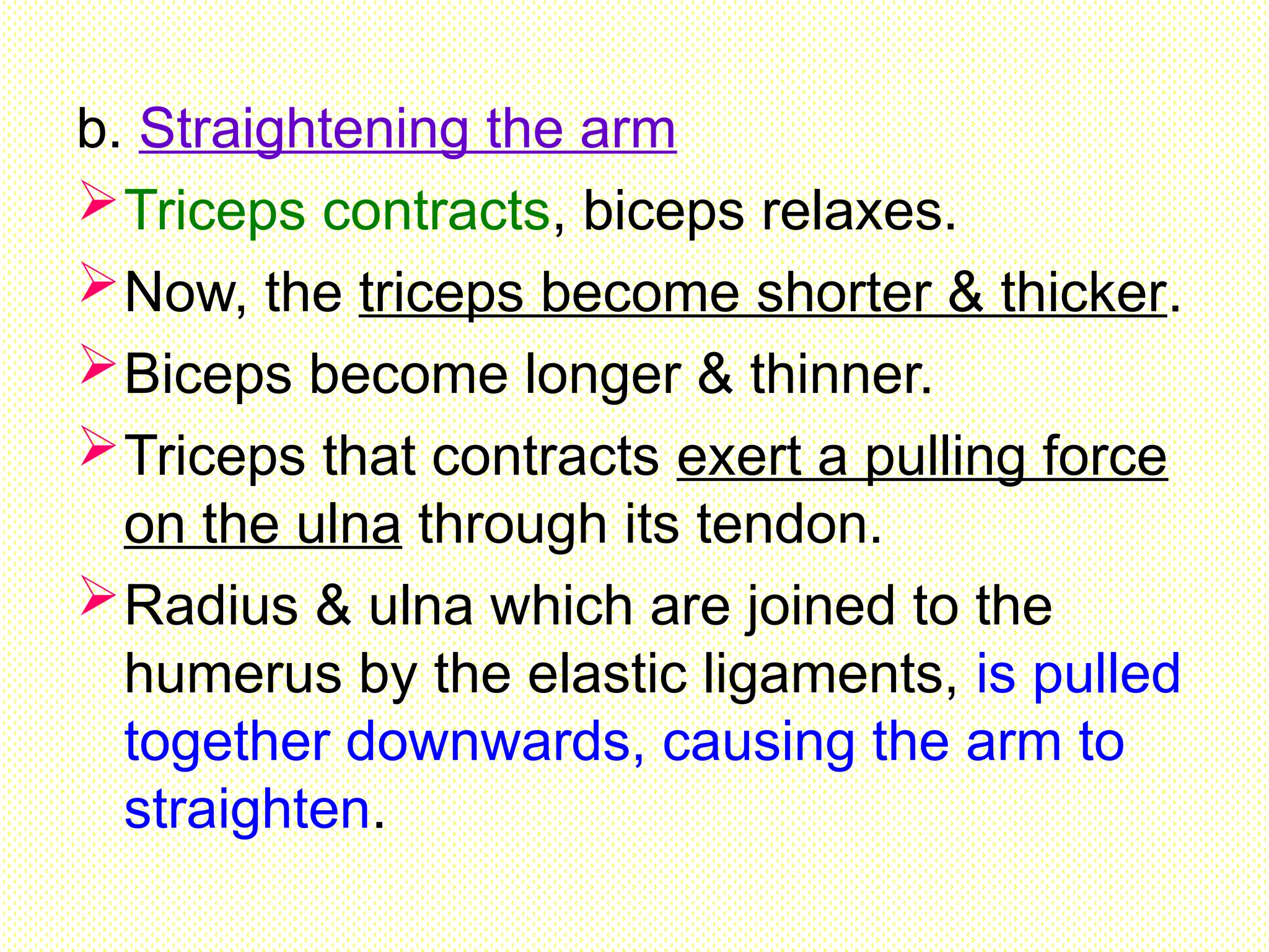
b. Straightening thearm
Triceps contracts, biceps relaxes.
Now, the triceps become shorter & thicker.
Biceps become longer & thinner.
Triceps that contracts exert a pulling force
on the ulna through its tendon.
Radius & ulna which are joined to the
humerus by the elastic ligaments, is pulled
together downwards, causing the arm to
straighten.
54.
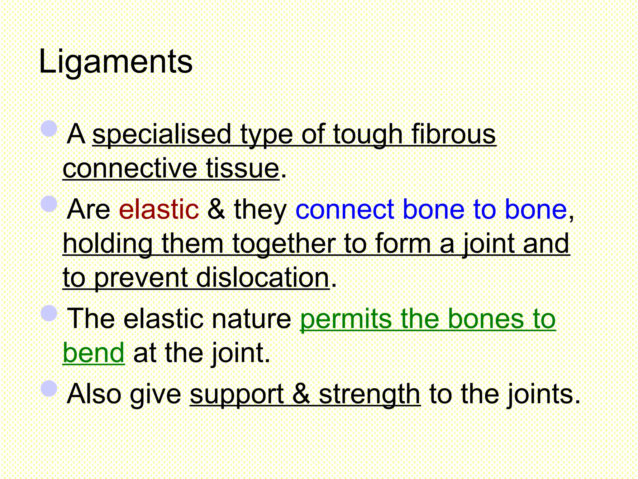
Ligaments
A specialised typeof tough fibrous
connective tissue.
Are elastic & they connect bone to bone,
holding them together to form a joint and
to prevent dislocation.
The elastic nature permits the bones to
bend at the joint.
Also give support & strength to the joints.
55.
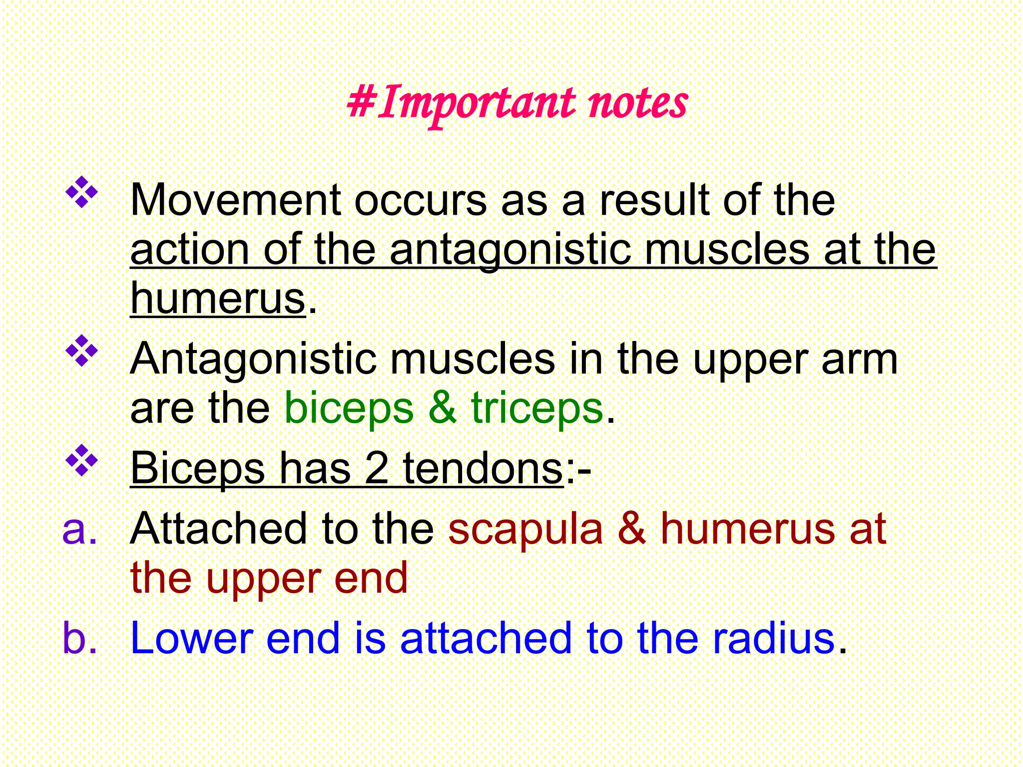
#Important notes
Movementoccurs as a result of the
action of the antagonistic muscles at the
humerus.
Antagonistic muscles in the upper arm
are the biceps & triceps.
Biceps has 2 tendons:-
a. Attached to the scapula & humerus at
the upper end
b. Lower end is attached to the radius.
56.
Triceps has3 tendons :-
a. One of the upper ends attached to the
scapula
b. One attached to the humerus
c. Lower end is attached to the ulna
Ligaments attach
a. the upper end of the humerus to the
scapula
b. The lower end of the humerus to the
radius & ulna
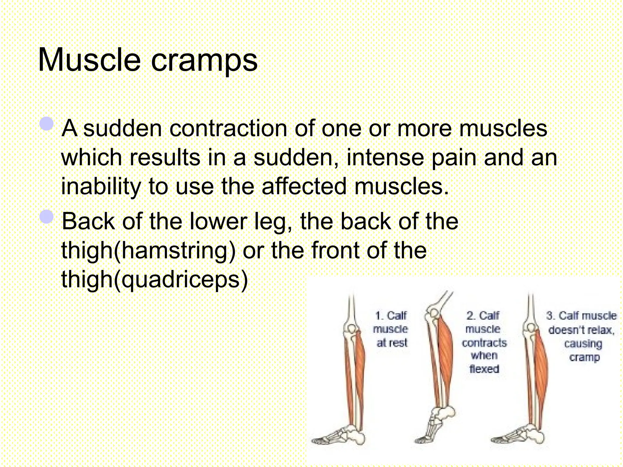
Muscle cramps
A suddencontraction of one or more muscles
which results in a sudden, intense pain and an
inability to use the affected muscles.
Back of the lower leg, the back of the
thigh(hamstring) or the front of the
thigh(quadriceps)
61.
Muscular dystrophy
Caused bythe progressive
degeneration and weakening
of the skeletal muscles that
control movement.
Caused by mutated gene in
X chromosome and mainly
affects boys.
The body muscles gradually
become weak as they are
replaced by fibrous tissue.
62.
Osteoporosis
Cause the bonesto become thinner, more brittle and
more porous.
Common among elderly woman-particularly affects
woman after menopause when changes in hormone
levels(especially the oestrogen level) reduce the bone
strength.
The loss of bone mass normally begins after the age of 30
years and accelerate greatly after the age of 45 years.
The body needs phosphate and calcium to build bones.
Prevention-taking sufficient amounts of calcium,
phosphorus, vitamin D and regular exercise.
65.
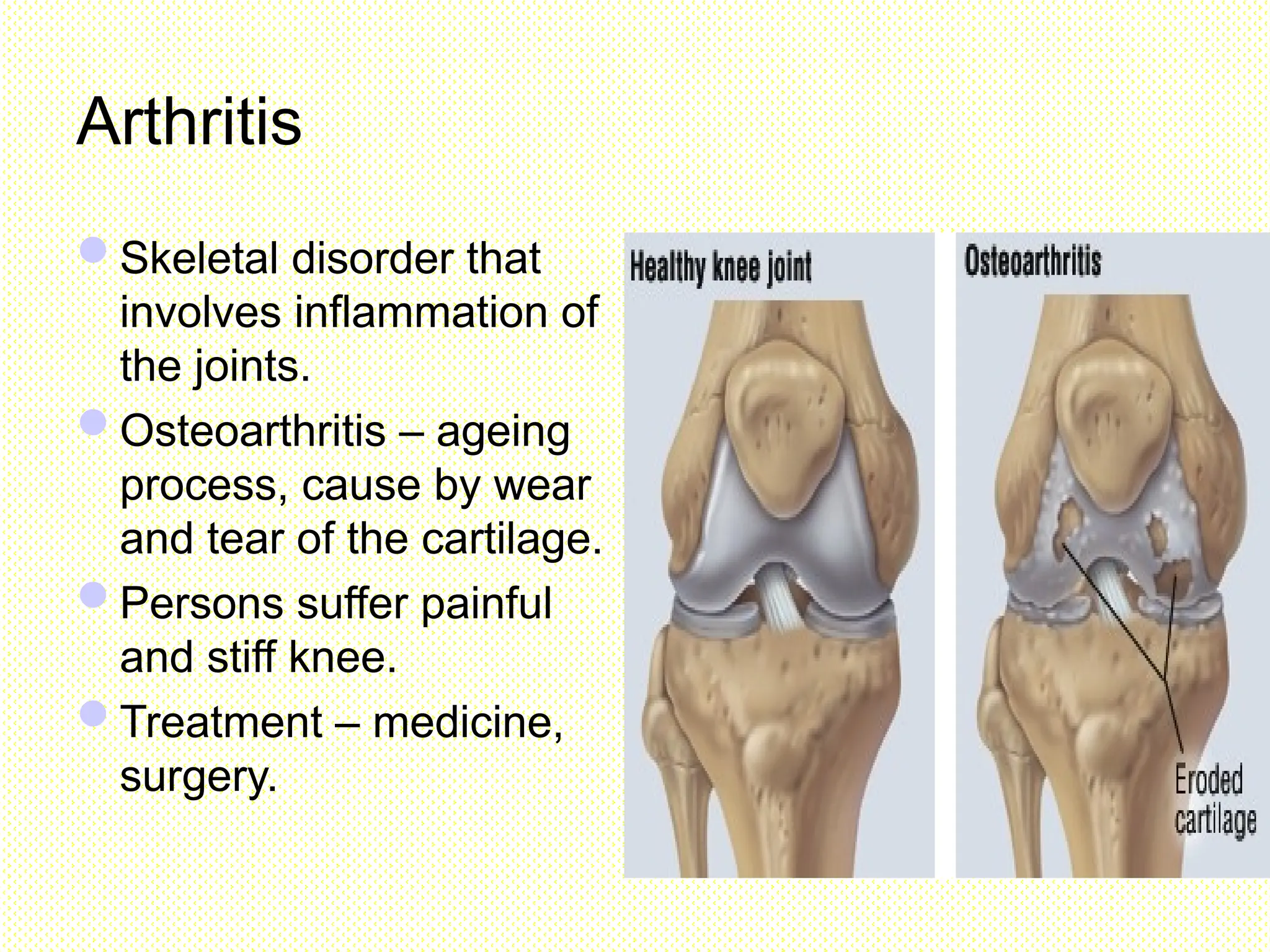
Arthritis
Skeletal disorder that
involvesinflammation of
the joints.
Osteoarthritis – ageing
process, cause by wear
and tear of the cartilage.
Persons suffer painful
and stiff knee.
Treatment – medicine,
surgery.
67.
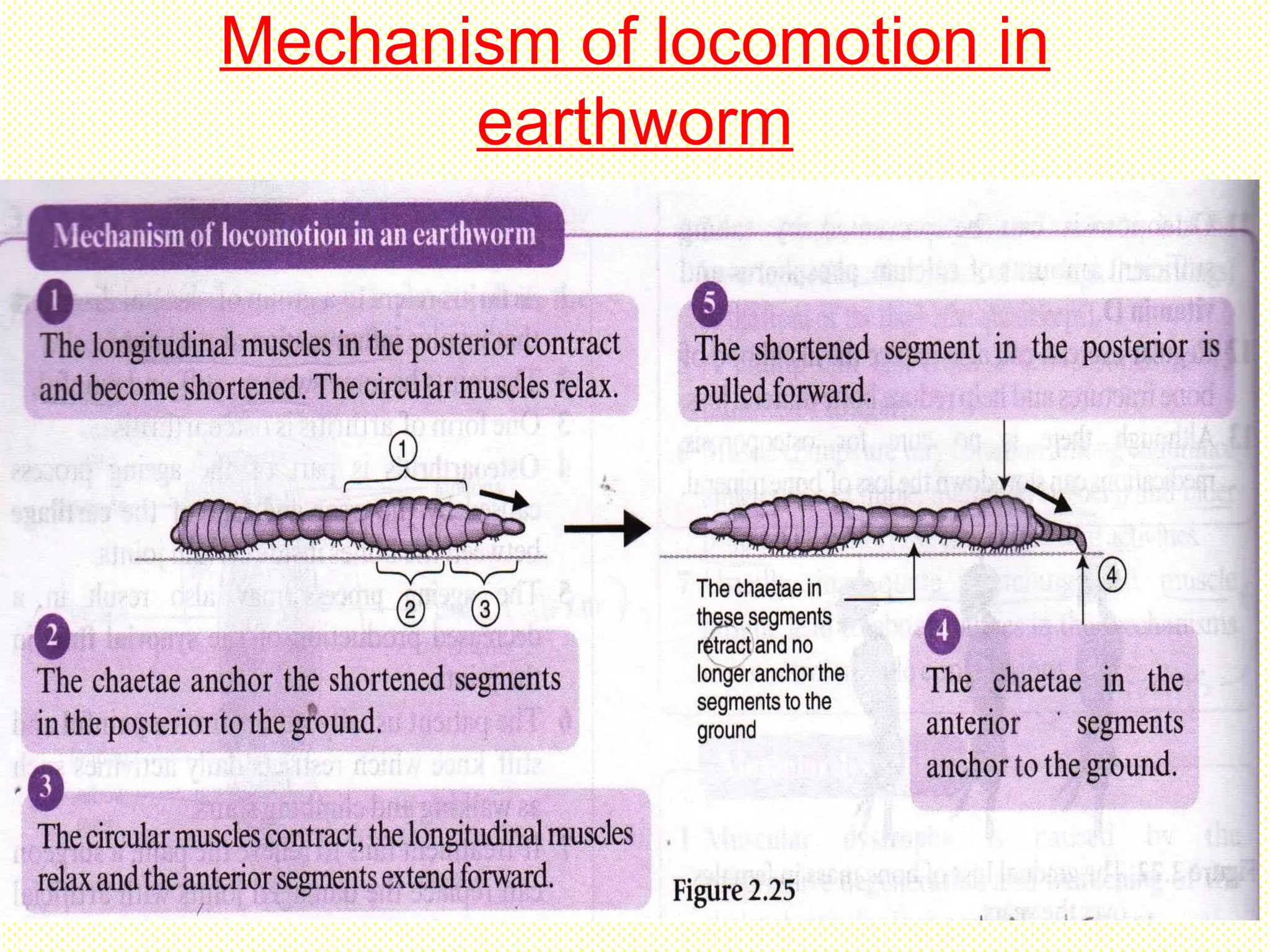
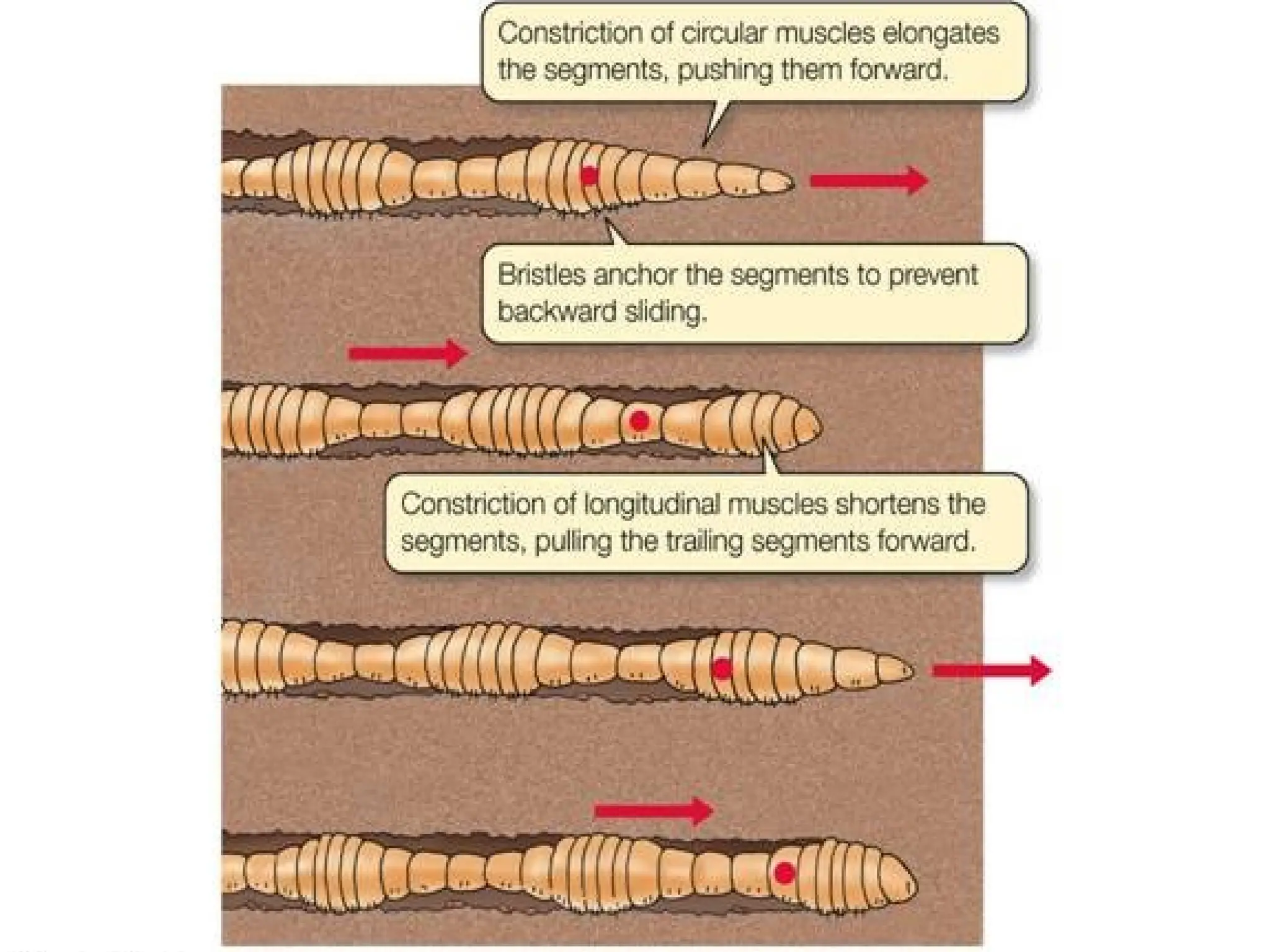
Locomotion in Earthworm
Earthwormhas a
hydrostatic skeleton &
moves by crawling &
creeping.
The body wall
consists of outer
circular muscles &
inner longitudinal
muscles.
68.
On the ventralpart of the
body wall, there are
small bristles called
chaetae which act as
hooks during movement.
The movement is
caused by a series of
contraction & relaxation
of the circular and
longitudinal muscles on
the body wall.
68
69.
The antagonistic actionof the muscles in
the earthworm during locomotion produces
a hydrostatic pressure in the body fluid &
forms the peristaltic waves, moving from
the anterior end to the posterior end of the
body.
Mechanism of locomotionin earthworm
a) When the earthworm is crawling over the
surface, the chaetae in the posterior end of
the body are pushed into the ground to
anchor it.
b) The circular muscles in the anterior end of
the body contract, while the longitudinal
muscles relax.
72.
Mechanism of locomotionin earthworm
a) Hence, the anterior end of the body
elongates (longer & thinner).
b) The hydrostatic pressure builds up in the
body and, thus the body fluid is pushed
backward.
74.
e) The chaetaein the posterior end of the
body are withdrawn, while the chaetae in
the anterior end of the body are pushed
into the ground.
f) The longitudinal muscle in the anterior end
of the body contract, while the circular
muscles relax.
g) Thus, the anterior end of the body becomes
short & thick.
75.
e) The bodyfluid flows into the
anterior end of the body, causing the
posterior end of the body to be
pulled forward.
f) The earthworm moves on the ground
by alternately lengthening &
shortening its body, assisted by the
chaetae.
76.
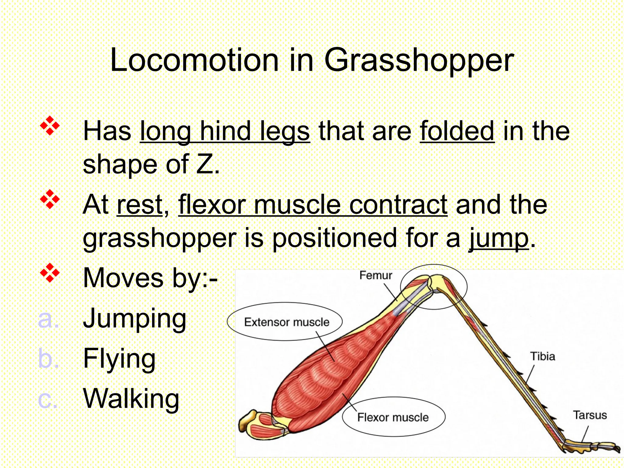
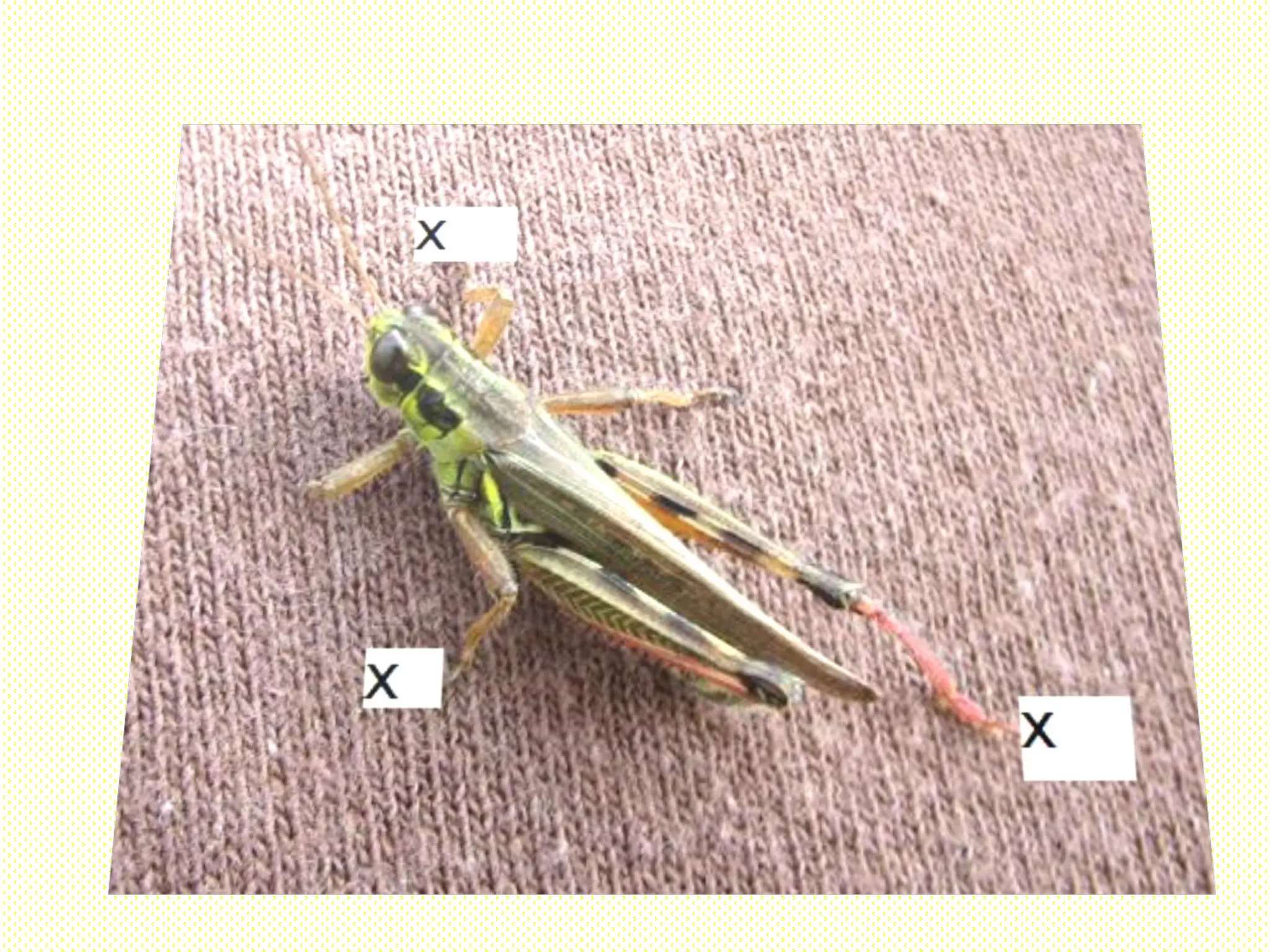
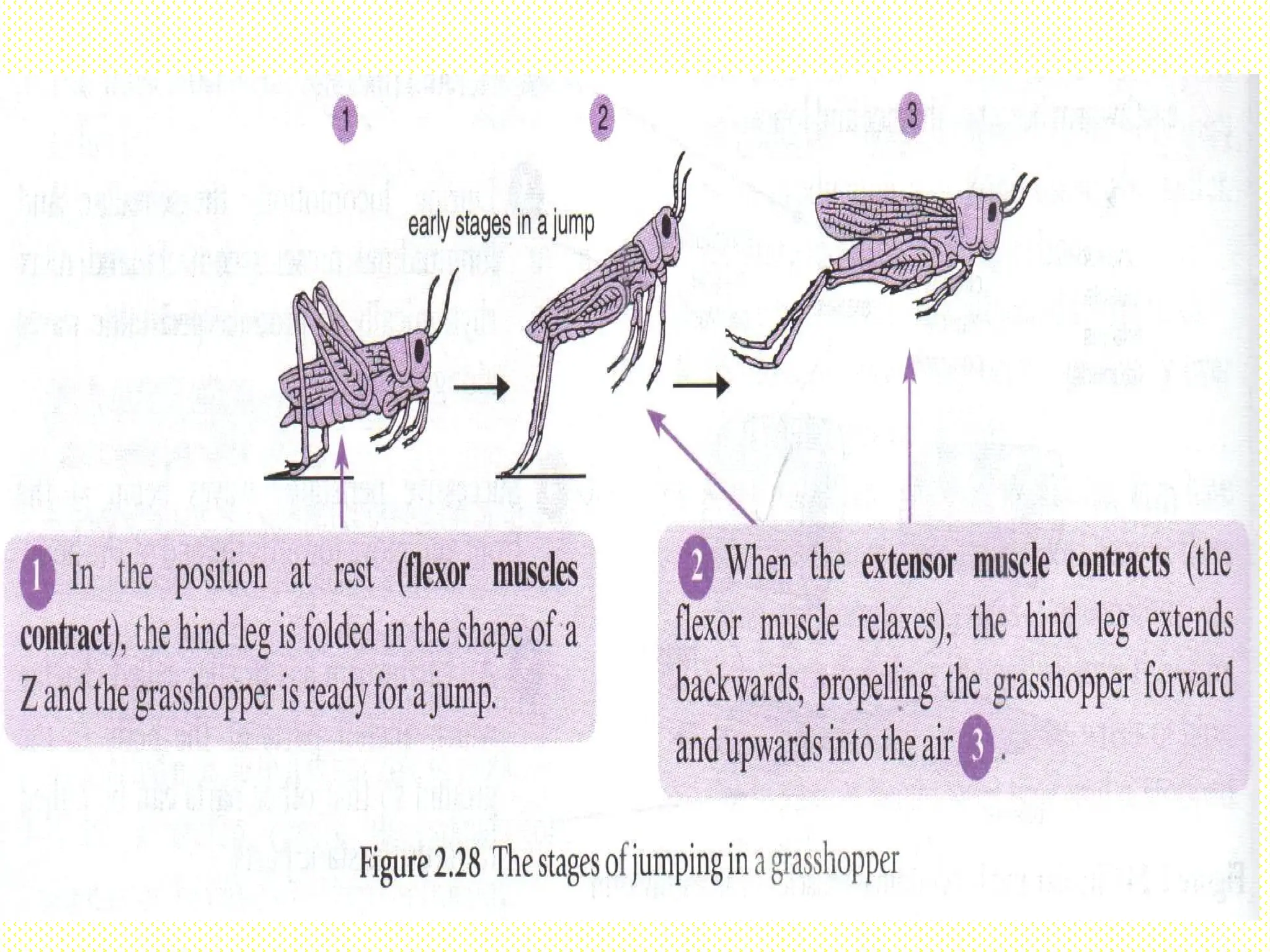
Locomotion in Grasshopper
Has long hind legs that are folded in the
shape of Z.
At rest, flexor muscle contract and the
grasshopper is positioned for a jump.
Moves by:-
a. Jumping
b. Flying
c. Walking
77.
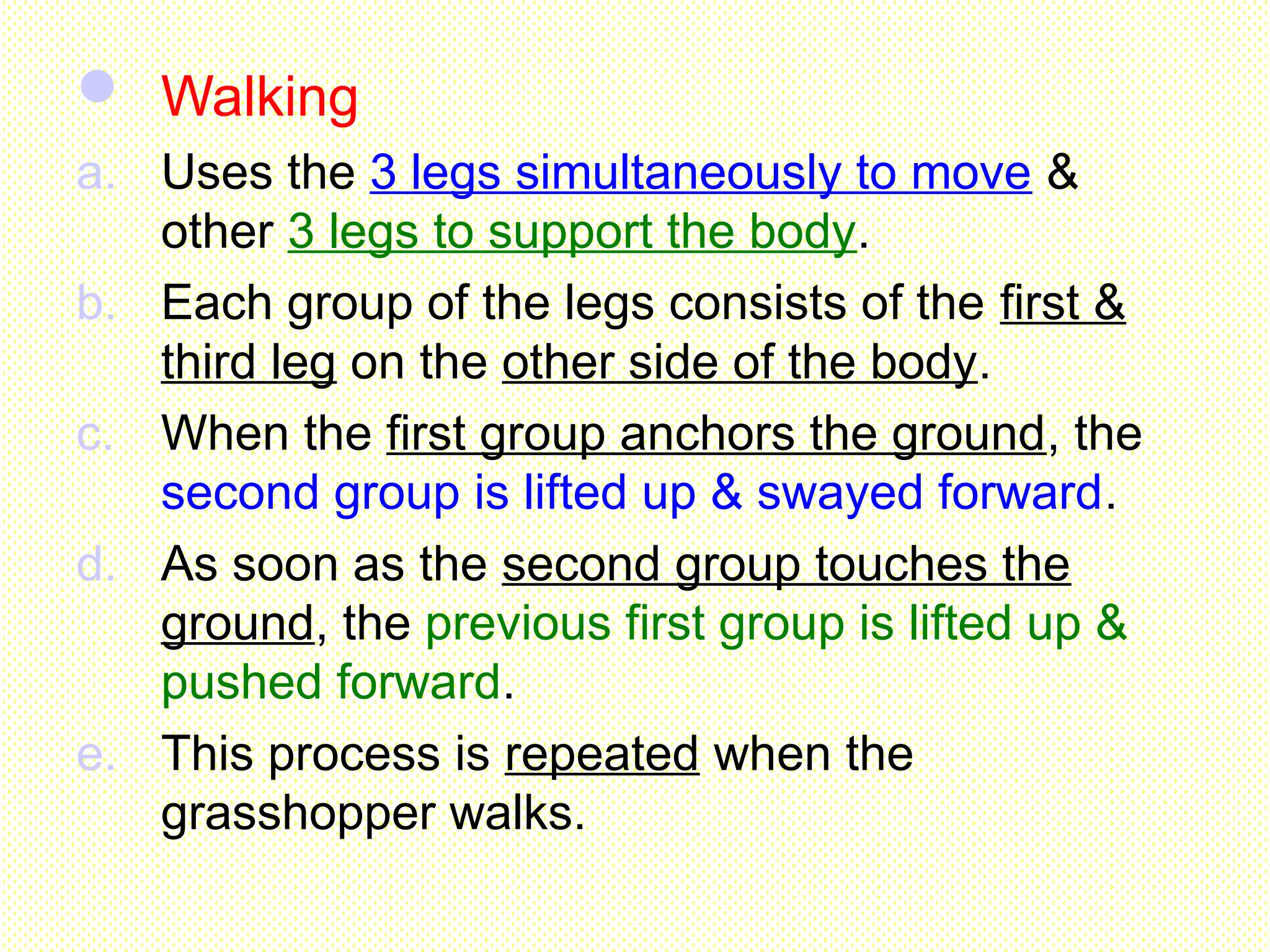
Walking
a. Usesthe 3 legs simultaneously to move &
other 3 legs to support the body.
b. Each group of the legs consists of the first &
third leg on the other side of the body.
c. When the first group anchors the ground, the
second group is lifted up & swayed forward.
d. As soon as the second group touches the
ground, the previous first group is lifted up &
pushed forward.
e. This process is repeated when the
grasshopper walks.
80.
Jumping
a. Tojump, grasshopper extends its hind
legs to the back when the extensor
muscle contracts.
b. The legs pressing against a resistant
surface produces a force which propels
the body forwards & upwards.
c. The movement is due to the antagonistic
contraction of the muscles in the leg to
fold & straighten the legs.
81.
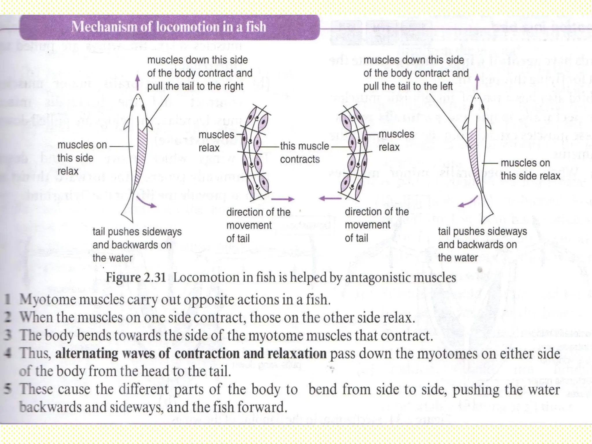
Locomotion in fish
-toreduce water resistance while
swimming-
Fish swims actively in the water has a
streamlined body shape which allows it to move
easily through the water with minimal frictional
drag.
82.
The body ofthe fish is
covered with scales
that overlap one
another with the free
ends pointing
backwards to reduce
the frictional drag in
the water.
Locomotion in fish
-to reduce water resistance while
swimming-
83.
Slimy coatings on
theirbodies to
minimise frictional
drag and maintain a
smooth flow of water
over the body
Locomotion in fish
-to reduce water resistance while
swimming-
Since body tissueis denser than water, fish must compensate for the
difference or they will sink.
Many bony fishes have an internal organ called a swim bladder that adjusts
their buoyancy through manipulation of gases.
The swim bladder is a sac inside the abdomen that contains gas.
The pressure in the bladder can be increased or decreased by gulping or
releasing air through the mouth.
This keep the fish buoyant when stop swimming
88.
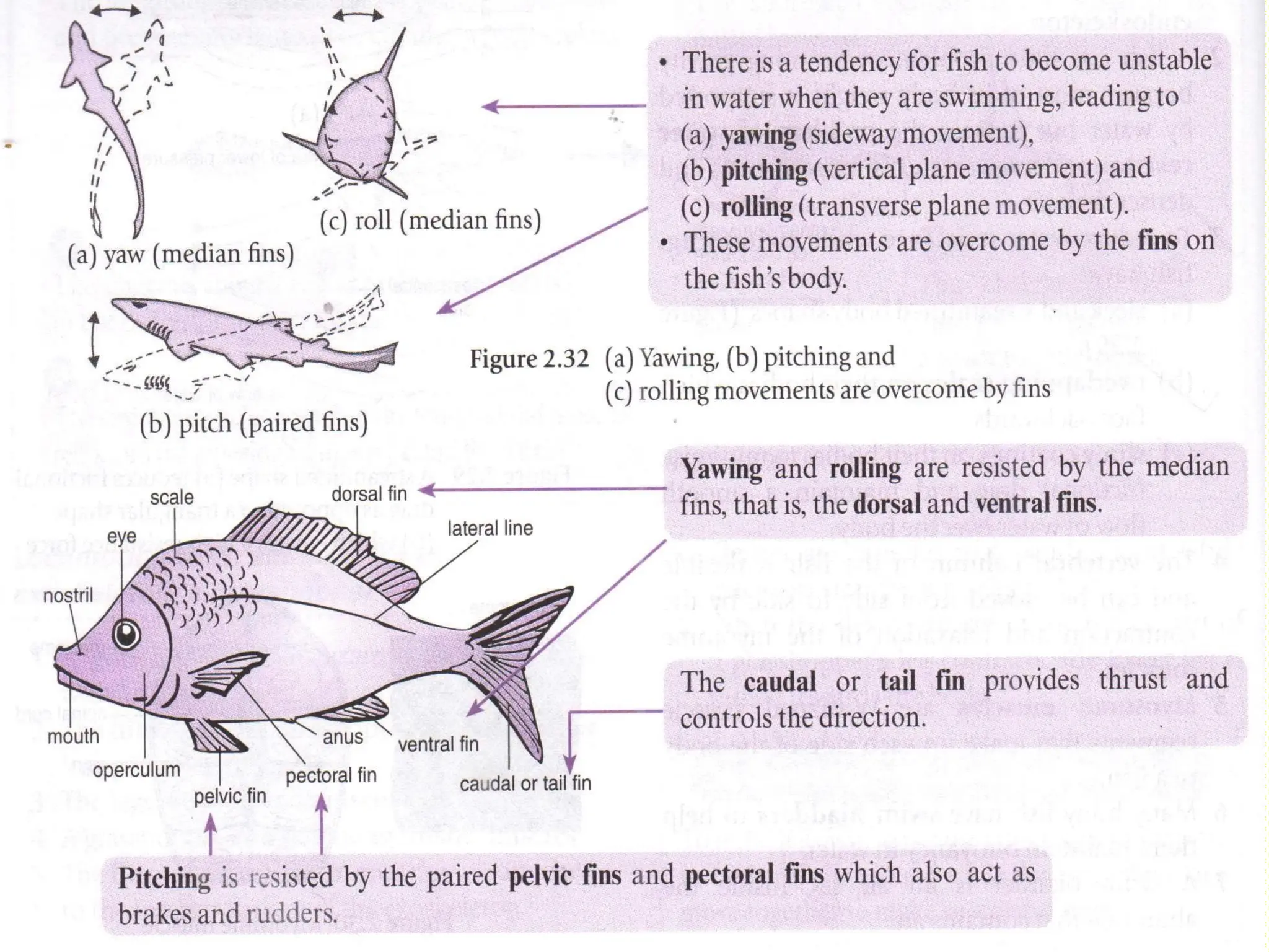
Pectoral fins
Pectoral fins
•Controlthe pitch of the
fish, causing it to swim
upward or downward.
•Help in slowing down
or stopping.
Pelvic fins
Pelvic fins
•Control the
pitch of the fish
•Control rolling
of the fish.
Ventral fins
Ventral fins
•Controls yawning
and rolling of fish.
Dorsal fins
Dorsal fins
•Controls
yawning and
rolling of fish
Caudal fins
Caudal fins
•Increase the
surface area of
the tail, allowing
for an extra
boost in speed.
2.2 Appreciating aHealthy
Musculoskeletal System
Ways to care for the
musculoskeletal system
92.
Following a balanced
diet
Havinga good
posture-sitting, lifting
or carrying
objects,standing
Using proper attire for
daily activities.
Taking appropiate
precautions during
vigorous activities.
Practising the correct
and safe exercise
technique
94.
Do plants needa support?
enable the plants to stay upright
enable the plants to obtain sufficient
sunlight
bear the weight of the plants
provide strength to withstand wind
resistance
Support in plants is provided by the
turgidity of cell, vascular tissue &
buoyancy of water.
95.
Aquatic plants
Dividedinto 2 types:-
a. Submerged water plants: Hydrilla sp.,
Elodea sp., Utricularia sp. (bladderwort)
b. Floating water plants: Nelumbium sp.
(lotus), Eichhornia sp. (water hyacinth)
Obtain support from the buoyancy of
water or upthrust of the water.
Buoyancy of water is greater than the
pull of gravity.
96.
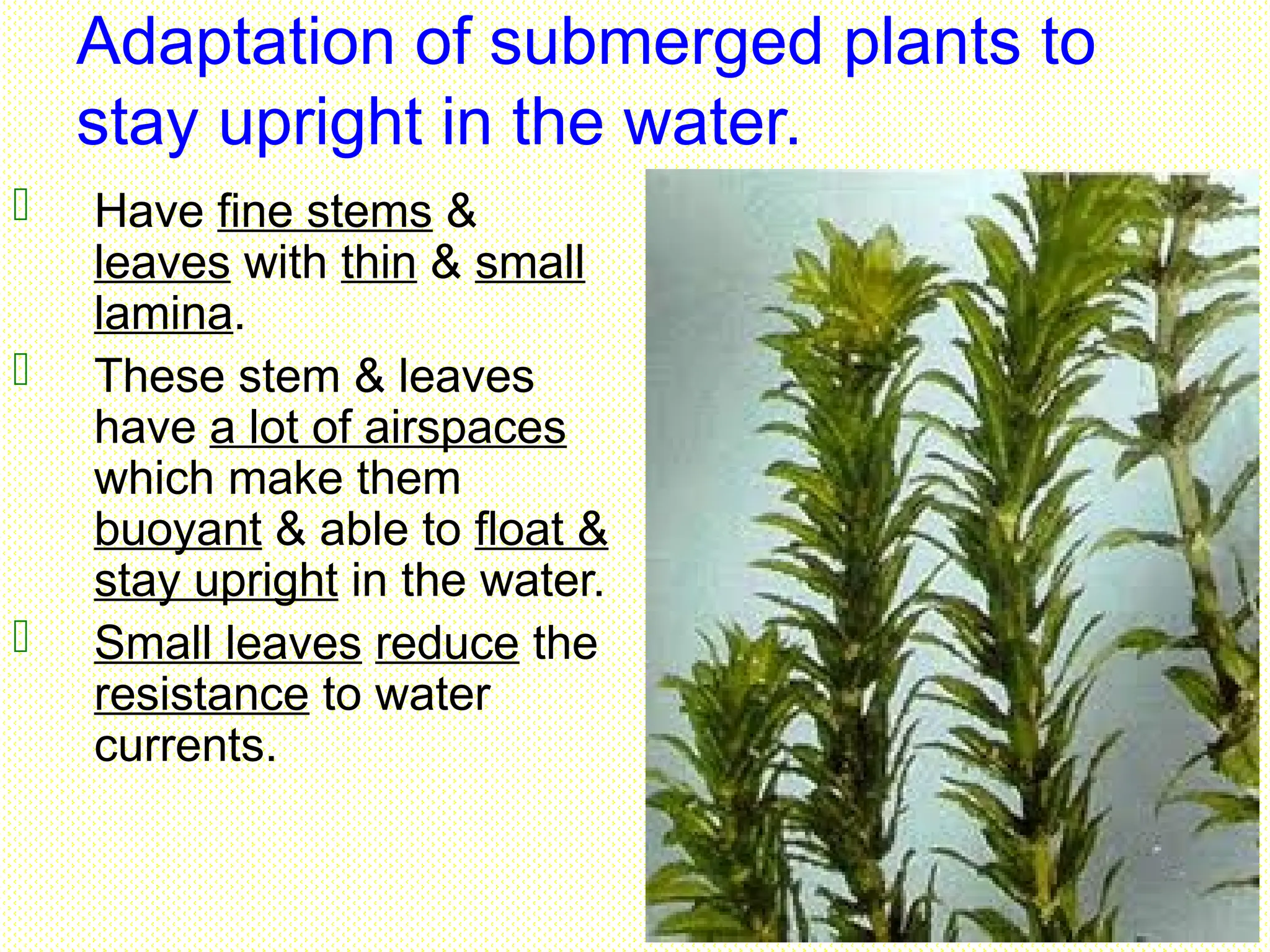
Adaptation of submergedplants to
stay upright in the water.
Have fine stems &
leaves with thin & small
lamina.
These stem & leaves
have a lot of airspaces
which make them
buoyant & able to float &
stay upright in the water.
Small leaves reduce the
resistance to water
currents.
97.
Do nothave roots & woody tissues as the
cuticle of the plant is thin & easily
permeable to water.
These plants can absorb water, mineral,
carbon dioxide & oxygen over its whole
surface.
98.
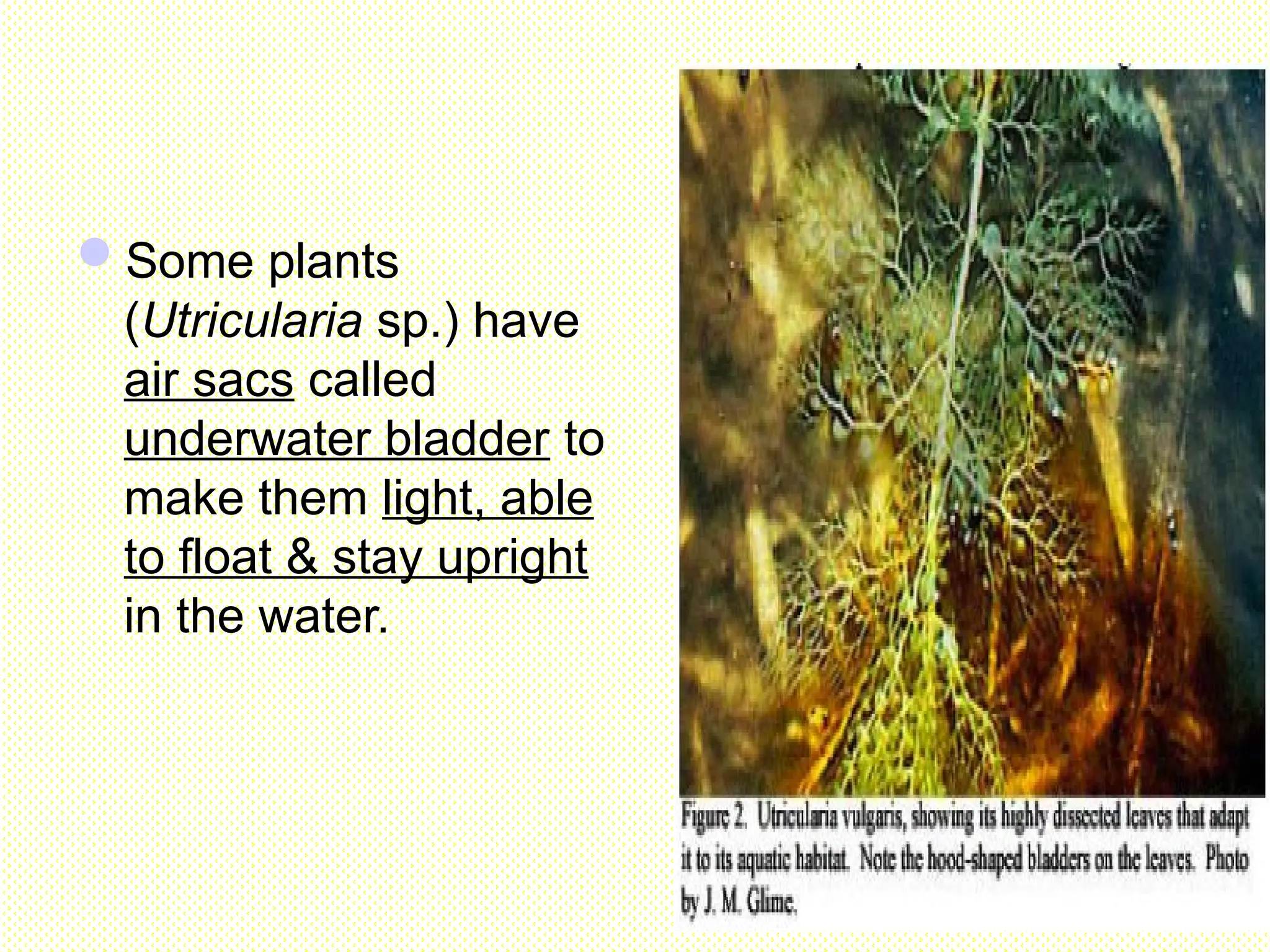
Some plants
(Utricularia sp.)have
air sacs called
underwater bladder to
make them light, able
to float & stay upright
in the water.
99.
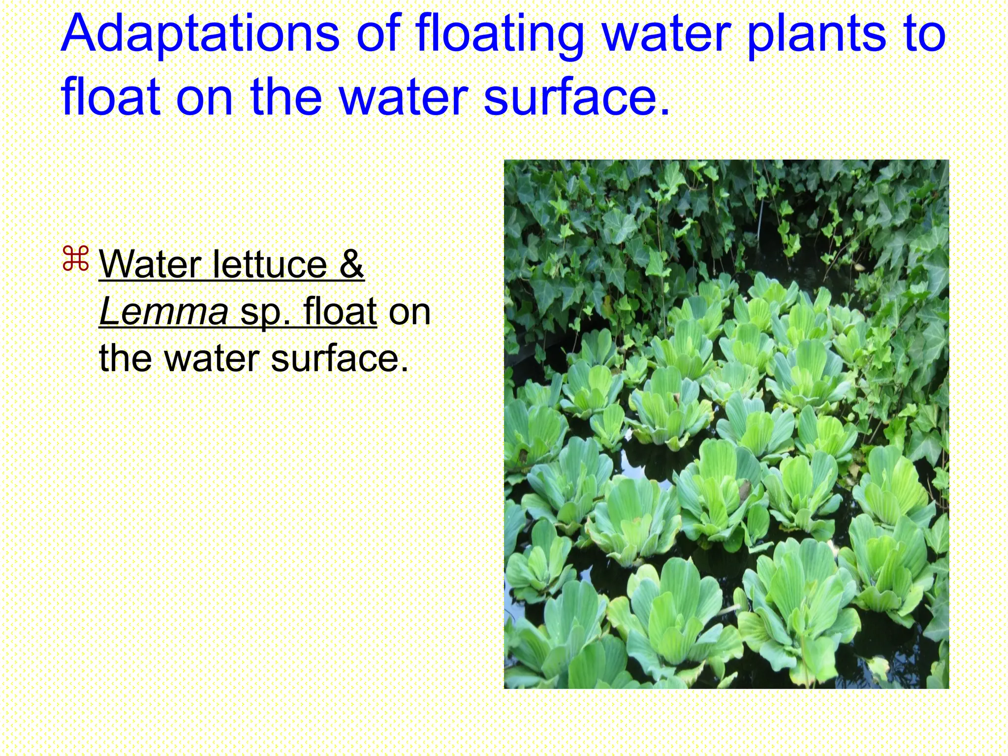
Adaptations of floatingwater plants to
float on the water surface.
Water lettuce &
Lemma sp. float on
the water surface.
100.
Adaptations of floatingwater plants to
float on the water surface.
Water lily has roots
embedded in the mud
at the bottom of the
pond, while the
leaves & floral parts
are floating.
101.
Adaptations of floatingwater plants to
float on the water surface.
Lotus plant & the
water hyacinth have
leaves & floral parts
that stick out of the
water into the air.
Water hyacinth have
stem & enlarged
petiole with many air
sacs to provide more
buoyancy.
102.
The surface ofthe floating leaf is covered
with a waxy cuticle to prevent the stomata
from being blocked by the water.
The stem & leaf have special tissue called
aerenchyma tissue which consists of air sac
cells with thin walls & many air spaces in
between the cells.
The aerenchyma tissue make the plant light.
The air spaces provide buoyancy & enable
the plant to float & facilitate the diffusion of
gaseous.
Numerous fine fibrous roots can trap air
bubbles to allow the plant to float easily.
103.
Terrestrial
plants
Support areprovided by the turgidity of
cells & woody tissues.
Divided into :-
a. Herbaceous plants
b. Terrestrial woody plants
104.
Herbaceous plants
Non-woody plants depend on
the turgidity of the plant cells
for support.
Cells in the stem consists of
parenchyma cells with thin
walls.
These cells absorb water, the
turgor pressure that exist in
the cells causes the cells to
become turgid.
The turgidity causes the stem
to be upright & maintains the
shape of the plants.
105.
Terrestrial woody plants
Supportis achieved through
tissue modifications.
Vascular tissues & plant cells
are modified to have thick walls
strengthened with cellulose or
lignin to provide support.
Woody plants have hard woody
stems.
Large mechanical strength is
required to support these
plants.
106.
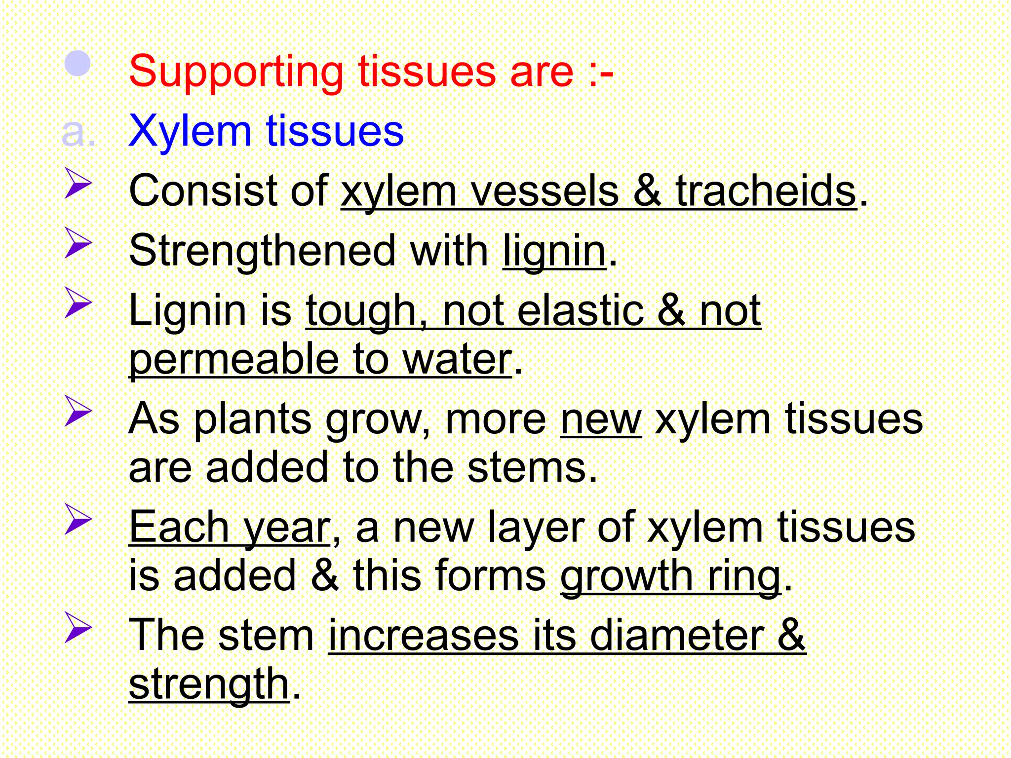
Supporting tissuesare :-
a. Xylem tissues
Consist of xylem vessels & tracheids.
Strengthened with lignin.
Lignin is tough, not elastic & not
permeable to water.
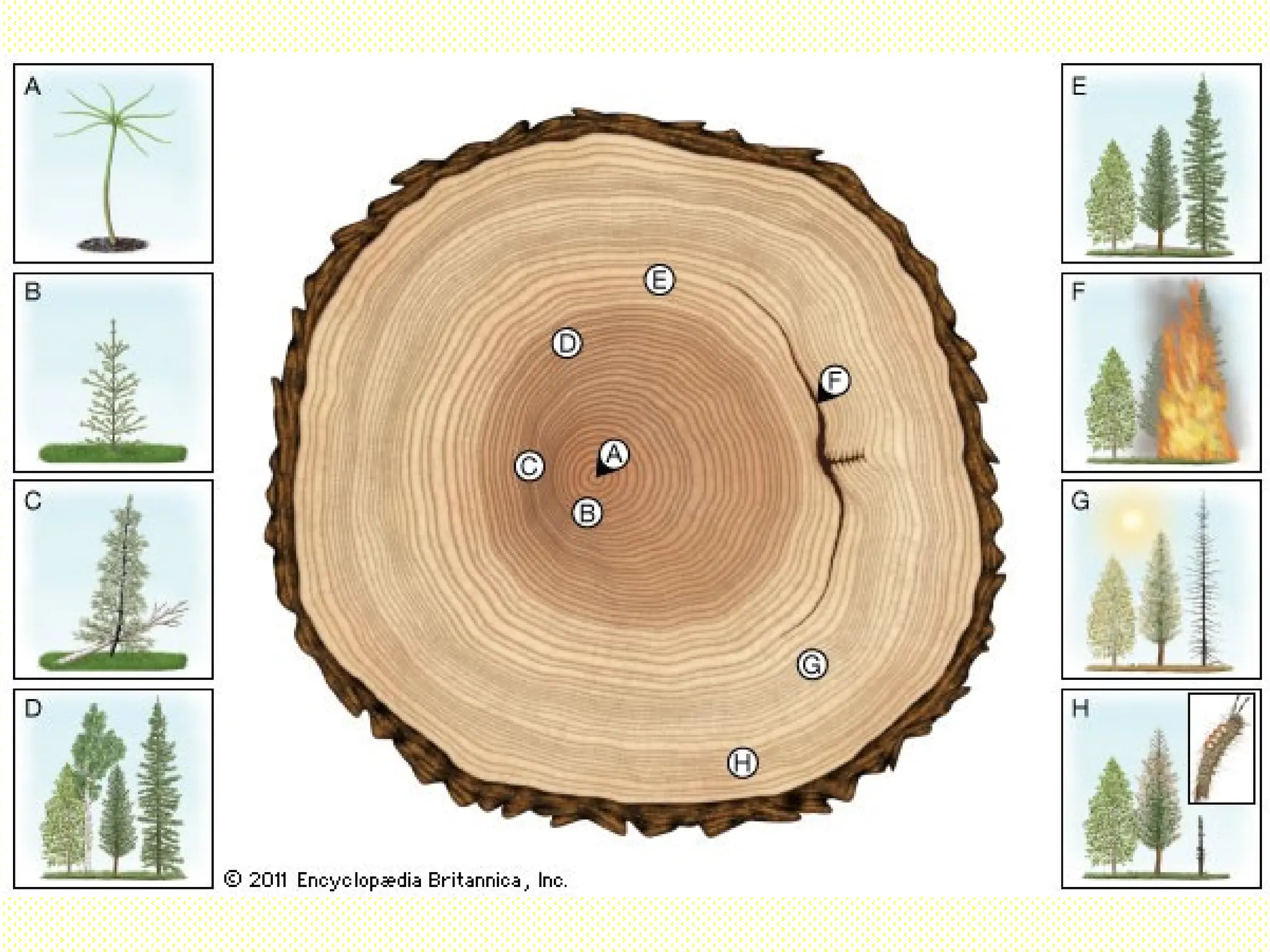
As plants grow, more new xylem tissues
are added to the stems.
Each year, a new layer of xylem tissues
is added & this forms growth ring.
The stem increases its diameter &
strength.
108.
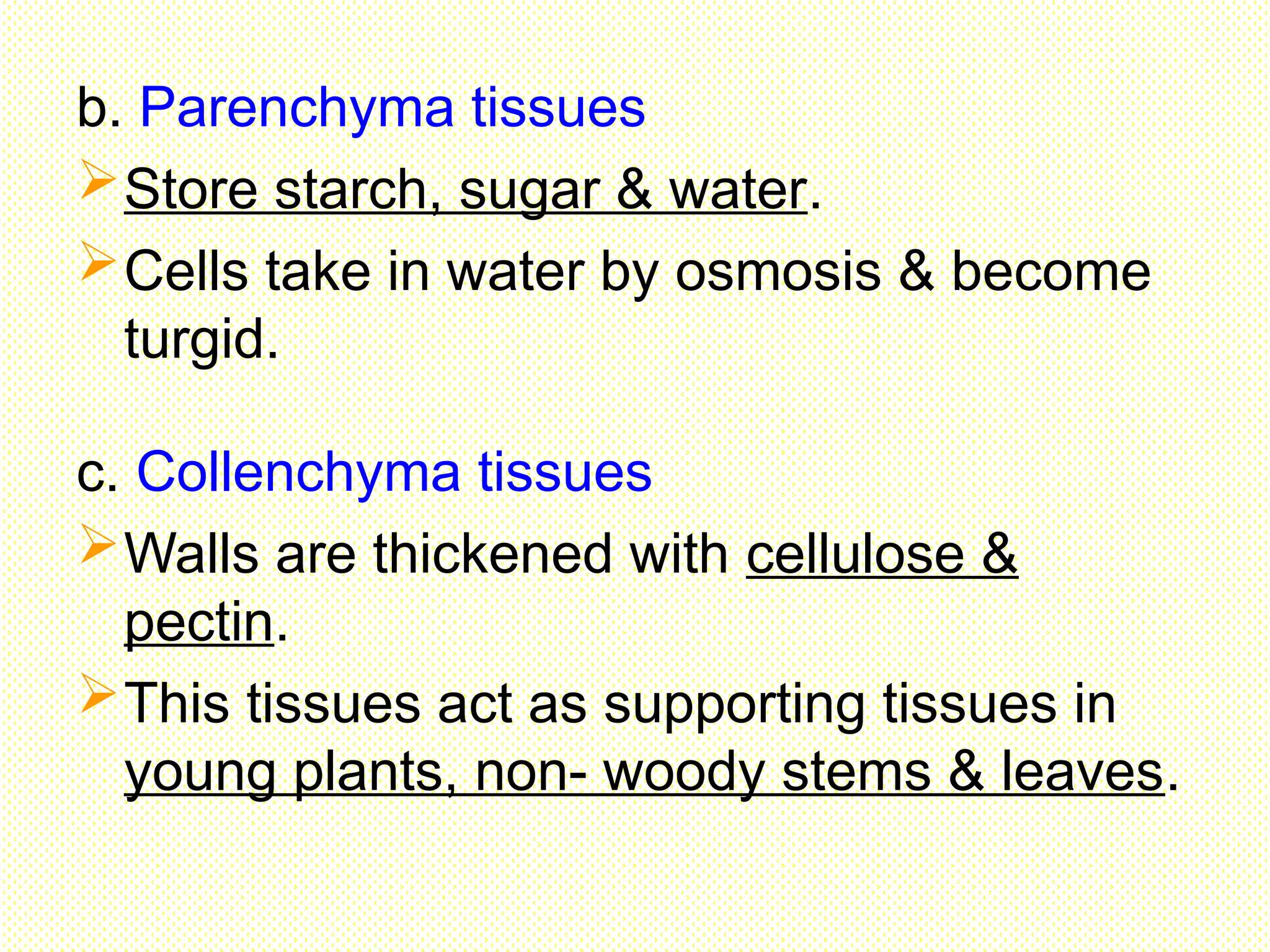
b. Parenchyma tissues
Storestarch, sugar & water.
Cells take in water by osmosis & become
turgid.
c. Collenchyma tissues
Walls are thickened with cellulose &
pectin.
This tissues act as supporting tissues in
young plants, non- woody stems & leaves.
109.
d. Sclerenchyma tissues
Wallsare thickened with lignin to provide
support.
Examples : sugarcane, stems, coconut
leaves, fruits & seeds with hard coverings.