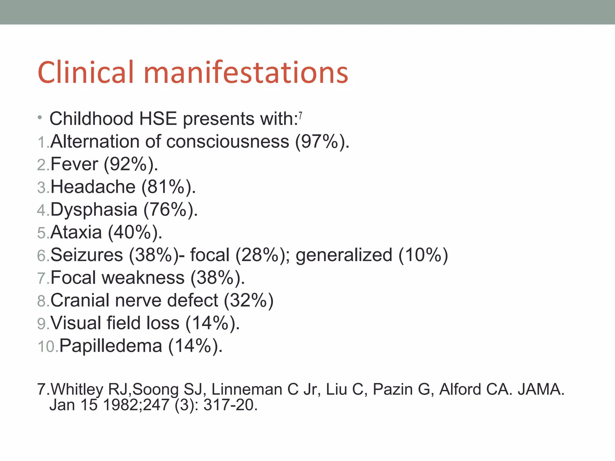
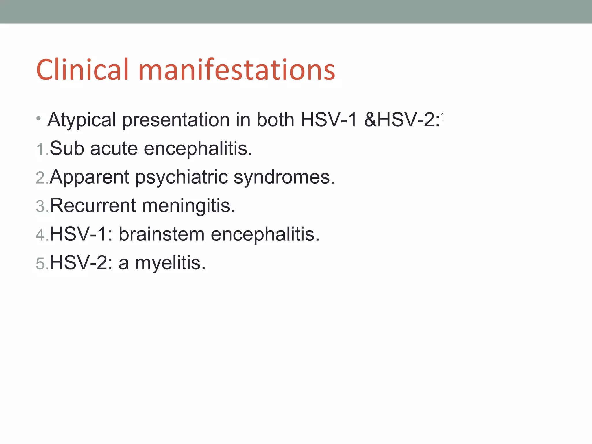
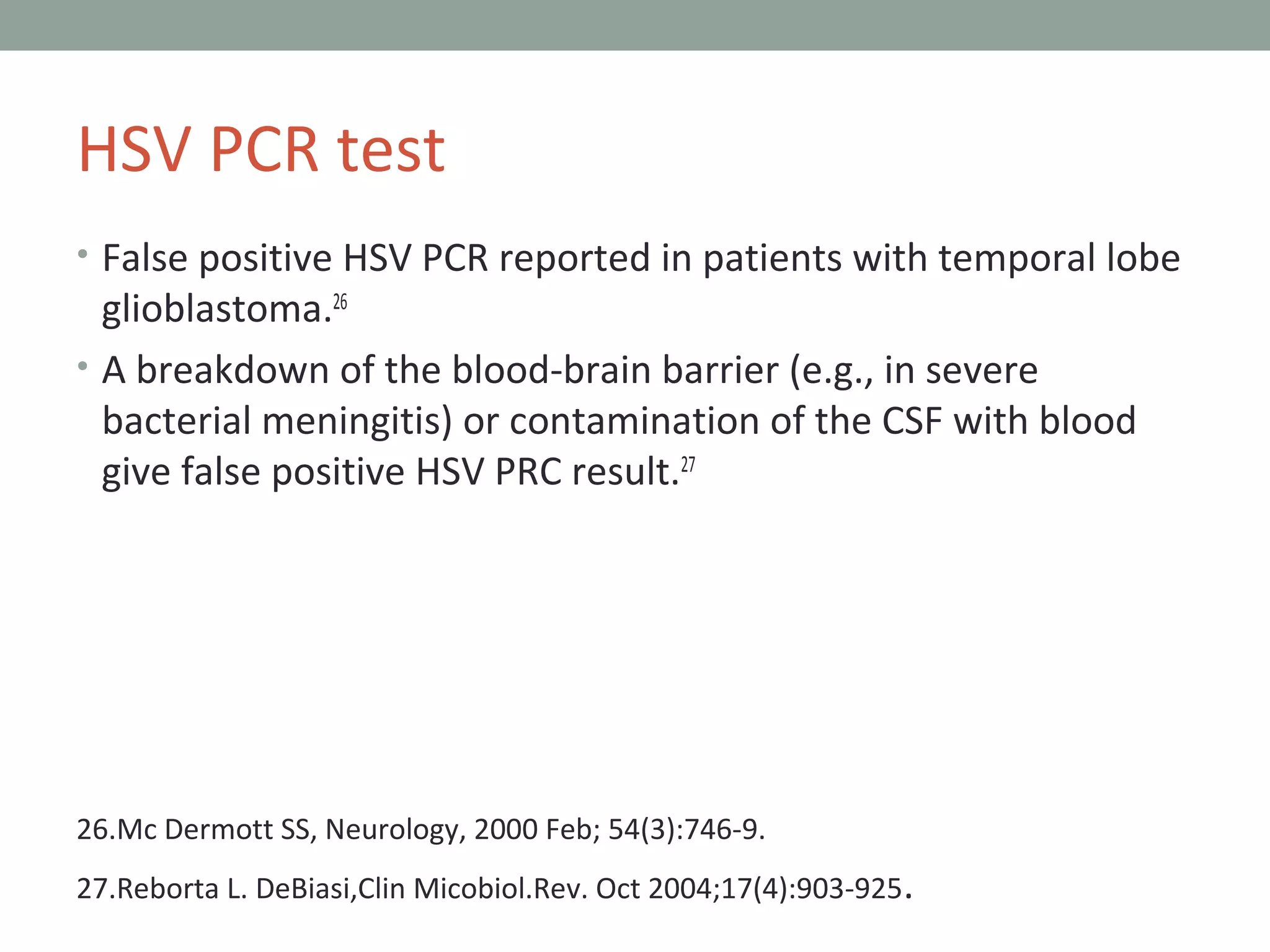
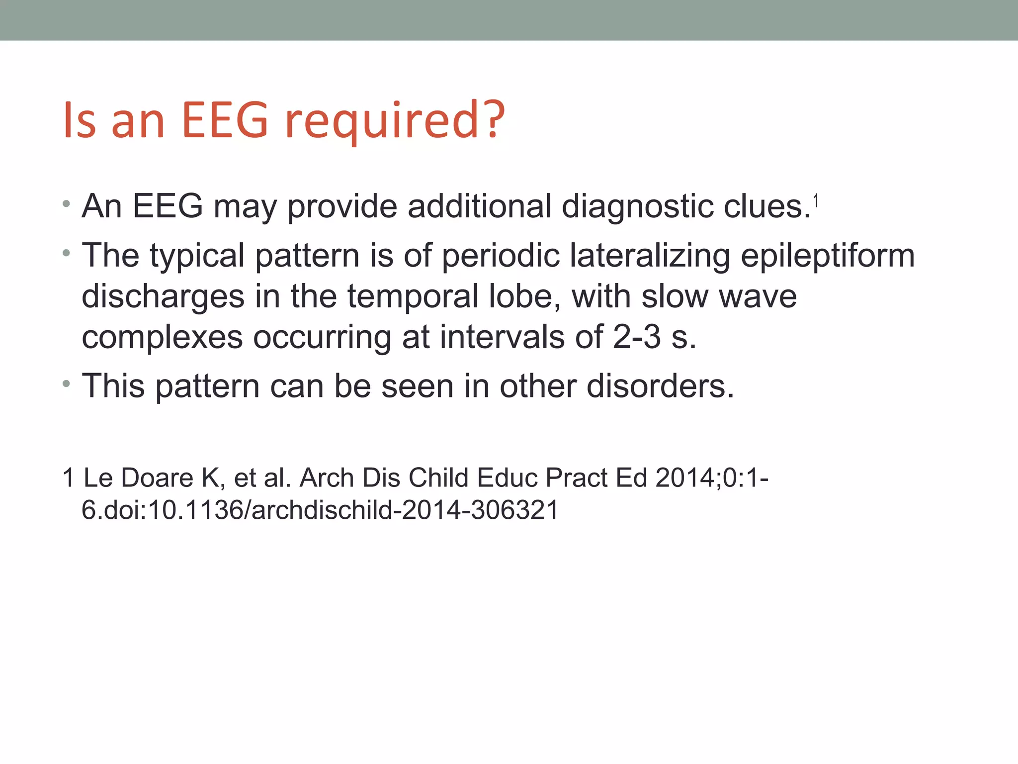
This document provides an overview of herpes simplex encephalitis (HSE). It discusses the etiology, epidemiology, pathophysiology, clinical manifestations, diagnosis and treatment of HSE. Key points include: HSE is most commonly caused by HSV-1 and is a leading cause of viral encephalitis in children; presentation varies from non-specific symptoms to seizures, altered mental status and focal neurological deficits; diagnosis involves CSF analysis, neuroimaging and PCR testing; and treatment involves high-dose intravenous acyclovir administered for at least 21 days. Recurrence of HSE is possible due to viral reactivation or immune-mediated processes.
This presentation briefly summarizes pathophysiology, clinical features, diagnosis and treatment of different types of tuberculosis of brain and spinal cord.
IoT platform as a regular server software. Types of platforms and its' communicaion with devices. Data normalization, storage, processing and visualization. IoT Platform Enterprise Integration. AggreGate Platform.
herpes simplex virus is a double stranded DNA virus causing many symptoms all over the body. it affects globally all over the world .
neonatal hsv attacks even the baby and made them to a fatal conditions.
Bacteriological profile of childhood sepsis at a tertiary health centre in so...QUESTJOURNAL
Introduction: Sepsis is a leading cause of morbidity and mortality in children worldwide, even more so in developing countries. Knowledge of common pathogens and their antibiotic susceptibility pattern is useful for guiding initial treatment while awaiting blood culture results. Objective:To determine the major causative organisms and their antibiotic sensitivity pattern of childhood sepsis at the Niger Delta University TeachingHospital (NDUTH), with the aim of revising existing treatment protocols. Methods: Within a 2 year period (1st January 2014 to 31st December 2015) blood culture results of children with clinical suspicion of sepsis were retrospectively studied. Results:During the study period, 116 (12.11%) of the 958 children admitted into the Children Emergency Ward had blood culture tests. Thirty one (26.72%) had positive blood cultures.Eighteen (58.06%) of the organisms were gram positive while thirteen (41.93%) were gram negative. The predominant organism was Staphylococcus aureus in 16 (51.61%) followed by Klebsiella pneumoniae in 5 (16.13%) patients. The bacterial isolates demonstrated the highest sensitivity to the quinolones. Conclusion:There is need for periodic surveillance of the causative organisms and antibiotic susceptibility pattern of childhood sepsis to guide effective management of patients.
Childhood demyelinating syndromes
In the past decade, the number of studies related to demyelinating diseases in children has exponentially increased. Demyelinating disease in children may be monophasic or chronic. Typical monophasic disorders in children are acute disseminated encephalomyelitis and clinically isolated syndromes, including optic neuritis and transverse myelitis. However, some cases of acute disseminated encephalomyelitis or clinically isolated syndrome progress to become chronic disorders, including multiple sclerosis and neuromyelitis optica. This review summarizes the current knowledge on monophasic and chronic demyelinating disorders in children, focusing on an approach to diagnosis and management.
Nipah Virus (NiV) is a negative sense, single stranded, enveloped RNA virus.
Zoonotic virus
Family – Paramyxoviridae
Genus - Henipavirus
It is a BSL-4 pathogen.
The name "Nipah" refers to the place, Sungai Nipah (literally 'nipah river') in Malaysia, the source of the human case from which Nipah virus was first isolated.Nipah virus can remain viable for a few days in few fruit juices or mango fruits, and at least 7 days in palm milk.
Bats act as a breeding ground for many dangerous viruses, including Nipah, rabies, and Marburg viruses. Such viruses are not associated with any major pathological changes within the bat population.
Transmission of NiV occurs by eating contaminated food. Risks include contact, touch, breastfeeding, or exposure to an infected person, thereby making it easier to come in contact with a droplet of NiV infection.
More recently, experimental studies with aerosolized NiV in Syrian hamsters have found that NiV droplets (aerosol distribution) may cause NiV transmission during close contact. Drinking fresh palm milk is a very common method, and the use of Tari (ripe palm juice) is a powerful way to transmit the virus.
Tom Selleck Health: A Comprehensive Look at the Iconic Actor’s Wellness Journeygreendigital
Tom Selleck, an enduring figure in Hollywood. has captivated audiences for decades with his rugged charm, iconic moustache. and memorable roles in television and film. From his breakout role as Thomas Magnum in Magnum P.I. to his current portrayal of Frank Reagan in Blue Bloods. Selleck's career has spanned over 50 years. But beyond his professional achievements. fans have often been curious about Tom Selleck Health. especially as he has aged in the public eye.
Follow us on: Pinterest
Introduction
Many have been interested in Tom Selleck health. not only because of his enduring presence on screen but also because of the challenges. and lifestyle choices he has faced and made over the years. This article delves into the various aspects of Tom Selleck health. exploring his fitness regimen, diet, mental health. and the challenges he has encountered as he ages. We'll look at how he maintains his well-being. the health issues he has faced, and his approach to ageing .
Early Life and Career
Childhood and Athletic Beginnings
Tom Selleck was born on January 29, 1945, in Detroit, Michigan, and grew up in Sherman Oaks, California. From an early age, he was involved in sports, particularly basketball. which played a significant role in his physical development. His athletic pursuits continued into college. where he attended the University of Southern California (USC) on a basketball scholarship. This early involvement in sports laid a strong foundation for his physical health and disciplined lifestyle.
Transition to Acting
Selleck's transition from an athlete to an actor came with its physical demands. His first significant role in "Magnum P.I." required him to perform various stunts and maintain a fit appearance. This role, which he played from 1980 to 1988. necessitated a rigorous fitness routine to meet the show's demands. setting the stage for his long-term commitment to health and wellness.
Fitness Regimen
Workout Routine
Tom Selleck health and fitness regimen has evolved. adapting to his changing roles and age. During his "Magnum, P.I." days. Selleck's workouts were intense and focused on building and maintaining muscle mass. His routine included weightlifting, cardiovascular exercises. and specific training for the stunts he performed on the show.
Selleck adjusted his fitness routine as he aged to suit his body's needs. Today, his workouts focus on maintaining flexibility, strength, and cardiovascular health. He incorporates low-impact exercises such as swimming, walking, and light weightlifting. This balanced approach helps him stay fit without putting undue strain on his joints and muscles.
Importance of Flexibility and Mobility
In recent years, Selleck has emphasized the importance of flexibility and mobility in his fitness regimen. Understanding the natural decline in muscle mass and joint flexibility with age. he includes stretching and yoga in his routine. These practices help prevent injuries, improve posture, and maintain mobilit
Ethanol (CH3CH2OH), or beverage alcohol, is a two-carbon alcohol
that is rapidly distributed in the body and brain. Ethanol alters many
neurochemical systems and has rewarding and addictive properties. It
is the oldest recreational drug and likely contributes to more morbidity,
mortality, and public health costs than all illicit drugs combined. The
5th edition of the Diagnostic and Statistical Manual of Mental Disorders
(DSM-5) integrates alcohol abuse and alcohol dependence into a single
disorder called alcohol use disorder (AUD), with mild, moderate,
and severe subclassifications (American Psychiatric Association, 2013).
In the DSM-5, all types of substance abuse and dependence have been
combined into a single substance use disorder (SUD) on a continuum
from mild to severe. A diagnosis of AUD requires that at least two of
the 11 DSM-5 behaviors be present within a 12-month period (mild
AUD: 2–3 criteria; moderate AUD: 4–5 criteria; severe AUD: 6–11 criteria).
The four main behavioral effects of AUD are impaired control over
drinking, negative social consequences, risky use, and altered physiological
effects (tolerance, withdrawal). This chapter presents an overview
of the prevalence and harmful consequences of AUD in the U.S.,
the systemic nature of the disease, neurocircuitry and stages of AUD,
comorbidities, fetal alcohol spectrum disorders, genetic risk factors, and
pharmacotherapies for AUD.
These lecture slides, by Dr Sidra Arshad, offer a quick overview of physiological basis of a normal electrocardiogram.
Learning objectives:
1. Define an electrocardiogram (ECG) and electrocardiography
2. Describe how dipoles generated by the heart produce the waveforms of the ECG
3. Describe the components of a normal electrocardiogram of a typical bipolar leads (limb II)
4. Differentiate between intervals and segments
5. Enlist some common indications for obtaining an ECG
Study Resources:
1. Chapter 11, Guyton and Hall Textbook of Medical Physiology, 14th edition
2. Chapter 9, Human Physiology - From Cells to Systems, Lauralee Sherwood, 9th edition
3. Chapter 29, Ganong’s Review of Medical Physiology, 26th edition
4. Electrocardiogram, StatPearls - https://www.ncbi.nlm.nih.gov/books/NBK549803/
5. ECG in Medical Practice by ABM Abdullah, 4th edition
6. ECG Basics, http://www.nataliescasebook.com/tag/e-c-g-basics
Title: Sense of Taste
Presenter: Dr. Faiza, Assistant Professor of Physiology
Qualifications:
MBBS (Best Graduate, AIMC Lahore)
FCPS Physiology
ICMT, CHPE, DHPE (STMU)
MPH (GC University, Faisalabad)
MBA (Virtual University of Pakistan)
Learning Objectives:
Describe the structure and function of taste buds.
Describe the relationship between the taste threshold and taste index of common substances.
Explain the chemical basis and signal transduction of taste perception for each type of primary taste sensation.
Recognize different abnormalities of taste perception and their causes.
Key Topics:
Significance of Taste Sensation:
Differentiation between pleasant and harmful food
Influence on behavior
Selection of food based on metabolic needs
Receptors of Taste:
Taste buds on the tongue
Influence of sense of smell, texture of food, and pain stimulation (e.g., by pepper)
Primary and Secondary Taste Sensations:
Primary taste sensations: Sweet, Sour, Salty, Bitter, Umami
Chemical basis and signal transduction mechanisms for each taste
Taste Threshold and Index:
Taste threshold values for Sweet (sucrose), Salty (NaCl), Sour (HCl), and Bitter (Quinine)
Taste index relationship: Inversely proportional to taste threshold
Taste Blindness:
Inability to taste certain substances, particularly thiourea compounds
Example: Phenylthiocarbamide
Structure and Function of Taste Buds:
Composition: Epithelial cells, Sustentacular/Supporting cells, Taste cells, Basal cells
Features: Taste pores, Taste hairs/microvilli, and Taste nerve fibers
Location of Taste Buds:
Found in papillae of the tongue (Fungiform, Circumvallate, Foliate)
Also present on the palate, tonsillar pillars, epiglottis, and proximal esophagus
Mechanism of Taste Stimulation:
Interaction of taste substances with receptors on microvilli
Signal transduction pathways for Umami, Sweet, Bitter, Sour, and Salty tastes
Taste Sensitivity and Adaptation:
Decrease in sensitivity with age
Rapid adaptation of taste sensation
Role of Saliva in Taste:
Dissolution of tastants to reach receptors
Washing away the stimulus
Taste Preferences and Aversions:
Mechanisms behind taste preference and aversion
Influence of receptors and neural pathways
Impact of Sensory Nerve Damage:
Degeneration of taste buds if the sensory nerve fiber is cut
Abnormalities of Taste Detection:
Conditions: Ageusia, Hypogeusia, Dysgeusia (parageusia)
Causes: Nerve damage, neurological disorders, infections, poor oral hygiene, adverse drug effects, deficiencies, aging, tobacco use, altered neurotransmitter levels
Neurotransmitters and Taste Threshold:
Effects of serotonin (5-HT) and norepinephrine (NE) on taste sensitivity
Supertasters:
25% of the population with heightened sensitivity to taste, especially bitterness
Increased number of fungiform papillae
These simplified slides by Dr. Sidra Arshad present an overview of the non-respiratory functions of the respiratory tract.
Learning objectives:
1. Enlist the non-respiratory functions of the respiratory tract
2. Briefly explain how these functions are carried out
3. Discuss the significance of dead space
4. Differentiate between minute ventilation and alveolar ventilation
5. Describe the cough and sneeze reflexes
Study Resources:
1. Chapter 39, Guyton and Hall Textbook of Medical Physiology, 14th edition
2. Chapter 34, Ganong’s Review of Medical Physiology, 26th edition
3. Chapter 17, Human Physiology by Lauralee Sherwood, 9th edition
4. Non-respiratory functions of the lungs https://academic.oup.com/bjaed/article/13/3/98/278874
Couples presenting to the infertility clinic- Do they really have infertility...Sujoy Dasgupta
Dr Sujoy Dasgupta presented the study on "Couples presenting to the infertility clinic- Do they really have infertility? – The unexplored stories of non-consummation" in the 13th Congress of the Asia Pacific Initiative on Reproduction (ASPIRE 2024) at Manila on 24 May, 2024.
Factory Supply Best Quality Pmk Oil CAS 28578–16–7 PMK Powder in Stockrebeccabio
Factory Supply Best Quality Pmk Oil CAS 28578–16–7 PMK Powder in Stock
Telegram: bmksupplier
signal: +85264872720
threema: TUD4A6YC
You can contact me on Telegram or Threema
Communicate promptly and reply
Free of customs clearance, Double Clearance 100% pass delivery to USA, Canada, Spain, Germany, Netherland, Poland, Italy, Sweden, UK, Czech Republic, Australia, Mexico, Russia, Ukraine, Kazakhstan.Door to door service
Hot Selling Organic intermediates
Title: Sense of Smell
Presenter: Dr. Faiza, Assistant Professor of Physiology
Qualifications:
MBBS (Best Graduate, AIMC Lahore)
FCPS Physiology
ICMT, CHPE, DHPE (STMU)
MPH (GC University, Faisalabad)
MBA (Virtual University of Pakistan)
Learning Objectives:
Describe the primary categories of smells and the concept of odor blindness.
Explain the structure and location of the olfactory membrane and mucosa, including the types and roles of cells involved in olfaction.
Describe the pathway and mechanisms of olfactory signal transmission from the olfactory receptors to the brain.
Illustrate the biochemical cascade triggered by odorant binding to olfactory receptors, including the role of G-proteins and second messengers in generating an action potential.
Identify different types of olfactory disorders such as anosmia, hyposmia, hyperosmia, and dysosmia, including their potential causes.
Key Topics:
Olfactory Genes:
3% of the human genome accounts for olfactory genes.
400 genes for odorant receptors.
Olfactory Membrane:
Located in the superior part of the nasal cavity.
Medially: Folds downward along the superior septum.
Laterally: Folds over the superior turbinate and upper surface of the middle turbinate.
Total surface area: 5-10 square centimeters.
Olfactory Mucosa:
Olfactory Cells: Bipolar nerve cells derived from the CNS (100 million), with 4-25 olfactory cilia per cell.
Sustentacular Cells: Produce mucus and maintain ionic and molecular environment.
Basal Cells: Replace worn-out olfactory cells with an average lifespan of 1-2 months.
Bowman’s Gland: Secretes mucus.
Stimulation of Olfactory Cells:
Odorant dissolves in mucus and attaches to receptors on olfactory cilia.
Involves a cascade effect through G-proteins and second messengers, leading to depolarization and action potential generation in the olfactory nerve.
Quality of a Good Odorant:
Small (3-20 Carbon atoms), volatile, water-soluble, and lipid-soluble.
Facilitated by odorant-binding proteins in mucus.
Membrane Potential and Action Potential:
Resting membrane potential: -55mV.
Action potential frequency in the olfactory nerve increases with odorant strength.
Adaptation Towards the Sense of Smell:
Rapid adaptation within the first second, with further slow adaptation.
Psychological adaptation greater than receptor adaptation, involving feedback inhibition from the central nervous system.
Primary Sensations of Smell:
Camphoraceous, Musky, Floral, Pepperminty, Ethereal, Pungent, Putrid.
Odor Detection Threshold:
Examples: Hydrogen sulfide (0.0005 ppm), Methyl-mercaptan (0.002 ppm).
Some toxic substances are odorless at lethal concentrations.
Characteristics of Smell:
Odor blindness for single substances due to lack of appropriate receptor protein.
Behavioral and emotional influences of smell.
Transmission of Olfactory Signals:
From olfactory cells to glomeruli in the olfactory bulb, involving lateral inhibition.
Primitive, less old, and new olfactory systems with different path
- Video recording of this lecture in English language: https://youtu.be/lK81BzxMqdo
- Video recording of this lecture in Arabic language: https://youtu.be/Ve4P0COk9OI
- Link to download the book free: https://nephrotube.blogspot.com/p/nephrotube-nephrology-books.html
- Link to NephroTube website: www.NephroTube.com
- Link to NephroTube social media accounts: https://nephrotube.blogspot.com/p/join-nephrotube-on-social-media.html
3. Herpes Simplex Encephalitis
• Herpes simplex encephalitis is the most common
single cause of viral encephalitis in infants and
children.1
• It is important due to its frequency, high mortality
reaching 20% of patients and its long term
morbidity.2
1.Le Doare K, et al, Child Educ Pract Ed August 2014,BMJ.
2.Javier Riancho,Neurol Sci 2013.
4. Etiology
• Herpes simplex viruses are members of herpes virus
family.3
• Genome consist of a single double stranded DNA
molecule.
• Type 1 virus associated with orofacial herpes infections.
• Type 2 virus identified in genital herpes and causes
most of the congenital or perinatally acquired infections.
3.John H.Menkes, child neurology, 7th
edition, chapter 7.
5. Epidemiology
• Neonatal disease is more common than childhood
disease.
• The incidence of severe disease in infants is 1 in 64 000
infants per year.
• In children over 1 year, HSE is nearly four times less
common, occurring in 1 in 230 000 children per year.4
4.Ward KN,Ohrling A, Bryant NJ, et al. Arch Dis Child 2012;97:162-5.
6. Pathophysiology
• The pathogenesis of HSE in humans is poorly
understood.
• The exact mechanism of cellular damage is unclear, but it
may involve both direct virus-mediated and indirect
immune-mediated processes.28
28.http://pmj.bmj.com/ on January 12, 2015.
7. Pathophysiology
• The ability of HSV-1 to induce apoptosis in neuronal cells,
a property not shared by HSV-2, might explain why the
former causes virtually all cases of herpes simplex
encephalitis in immunocompetent older children and
adults.
• Brain infection is thought to occur by means of direct
neuronal transmission of the virus from a peripheral site to
the brain via the trigeminal or olfactory nerve.
8.
9. What are the presenting features of
neonatal and childhood HSV
encephalitis?
• Neonatal HSV disease presents in the first 4 weeks of
life.1
• Almost always acquired by perinatal exposure to HSV.
• Illness symptoms often begin between the first 7-21
days of life.
1 Le Doare K, et al. Arch Dis Child Educ Pract Ed 2014;0:1-
6.doi:10.1136/archdischild-2014-306321
10. What are the presenting features of
neonatal and childhood HSV encephalitis?
• Neonatal HSV infection categorized as:5
1.Skin,eyes and mouth disease43%.
2.Disseminated disease 23%.
3.Encephalitis 34%
5 Kenneth F. Swaiman, Pediatric neurology principles and practice, fifth
edition, chapter 81, p 1271-1273.
11. What are the presenting features of
neonatal and childhood HSV encephalitis?
• Non specific clinical features as poor oral intake,
behavioral changes or fever.
• Focal or generalized seizures, apnea, lethargy or coma.
• Can accompanied by pneumonitis,hepatitis or
disseminated intravascular coagulopathy.
• 60 to 70% have associated skin vesicles.1
1 Le Doare K, et al. Arch Dis Child Educ Pract Ed 2014;0:1-
12. 6-AAP 2005, Herpes Simplex, p 309-318. In L.K. Pickering (ed.) 2005 Red Book.
13. Clinical manifestations
• Childhood HSE presents with:7
1.Alternation of consciousness (97%).
2.Fever (92%).
3.Headache (81%).
4.Dysphasia (76%).
5.Ataxia (40%).
6.Seizures (38%)- focal (28%); generalized (10%)
7.Focal weakness (38%).
8.Cranial nerve defect (32%)
9.Visual field loss (14%).
10.Papilledema (14%).
7.Whitley RJ,Soong SJ, Linneman C Jr, Liu C, Pazin G, Alford CA. JAMA.
Jan 15 1982;247 (3): 317-20.
14. Clinical manifestations
• Atypical presentation in both HSV-1 &HSV-2:1
1.Sub acute encephalitis.
2.Apparent psychiatric syndromes.
3.Recurrent meningitis.
4.HSV-1: brainstem encephalitis.
5.HSV-2: a myelitis.
15. Clinical manifestations
6. Global aphasia for 1 language but retained most of his birth
language ability.8
7. Anterior opercular syndrome.9
8. Lingual epilepsia partialis continua.10
8- Ku A,Lachmann EA, Nagler W. Pediatr Neurol.Sep 1996;15(2):169-71.
9-De Kleermaeker FG, J Child Neurol, 2014 Apr; 29(4):560-3.
10-Iyer RS1, Ramalingam Ramakrishnan TCNeurol India. 2014 Jul-Aug;62(4):439-41.
doi: 10.4103/0028-3886.141230.
16. Clinical manifestations
9. Extrapyramidal symptoms and basal ganglia
involvement.11
10. HSE- associated cerebral hemorrhage in an HIV-
positive person.12
11- Mondal G, Kumar R, Ghosh JK, Basu K, Chatterejee S. India J Pediatr. Jul
2009;76(7): 749-50.
12- Li JZ,Sax , AIDS Read. Apr 2009;19(4): 153-5.
18. Does a child with HSE have to have
external lesions for the diagnosis to be
likely?
• In neonates, skin lesions may be present, it may appear later
in clinical course.13
13-Pediatric in review, volume 17 number 12,Herpes simplex virus infections by: Paula W.
Annunziato, MD and Anne Gershon.
19. Does a child with HSE have to have
external lesions for the diagnosis to be
likely?
• In childhood HSE, only 10% have a prior history of
mucosal symptoms such as cold sores or conjunctivitis.1
1 Le Doare K, et al. Arch Dis Child Educ Pract Ed 2014;0:1-
6.doi:10.1136/archdischild-2014-306321
20. Should a lumbar puncture always be
attempted in HSE?
• CSF is an essential component of the diagnostic work up.
• Patients typically have mononuclear pleocytosis.
• RBC count may be elevated.
• Protein levels are elevated.
• Glucose values may be normal or mildly decreased.
• In about 5-10% of patients, especially children, initial CSF
results may be normal.5
5.Kenneth F. Swaiman, Pediatric neurology principles and practice, fifth
edition,chapter 81, p 1271-1273.
21. Does it make a difference if the PCR is
positive for HSV-1 or for HSV-2?
• No, not really.
• HSV-1 is more common in childhood HSE.1
• HSV-2 tends to be associated with genital herpes and is
more common in neonatal HSE.
1 Le Doare K, et al. Arch Dis Child Educ Pract Ed 2014;0:1-
6.doi:10.1136/archdischild-2014-306321
22. HSV PCR test
• PCR is highly sensitive (94-98%) and specific (98-100%)26
• Results become positive within 24 hours of the onset of
symptoms and remain positive for at least 5-7 days after
the start of antiviral therapy.
26.Mc Dermott SS, Neurology, 2000 Feb;54(3):746-9.
23. HSV PCR test
• Negative CSF HSV PCR tests can occur within first 72 h of
illness.26
• Patients with HSV encephalitis will typically have a negative
CSF HSV PCR after 14 days of acyclovir treatment.
26.Mc Dermott SS, Neurology, 2000 Feb;54(3):746-9.
24. HSV PCR test
• False positive HSV PCR reported in patients with temporal lobe
glioblastoma.26
• A breakdown of the blood-brain barrier (e.g., in severe
bacterial meningitis) or contamination of the CSF with blood
give false positive HSV PRC result.27
26.Mc Dermott SS, Neurology, 2000 Feb; 54(3):746-9.
27.Reborta L. DeBiasi,Clin Micobiol.Rev. Oct 2004;17(4):903-925.
25. What is the best neuroimaging
modality?
• CT scan with contrast.
• MRI is better than CT for the diagnosis of encephalitis.
26. Neuroimaging
• In neonate:5
• MRI and CT usually reveal diffuse edema during the acute
stage and later exhibit atrophy, parynchymal calcification
or cystic encephalomalacia.
• MRI can reveal a watershed distribution of abnormalities.
5- Kenneth F. Swaiman, Pediatric neurology princeples and practice, fifth
edition,chapter 81, p 1271-1273.
27. • A 2 weeks old baby with HSV-2 encephalitis.
14-http://www.hawaii.edu/medicine/pediatrics/pemxray/v7c09.html
28.
29.
30.
31. 24.Okanishi T et al. Diffusion-weighted MRI for early dignosis of neonatal herpes simplex
encephalitis. Brain Dev (2014).
32. Neuroimaging
• In children:5
• Edema and hemorrhages may be present. After 1 week,
contrast enhancement may be detectable.
• Unilateral or bilateral abnormalities consisting of T2
prolongation in the temporal lobe or insula .
• Enhancement of the insula, temporal cortex and cingulate
gyrus on T1- weighted, gadolinum –enhanced images.
5- Kenneth F. Swaiman, Pediatric neurology princeples and practice, fifth
edition,chapter 81, p 1271-1273.
35. Is an EEG required?
• An EEG may provide additional diagnostic clues.1
• The typical pattern is of periodic lateralizing epileptiform
discharges in the temporal lobe, with slow wave
complexes occurring at intervals of 2-3 s.
• This pattern can be seen in other disorders.
1 Le Doare K, et al. Arch Dis Child Educ Pract Ed 2014;0:1-
6.doi:10.1136/archdischild-2014-306321
37. What therapy is recommended while
waiting for the investigation results?
• Clinical overlap between patients with encephalitis,
meningitis and septic shock.
• Broad spectrum antibiotics and high dose acyclovir until
test results are available.1
1 Le Doare K, et al. Arch Dis Child Educ Pract Ed 2014;0:1-
6.doi:10.1136/archdischild-2014-306321
38. What is adequate treatment?
• High dose intravenous acyclovir is most effective when
started early.
• Neonatal HSE: 60mg/kg/day in three divided doses every
8 h,assuming that renal function is normal.
• Treatment should be 14 days if the disease is limited to
the skin, eyes or mouth and a minimum of 21 days if the
infection involved the CNS or is disseminated.15
15. Upton D Allen,Joan L Robinson; Canadian Paediatric
Society,pediatric child health Vol 19 No 4 April 2014.
39. Treatment
• In 2006, the American Academy of Pediatrics updated its
dosing recommendations for children aged 3 months to
12 years to receive high-dose acyclovir (60 mg/kg/day).
• The incidence of renal injury or failure was similar
between children who received standard- dose and high-
dose acyclovir.16
40. Is acyclovir resistance an issue?
• In immunocompetent patients, viral resistance to acyclovir
occur rarely , the reported prevalence less than 1 %.17
• In immunocompromised patients, raises to 6%.
17-Piret J, Boivin G. Antimicrob agents chemother 2011;55:459-72.
41. If HSE is unlikely, when is it safe to
stop acyclovir?
• Where clinical suspicion of HSE is low, it is usually
appropriate to stop acyclovir if:
1.Negative CSF HSV PCR and cell count (<5cells/mm3)
and.
2.Normal brain MRI and.
3.Normal EEG and.
4.A full and rapid clinical recovery with normal level of
consciousness or
5.An alternative diagnosis becomes apparent.1
1 Le Doare K, et al. Arch Dis Child Educ Pract Ed 2014;0:1-
6.doi:10.1136/archdischild-2014-306321
42. Does a negative LP rule-out HSE?
• Acyclovir should not be stopped if the CSF is negative on
PCR for HSV when other features are consistent with
HSE.
• HSV PCR is a highly sensitive and specific test (94 and
96%), except during the first 24 h of the disease, when
as many as 10% of HSE CSF samples can be falsely
negative.18
18-Lakeman FD, Whitley RJ. National institute of allergy and infectious
diseases collaborative antiviral study group. J infect Dis 1995; 171:857-63.
43. Can neonatal and childhood HSE
recur?
• Children presenting with features suggestive of HSV relapse
need repeat cultures of blood and CSF, including CSF PCR for
HSV and autoantibody quantification.
• After neonatal HSE, HSV can reactivate months to years after
the initial infection.
• Presenteing as dermal flares or an encephalitis as the initial
illness.
44.
45. HSV relapse
•Abnormal movements as chorea, seizures, focal neurological
deficits and psychiatric symptoms.
•May be immune mediated.
•Autoantibodies against N-methyl-D-aspartate receptor
(NMDAR).
46. Genetic factors
• Childhood-onset HSE is due to TLR3 deficiency in a
traceable fraction of patients, in particular the ones with HSE
recurrence.29
• TLR3 deficiency is a ssociated with insufficient virus
control in the brain, leading to incomplete viral latency in the
CNS itself in turn leading to a high rate of HSE recurrence
due to virus reactivation.
29.Hye Kyung Lim,neurology 83 Nov 2014, 1888-1897
47. HSV relapse
• Both neonatal and childhood HSE predispose to
subsequent autoimmune encephalitis with new
neurological symptoms including:
• Aphasia.
• Behavioral changes.
• Choreoathetoid movements.
• Opercular syndrome.19
19 De Tiege X,Rozenberg F,Heron B. Europ J Paediatr Neurol
2008; 12:72-81.
48. HSV recurrence
• Individuals with HSV-2 infection generally have high rates of
recurrence in the first and second years followed by a
substantial decrease in subsequent years.25
25.Kimberlin DW. Semin Pediatr Infect Dis. Oct 2005;16(4):271-81.
49. What about prophylaxis after HSE?
• A small patient series proposed doses of 1340 mg/m2
acyclovir given twice daily (total daily dose 2680 mg/m2),
based on CSF penetrating data.20
• Placebo-controlled RCT validated oral acyclovir given at
300 mg/m2 three times daily (900 mg/m2) as adequate at
improving neurodevelopmental outcome.21
20- Rudd C, Rivadeneira ED, Gutman LT. Acta Pediatrica
1994;83:1237-43.
21-Kimkberlin DW. J Paeditr Infect Dis 2013;2:179-82.
50. Prophlaxis
• Oral acyclovir is recommended for neonates and
valacyclovir for older children requiring long-term
prophylaxis.
1 Le Doare K, et al. Arch Dis Child Educ Pract Ed 2014;0:1-
6.doi:10.1136/archdischild-2014-306321
51.
52. What onward referral is needed?
• If there is family history of invasive HSV disease and/or
consanguinity, children should be referred to an
immunology center.
• Children with HSE will require long-term multidisciplinary
neurodevelopmental follow up and possible psychological
and educational rehabilitation.
• Treatment of long term complications including epilepsy,
spasticity and dystonia.1
1 Le Doare K, et al. Arch Dis Child Educ Pract Ed 2014;0:1-
6.doi:10.1136/archdischild-2014-306321
53. Prognosis
• 70% mortality in untreated HSE patients.23
• Mortality in patients treated with acyclovir 11%.
• Mortality of neonatal HSE, 6% in patients with isolated HSE and
31% in patients with disseminated infection.
23.Whitley RJ, Cobbs CG,Alford CA Jr,Soong SJ, Hirsch MS, Connor JD, et al.
JAMA,Jul 14 1989;262(2):234-9.
54. Prognosis
• Neurologic outcomes in survivors treated with acyclovir
are as follows:23
1.No deficits or mild deficits-38%.
2.Moderate deficits -9%.
3.Severe deficit- 53%.
• Squeal among survivors depend on the patients age and
neurologic status at the time of diagnosis.
23-Whitley RJ, Cobbs CG,Alford CA Jr,Soong SJ, Hirsch MS, Connor JD, et al.
JAMA,Jul 14 1989;262(2):234-9.
55. Take home message
HSE represent medical emergency.
PCR is the golden standard diagnostic tool but
negative result can happen in early illness.
EARLY INITIATION of therapy within 3 days of
illnes improve the outcome with 21 days duration.
CHOREOATHETOTIC movement was reported
as first sign of HSE relapse.
Prophylaxis important in children less than 2
years.
Genetics play a role especially in recurrent
cases.
57. References
1.Le Doare K, et al, managing neonatal and childhood
herpes encephalitis, Child Educ Pract Ed August
2014,BMJ.
2.Javier Riancho,HSV encephalitis,Neurol Sci 2013.
3.John H.Menkes, child neurology, 7th
edition, chapter
7/infections of nervous system.
4.Ward KN,Ohrling A, Bryant NJ, et al. Herpes simplex
serious neurological disease in young children: incidence
and long term outcome. Arch Dis Child 2012;97:162-5.
58. References
5.Kenneth F. Swaiman, Pediatric neurology princeples and
practice, fifth edition,chapter 81, p 1271-1273.
6. AAP 2005, Herpes Simplex, p 309-318. In L.K. Pickering (ed.)
2005 Red Book.
7. Whitley RJ,Soong SJ, Linneman C Jr, Liu C, Pazin G,
Alford CA. Herpes simplex encephalitis. Clinical
assessment. JAMA. Jan 15 1982;247 (3): 317-20.
59. References
8. Ku A,Lachmann EA, Nagler W. Selective language
aphasia from herpes simplex encephalitis. Pediatr
Neurol.Sep 1996;15(2):169-71.
9.McGrath NM,Anderson NE,Hope JK,Croxson MC, Powell
KF. Anterior opercular syndrome,caused by herpes simplex
encephalitis. Neurology. Aug 1997;49(2):494-7.
10.Iyer RS1, Ramalingam Ramakrishnan TCNeurol India.
2014 Jul-Aug;62(4):439-41. doi: 10.4103/0028-
3886.141230.
60. References
11. Mondal G, Kumar R, Ghosh JK, Basu K, Chatterejee S.
Basal ganglia involvement in a child with herpes simplex
encephalitis. India J Pediatr. Jul 2009;76(7): 749-50.
12.Li JZ,Sax PE. HSV-1 encephalitis complicated by
cerebral hemorrhage in an HIV- positive person. AIDS
Read. Apr 2009;19(4): 153-5.
61. References
13.Pediatric in review, volume 17 number 12,Herpes
simplex virus infections by: Paula W. Annunziato, MD and
Anne Gershon.
14.http://www.hawaii.edu/medicine/pediatrics/pemxray/v7c
09.html
15. Upton D Allen,Joan L Robinson; Canadian Paediatric
Society,pediatric child health Vol 19 No 4 April 2014.
16.Jennifer G. et al, standerd dose versus high dose
acyclovir in children treated emprically for
encephalitis,Pediatr Drugs (2014) 16:229-234.
62. References
17.Piret J, Boivin G. Resistance of herpes simplex viruses to
nucleoside analogues: mechanisms, prevalence, and
management . Antimicrob agents chemother 2011;55:459-72.
18.Lakeman FD, Whitley RJ. Diagnosis of herpes simplex
encephalitis: application of polymerase chine reaction to
cerebrospinal fluid from brain-biopsaied patients and correlation
with disease. National institute of allergy and infectious diseases
collaborative antiviral study group. J infect Dis 1995; 171:857-63.
63. References
19. De Tiege X,Rozenberg F,Heron B. The spectrum of
herpes simplex encephalitis in children. Europ J Paediatr
Neurol 2008; 12:72-81.
20.Rudd C, Rivadeneira ED, Gutman LT.Dosing
considerations for oral acyclovir following neonatal herpes
disease. Acta Pediatrica 1994;83:1237-43.
21.Kimkberlin DW. Acyclovir Dosing in the neonatal period
and beyond. J Paeditr Infect Dis 2013;2:179-82.
64. References
22.Royal College of Obstetricians and Gynaecologists.
Management of genital herpes in pregnancy. 2007.
23.Whitley RJ, Cobbs CG,Alford CA Jr,Soong SJ, Hirsch
MS, Connor JD, et al. HSV encephalitis, diagnosis,
presentation and outcome.JAMA,Jul 14 1989;262(2):234-9.
24.Okanishi T et al. Diffusion-weighted MRI for early
dignosis of neonatal herpes simplex encephalitis. Brain Dev
(2014).
25.Kimberlin DW. Semin Pediatr Infect Dis. Oct
2005;16(4):271-81.
26.Mc Dermott SS, Neurology, 2000 Feb; 54(3):746-9.
27.Reborta L. DeBiasi,Clin Micobiol.Rev. Oct
2004;17(4):903-925.
Patients may have a prodrome of malaise, fever, headache, and nausea, followed by acute or subacute onset of an encephalopathy whose symptoms include lethargy, confusion, and delirium. The following are typically the most common symptoms of HSE[24] :
Cortical pseudobulbar
palsy (i.e. spastic anarthria and inability to swallow) with dissociation of automatic voluntary movements
in the affected muscles are the essential features of this syndrome
A- T2WI: asymmitrical hyperintense lesions n thalami.
B&C: asymetric extensive lesion with decrease ADC in thalamus, gray n white matter in both hemispheres.
CT without contrast show hypodensity in thalami and temporal lobes, indicative of cortical and subcortical edema.
Post contrast
TOL3: a member of Toll like receptor family which plays role in pathogen recognition and activation of innate system.