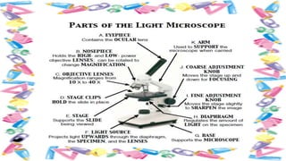
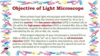
HOW TO USE LIGHT
MICROSCOPE
The microscope is a tool which can help you see
tiny objects and living organisms. It makes them look
bigger. This ability of the microscope is called its
magnifying power or magnification. The microscope
also has the capacity to distinguish small gaps between
two separate points which humans cannot distinguish.
It is called its resolving power or resolution.
The term light refers to
the method by which light
transmits the image to the eye.
The light microscope uses
diffused light from the sun or
artificial light to illuminate the
object to be observed. From its
source, visible light passes
through the small or thin
specimen to be observed
through the glass lenses. As light
passes through the lenses, it is
bent so specimen appears
bigger when it is projected to
the eye. The form and structure
of the specimen can then be
seen because some of their
parts reflect light
Objective of Light Microscope
Most schools have light microscopes with three objectives.
Others have four. Usually, the shortest one marked 3x, 4x or 5x is
called the scanner. The low power objective (LPO) is marked 10x or
12x while the high power objective (HPO) is marked 40x, 43x or 60x.
The objectives magnify the object to be observed to a certain size as
indicated by the 3x, 10x or 40x, etc. marks.
If the longest objective of your microscope is marked 97x or
100x or OIO or the word “oil” on it, then it has an oil immersion
objective (OIO). This objective is used to view bacteria, very small
protists and fungi. The OIO requires the use of a special oil such as
quality cedarwood oil or cargille’s immersion oil.
Activity 1
Objectives
After performing this activity, you should be able to:
1. Handle the microscope properly;
2. Identify the parts of the microscope;
3. Describe what parts of the microscope can do;
4. Prepare materials for microscope study;
5. Focus the microscope properly;
6. Compare the image of the object seen by the unaided eye and under
the microscope; and
7. Compute for the magnification of objects observed under the
microscope.
Field of View
The bright circle of light
Specimen
a part or sample of any material e.g. plant, animal,
paper or mineral, for study or examination under the
microscope. Specimens should be small and thin for light
to pass through them.
Parfocal
Current microscope models. This means the image
in clear focus under the low power objective, remains
focused after shifting to HPO. If the microscope you are
using is not parfocal, slightly turn the fine adjustment
knob in either direction to get a clear picture.
HOW TO USE LIGHT MICROSCOPE.pptx

HOW TO USE LIGHT MICROSCOPE.pptx

  • 1.
    HOW TO USELIGHT MICROSCOPE
  • 3.
    The microscope isa tool which can help you see tiny objects and living organisms. It makes them look bigger. This ability of the microscope is called its magnifying power or magnification. The microscope also has the capacity to distinguish small gaps between two separate points which humans cannot distinguish. It is called its resolving power or resolution.
  • 5.
    The term lightrefers to the method by which light transmits the image to the eye. The light microscope uses diffused light from the sun or artificial light to illuminate the object to be observed. From its source, visible light passes through the small or thin specimen to be observed through the glass lenses. As light passes through the lenses, it is bent so specimen appears bigger when it is projected to the eye. The form and structure of the specimen can then be seen because some of their parts reflect light
  • 7.
    Objective of LightMicroscope Most schools have light microscopes with three objectives. Others have four. Usually, the shortest one marked 3x, 4x or 5x is called the scanner. The low power objective (LPO) is marked 10x or 12x while the high power objective (HPO) is marked 40x, 43x or 60x. The objectives magnify the object to be observed to a certain size as indicated by the 3x, 10x or 40x, etc. marks. If the longest objective of your microscope is marked 97x or 100x or OIO or the word “oil” on it, then it has an oil immersion objective (OIO). This objective is used to view bacteria, very small protists and fungi. The OIO requires the use of a special oil such as quality cedarwood oil or cargille’s immersion oil.
  • 8.
    Activity 1 Objectives After performingthis activity, you should be able to: 1. Handle the microscope properly; 2. Identify the parts of the microscope; 3. Describe what parts of the microscope can do; 4. Prepare materials for microscope study; 5. Focus the microscope properly; 6. Compare the image of the object seen by the unaided eye and under the microscope; and 7. Compute for the magnification of objects observed under the microscope.
  • 9.
    Field of View Thebright circle of light Specimen a part or sample of any material e.g. plant, animal, paper or mineral, for study or examination under the microscope. Specimens should be small and thin for light to pass through them. Parfocal Current microscope models. This means the image in clear focus under the low power objective, remains focused after shifting to HPO. If the microscope you are using is not parfocal, slightly turn the fine adjustment knob in either direction to get a clear picture.