Download to read offline
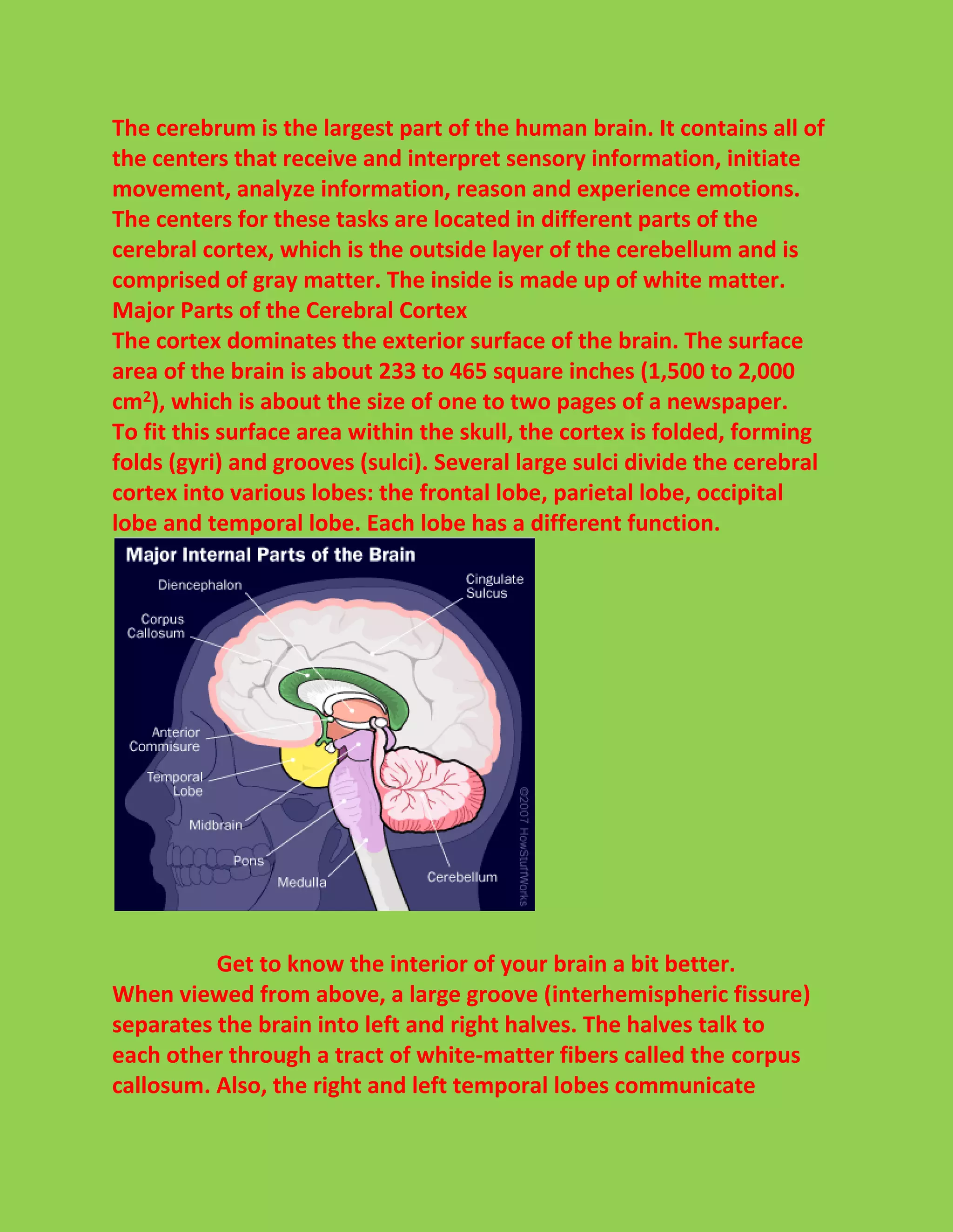
![Here we're looking at the underside of the brain, showing the brain
stem and cranial nerves.
Brains for Instinct
Lower animals, such as fish, amphibians, reptiles and birds, don't do
much "thinking," but instead concern themselves with the everyday
business of gathering food, eating, drinking, sleeping, reproducing and
defending themselves.
These are instinctual processes [source:National Geographic].
Therefore, their brains are organized along the major centers that
control these functions.
We humans perform these functions as well, and so have a "reptilian"
brain built into us. That means we have the same parts of the brain
found in reptiles, namely the brain stem and the cerebellum.
Ready to learn about the lower brain? We'll discuss that on the next
page.](https://image.slidesharecdn.com/howtobrainlearn-140928153312-phpapp01/75/How-to-brain-learn-7-2048.jpg)
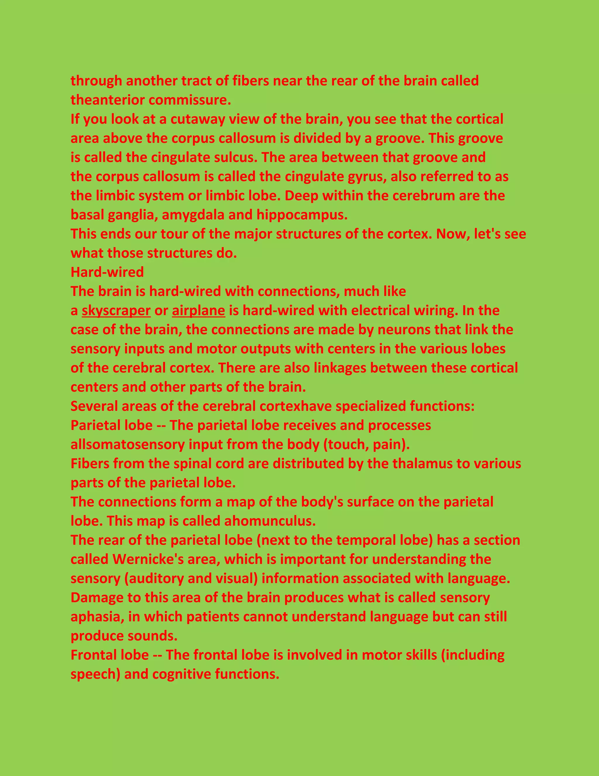
![Lower Brain
The basic lower brain consists of thespinal cord, brain
stem anddiencephalon (the cerebellum and cortex are also present,
but will be discussed in later sections). In turn, the brain stem
comprises the medulla, pons, midbrain, hypothalamus and thalamus
[source: Health Pages].
Within each of these structures are centers of neuronal cell bodies,
callednuclei, which are specialized for particular functions
(breathing, heart-rate regulation, sleep):
Medulla -- The medulla contains nuclei for regulating blood pressure
and breathing, as well as nuclei for relaying information from the
sense organs that comes in from the cranial nerves. It's also the most
ancient part of the brain.
Pons -- The pons contains nuclei that relay movement and position
information from the cerebellum to the cortex. It also contains
nuclei that are involved in breathing, taste and sleep, and physically
connects medulla to the midbrain.
Midbrain -- The midbrain contains nuclei that link the various sections
of the brain involved in motor functions (cerebellum, basal ganglia,](https://image.slidesharecdn.com/howtobrainlearn-140928153312-phpapp01/75/How-to-brain-learn-8-2048.jpg)


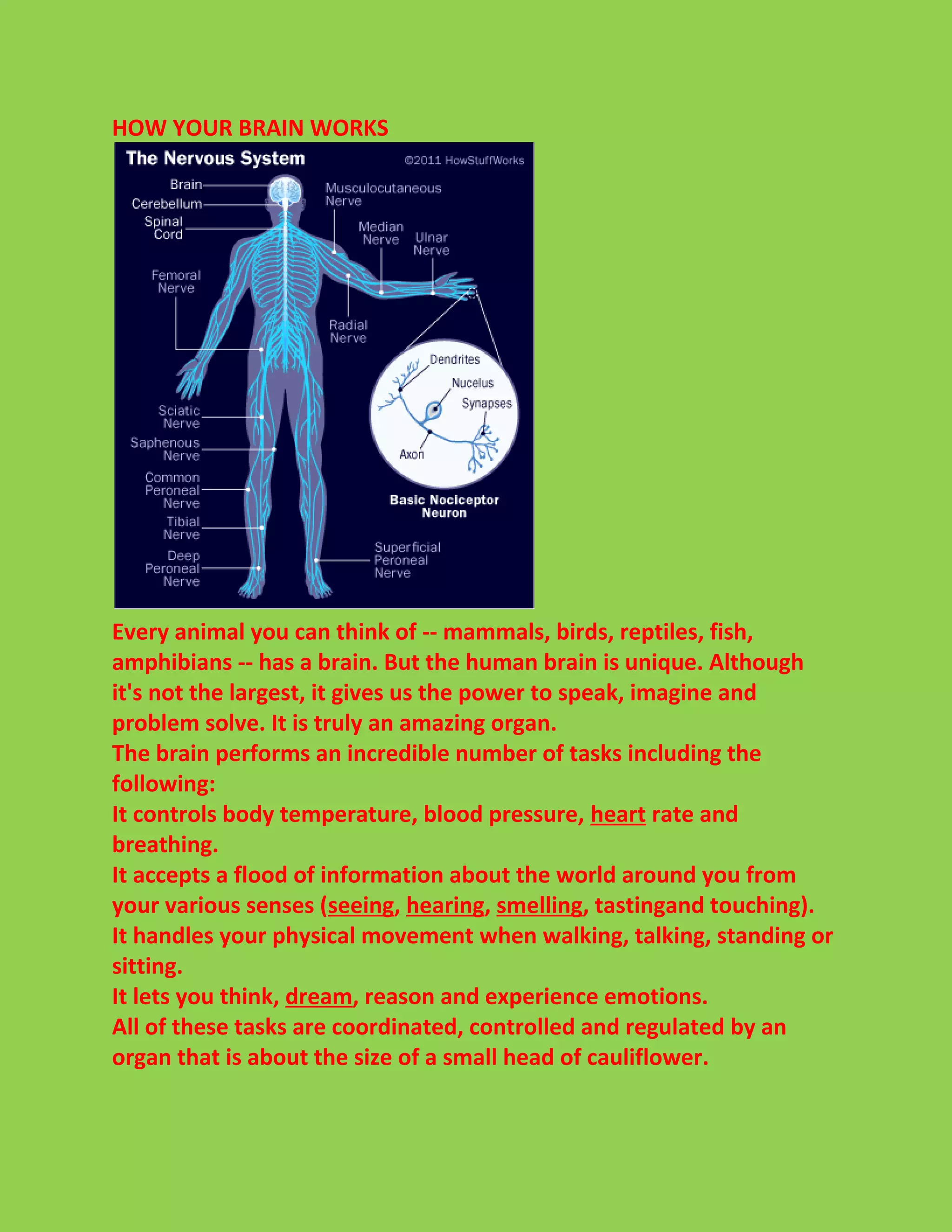
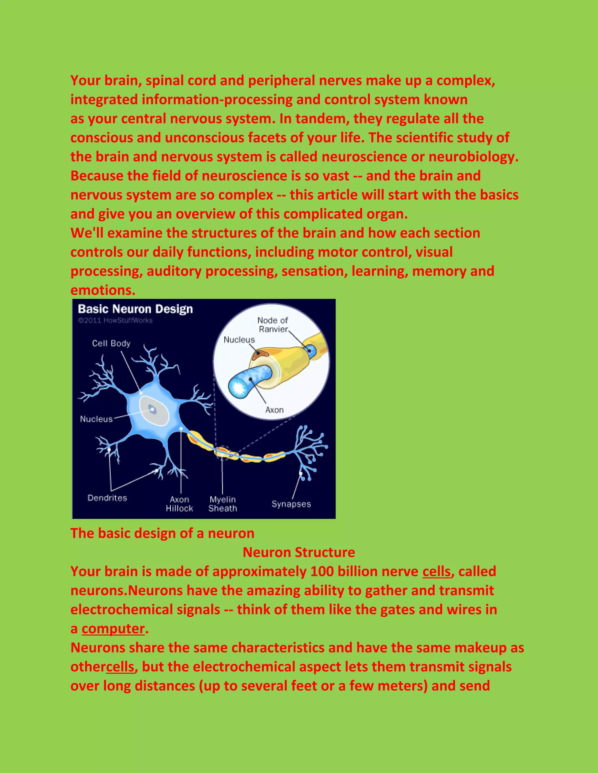
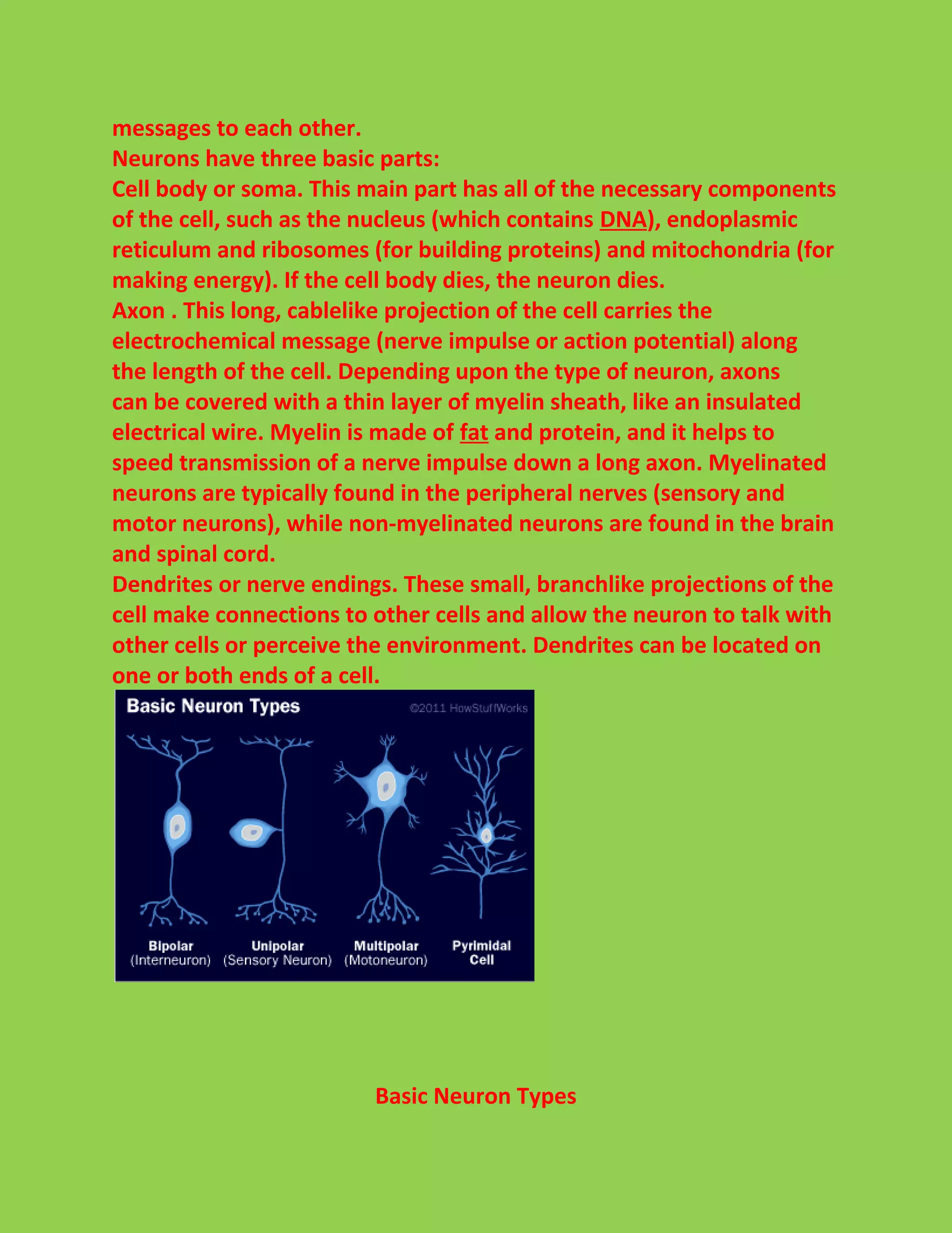
The document provides an overview of how the human brain works. It describes that the brain is made up of around 100 billion neurons that communicate via electrochemical signals. The brain is divided into sections that each control different functions - the lower brain controls basic instincts while the higher brain is involved in thinking. The cerebral cortex is folded and divided into four lobes (frontal, parietal, occipital, temporal) that process sensory information, initiate movement, analyze information, and experience emotions.