A brain isan organ that serves as the center of the nervous
system in all vertebrate and most invertebrate animals. It is located in the
head, usually close to the sensory organs for senses such as vision. It is the
most complex organ in a vertebrate's body. In a human, the cerebral cortex
contains approximately 14–16 billion neurons, and the estimated number of
neurons in the cerebellum is 55–70 billion. Each neuron is connected by
synapses to several thousand other neurons. These neurons communicate
with one another by means of long protoplasmic fibers called axons, which
carry trains of signal pulses called action potentials to distant parts of the
brain or body targeting specific recipient cells.
Physiologically, brains exert centralized control over a body's other organs.
They act on the rest of the body both by generating patterns of muscle
activity and by driving the secretion of chemicals called hormones. This
centralized control allows rapid and coordinated responses to changes in the
environment. Some basic types of responsiveness such as reflexes can be
mediated by the spinal cord or peripheral ganglia, but sophisticated
purposeful control of behavior based on complex sensory input requires the
information integrating capabilities of a centralized brain.
3.
The operations ofindividual brain cells are now understood in
considerable detail but the way they cooperate in ensembles of millions is
yet to be solved. Recent models in modern neuroscience treat the brain as
a biological computer, very different in mechanism from an electronic
computer, but similar in the sense that it acquires information from the
surrounding world, stores it, and processes it in a variety of ways.
Cellular structure:
The brains of all species are composed primarily of two broad classes of
cells: neurons and glial cells. Glial cells (also known as glia or neuroglia)
come in several types, and perform a number of critical functions,
including structural support, metabolic support, insulation, and guidance of
development. Neurons, however, are usually considered the most
important cells in the brain. The property that makes neurons unique is
their ability to send signals to specific target cells over long distances.
4.
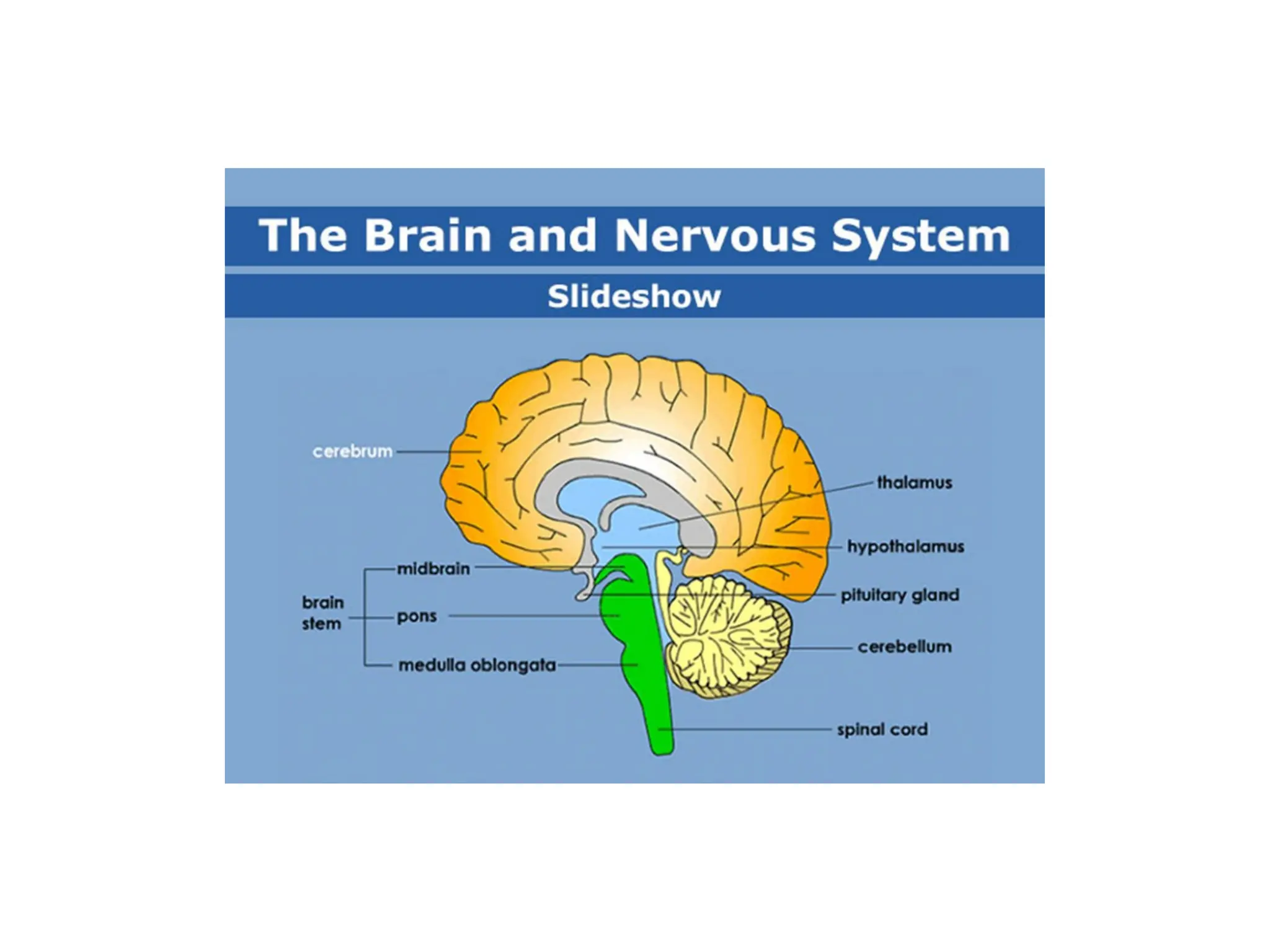
Neuroanatomists usually dividethe vertebrate brain into six main
regions: the telencephalon (cerebral hemispheres), diencephalon
(thalamus and hypothalamus), mesencephalon (midbrain), cerebellum,
pons, and medulla oblongata. Each of these areas has a complex
internal structure. Some parts, such as the cerebral cortex and the
cerebellar cortex, consist of layers that are folded or convoluted to fit
within the available space. Other parts, such as the thalamus and
hypothalamus, consist of clusters of many small nuclei. Thousands of
distinguishable areas can be identified within the vertebrate brain
based on fine distinctions of neural structure, chemistry, and
connectivity.
5.
Brainstem:
The brainstem consistsof the medulla, the pons and the midbrain. The medulla can be
referred to as an extension of the spinal cord, which both have similar organization and
functional properties. The tracts passing from the spinal cord to the brain pass through
here.
Regulatory functions of the medulla nuclei include control of blood pressure and
breathing. Other nuclei are involved in balance, taste, hearing, and control of muscles of
the face and neck.
The next structure rostral to the medulla is the pons, which lies on the ventral anterior
side of the brainstem. Nuclei in the pons include pontine nuclei which work with the
cerebellum and transmit information between the cerebellum and the cerebral cortex. In
the dorsal posterior pons lie nuclei that are involved in the functions of breathing, sleep,
and taste.
The midbrain, or mesencephalon, is situated above and rostral to the pons. It includes
nuclei linking distinct parts of the motor system, including the cerebellum, the basal
ganglia and both cerebral hemispheres, among others. Additionally, parts of the visual
and auditory systems are located in the midbrain, including control of automatic eye
movements.
6.
The brainstem atlarge provides entry and exit to the brain for a number of pathways
for motor and autonomic control of the face and neck through cranial nerves,
Autonomic control of the organs is mediated by the tenth cranial nerve. A large
portion of the brainstem is involved in such autonomic control of the body. Such
functions may engage the heart, blood vessels, and pupils, among others.
The brainstem also holds the reticular formation, a group of nuclei involved in both
arousal and alertness.
Cerebellum:
The cerebellum lies behind the pons. The cerebellum is composed of several dividing
fissures and lobes. Its function includes the control of posture and the coordination of
movements of parts of the body, including the eyes and head, as well as the limbs.
Further, it is involved in motion that has been learned and perfected through practice,
and it will adapt to new learned movements. Despite its previous classification as a
motor structure, the cerebellum also displays connections to areas of the cerebral
cortex involved in language and cognition. These connections have been shown by
the use of medical imaging techniques, such as functional MRI and Positron emission
tomography.
The body of the cerebellum holds more neurons than any other structure of the brain,
including that of the larger cerebrum, but is also more extensively understood than
other structures of the brain, as it includes fewer types of different neurons. It handles
and processes sensory stimuli, motor information, as well as balance information
from the vestibular organ.
7.
Diencephalon:
The two structuresof the diencephalon worth noting are the thalamus and the
hypothalamus. The thalamus acts as a linkage between incoming pathways from the
peripheral nervous system as well as the optical nerve (though it does not receive input
from the olfactory nerve) to the cerebral hemispheres. Previously it was considered only a
"relay station", but it is engaged in the sorting of information that will reach cerebral
hemispheres (neocortex).
Apart from its function of sorting information from the periphery, the thalamus also
connects the cerebellum and basal ganglia with the cerebrum. In common with the
aforementioned reticular system the thalamus is involved in wakefullness and
consciousness, such as though the SCN.
The hypothalamus engages in functions of a number of primitive emotions or feelings
such as hunger, thirst and maternal bonding. This is regulated partly through control of
secretion of hormones from the pituitary gland. Additionally the hypothalamus plays a
role in motivation and many other behaviors of the individual.
8.
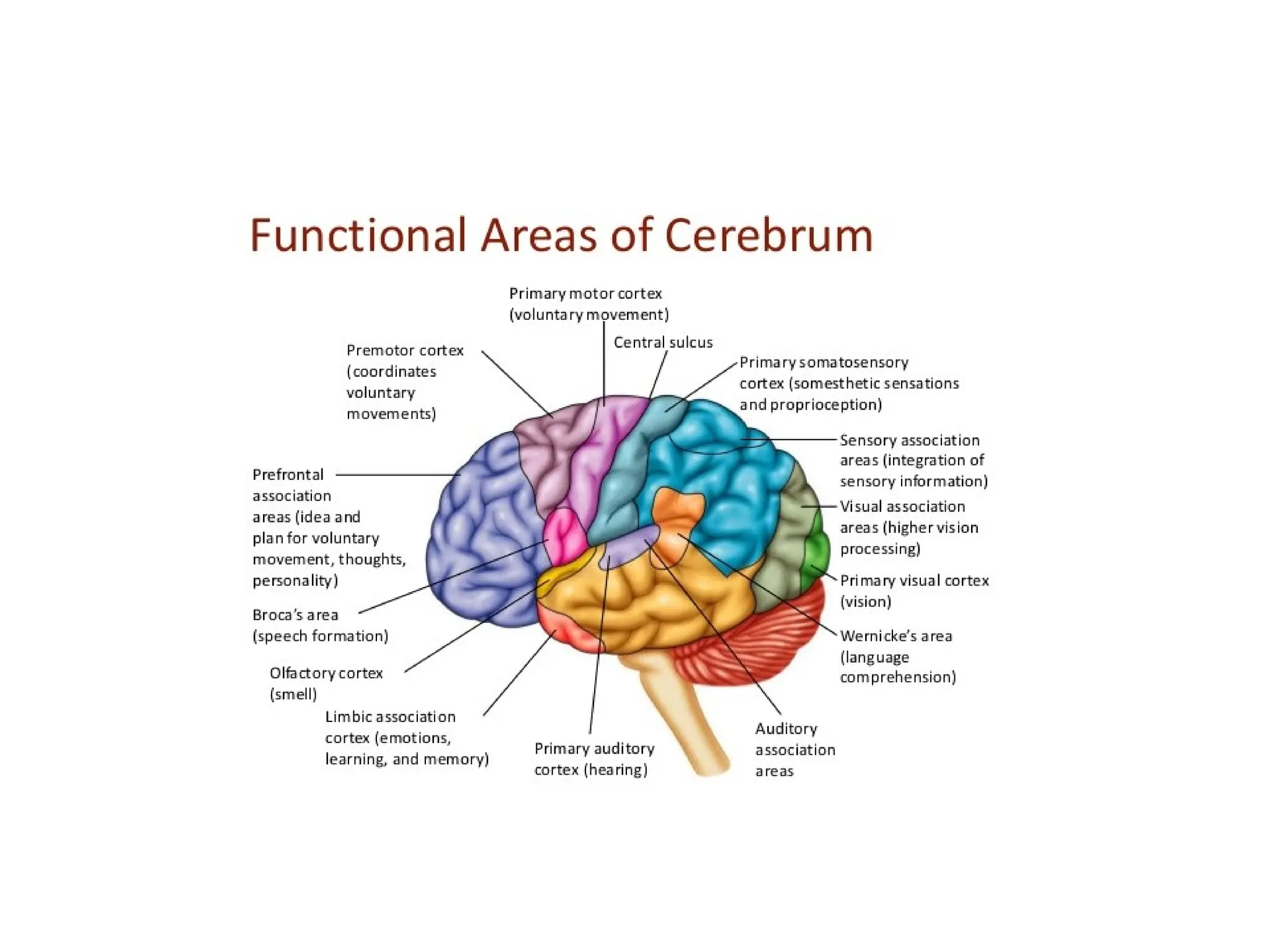
Cerebrum:
The cerebrum ofcerebral hemispheres make up the largest visual portion of
the human brain. Various structures combine to form the cerebral
hemispheres, among others: the cortex, basal ganglia, amygdala and
hippocampus. The hemispheres together control a large portion of the
functions of the human brain such as emotion, memory, perception and motor
functions. Apart from this the cerebral hemispheres stand for the cognitive
capabilities of the brain.
Connecting each of the hemispheres is the corpus callosum as well as several
additional commissures. One of the most important parts of the cerebral
hemispheres is the cortex, made up of gray matter covering the surface of the
brain. Functionally, the cerebral cortex is involved in planning and carrying
out of everyday tasks.
The hippocampus is involved in storage of memories, the amygdala plays a
role in perception and communication of emotion, while the basal ganglia
play a major role in the coordination of voluntary movement.
10.
Here is alist of some of the most important vertebrate brain components, along
with a brief description of their functions as currently understood:
• The medulla, along with the spinal cord, contains many small nuclei involved
in a wide variety of sensory and involuntary motor functions such as vomiting,
heart rate and digestive processes.
• The pons lies in the brainstem directly above the medulla. Among other things,
it contains nuclei that control often voluntary but simple acts such as sleep,
respiration, swallowing, bladder function, equilibrium, eye movement, facial
expressions, and posture.
• The hypothalamus is a small region at the base of the forebrain, whose
complexity and importance belies its size. It is composed of numerous small
nuclei, each with distinct connections and neurochemistry. The hypothalamus
is engaged in additional involuntary or partially voluntary acts such as sleep
and wake cycles, eating and drinking, and the release of some hormones.
11.
• The thalamusis a collection of nuclei with diverse functions: some are
involved in relaying information to and from the cerebral hemispheres,
while others are involved in motivation. The subthalamic area (zona
incerta) seems to contain action-generating systems for several types of
"consummatory" behaviors such as eating, drinking, defecation, and
copulation.
• The cerebellum modulates the outputs of other brain systems, whether
motor related or thought related, to make them certain and precise.
Removal of the cerebellum does not prevent an animal from doing
anything in particular, but it makes actions hesitant and clumsy. This
precision is not built-in, but learned by trial and error. The muscle
coordination learned while riding a bicycle is an example of a type of
neural plasticity that may take place largely within the cerebellum. 10%
of the brain's total volume consists of the cerebellum and 50% of all
neurons are held within its structure.
12.
• The optictectum allows actions to be directed toward points in space, most
commonly in response to visual input. In mammals it is usually referred to as
the superior colliculus, and its best-studied function is to direct eye
movements. It also directs reaching movements and other object-directed
actions. It receives strong visual inputs, but also inputs from other senses that
are useful in directing actions, such as auditory input in owls and input from
the thermosensitive pit organs in snakes. In some primitive fishes, such as
lampreys, this region is the largest part of the brain. The superior colliculus is
part of the midbrain.
• The pallium is a layer of gray matter that lies on the surface of the forebrain
and is the most complex and most recent evolutionary development of the
brain as an organ. In reptiles and mammals, it is called the cerebral cortex.
Multiple functions involve the pallium, including smell and spatial memory.
In mammals, where it becomes so large as to dominate the brain, it takes over
functions from many other brain areas. In many mammals, the cerebral
cortex consists of folded bulges called gyri that create deep furrows or
fissures called sulci. The folds increase the surface area of the cortex and
therefore increase the amount of gray matter and the amount of information
that can be stored and processed.
13.
• The hippocampus,strictly speaking, is found only in mammals. However,
the area it derives from, the medial pallium, has counterparts in all
vertebrates. There is evidence that this part of the brain is involved in
complex events such as spatial memory and navigation in fishes, birds,
reptiles, and mammals.
• The basal ganglia are a group of interconnected structures in the forebrain.
The primary function of the basal ganglia appears to be action selection:
they send inhibitory signals to all parts of the brain that can generate motor
behaviors, and in the right circumstances can release the inhibition, so that
the action-generating systems are able to execute their actions. Reward and
punishment exert their most important neural effects by altering
connections within the basal ganglia.
• The olfactory bulb is a special structure that processes olfactory sensory
signals and sends its output to the olfactory part of the pallium. It is a
major brain component in many vertebrates, but is greatly reduced in
humans and other primates (whose senses are dominated by information
acquired by sight rather than smell).
15.
References:
Saladin, Kenneth (2011).Human anatomy (3rd ed.). McGraw-Hill. p. 416. ISBN 978-
0-07-122207-5.
von Bartheld, CS; Bahney, J; Herculano-Houzel, S (15 December 2016). "The search
for true numbers of neurons and glial cells in the human brain: A review of 150
years of cell counting". The Journal of Comparative Neurology. 524 (18): 3865–
3895. doi:10.1002/cne.24040. PMC 5063692. PMID 27187682.
Yuste, Rafael; Church, George M. (March 2014). "The new century of the brain"
(PDF). Scientific American. 310 (3): 38–45. Bibcode:2014SciAm.310c..38Y.
doi:10.1038/scientificamerican0314-38. PMID 24660326. Archived from the
original (PDF) on 2014-07-14.
Singh, I (2006). "A Brief Review of the Techniques Used in the Study of
Neuroanatomy". Textbook of Human Neuroanatomy. Jaypee Brothers. p. 24. ISBN
978-81-8061-808-6.