Download as PDF, PPTX


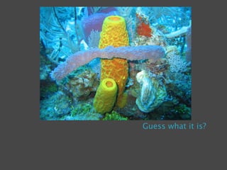


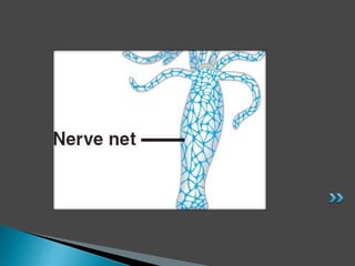


Multicellular organisms display complex life processes through chemical and electrical signaling. Plants utilize chemical signals while phototrophs, which rely on photosynthesis, are an example of one. Animals employ both chemical and electrical signals, with reflex actions serving as an example. The document then discusses the nervous systems of primitive multicellular organisms like hydra and their nerve nets, before exploring the structure and function of the central and peripheral nervous systems in more detail.


