Comparative Study of Visual Outcome between Femtosecond Lasik with Excimer La...iosrjce
IOSR Journal of Dental and Medical Sciences is one of the speciality Journal in Dental Science and Medical Science published by International Organization of Scientific Research (IOSR). The Journal publishes papers of the highest scientific merit and widest possible scope work in all areas related to medical and dental science. The Journal welcome review articles, leading medical and clinical research articles, technical notes, case reports and others.
Detection of Glaucoma using Optic Disk and Incremental Cup Segmentation from ...theijes
Medical researchers, detection of eye disease is very important because it may causes blindness. Glaucoma is one of the diseases that cause blindness. Standard procedure for detection glaucoma is to analysis of optic disk (OD) and cup region in retinal image. In this paper, introduce an automatic OD parameterized technique which is based on segmentation and Incremental Cup segmentation. The incremental cup segmentation method is based on anatomical evidence such as vessel bends at the cup boundary, considered relevant by glaucoma experts. Bends in a vessel are robustly detected using a region of support concept, which automatically selects the right scale for analysis. A multi-stage strategy is applied to derive a reliable subset of vessel bends called r-bends followed by a local 2-D spline fitting to derive the desired cup boundary. The results are compared with existing methods using different retinal images.
Timely analysis and constant intensive care of patient’spainsince eye ailmentsmustremainedmainworries in the computer-aided detection (CAD) ways and means. Intuitingsole or fewprecisestyles of retinal wrongs has setanessentialorigination in computed-aid screen in the first few dates. Thus far, in line for to range of retinal wrongs and multilayeredcommonusefulproductions, unplannedinnovation of wounds with unrevealed and wide-rangingstylesas retina leavingsanexcitingwork. In this pattern, a weakly supervised practice, demandingjustanorder of common and not level retinal picturesunderprivileged ofvital to justluster their places and multiplicities, is helpful for this charge. Specifically, a eye copy is considered as a superposition of related, blood vessels and related noise (lesions embraced for unequaldescriptions). Background is given as low-rank formfollowed by preprocessing steps, plusthree-Dplan, color regularization and blood vesselsrejection. Related noise is eyed as stochastic changeable and reputableover Gaussian for classicimageries and mixture of Gaussian (MoG) for asymmetricalpictures, harmoniously. The comingrun throughcodes together the relatedfacts of fundus pictures and the related noise into on its ownone and only ideal, and corporately enriches the idealbytypical and unequalpictures, which absolutelyexpose the low-rank subspace of the related and discriminate the wounds from related noise in unequal fundus pictures. Lastly, Ada boost classification process is castoff to increasesensing retinal injuries and to categorize the kinds of injuryusing a group of usual ones for exercisetopographies. Investigationalconsequencesprove that the future method is of wellskillscorrectness and outdoes the precedingconnectedapproaches.
FUZZY CLUSTERING BASED GLAUCOMA DETECTION USING THE CDR sipij
Glaucoma is a serious eye disease, overtime it will result in gradual blindness. Early detection of thedisease will help prevent against developing a more serious condition. A vertical cup-to-disc ratio which isthe ratio of the vertical diameter of the optic cup to that of the optic disc, of the fundus eye image is an important clinical indicator for glaucoma diagnosis. This paper presents an automated method for the extraction of optic disc and optic cup using Fuzzy C Means clustering technique combined with
thresholding. Using the extracted optic disc and optic cup the vertical cup-to-disc ratio was calculated.
The validity of this new method has been tested on 365 colour fundus images from two different publicly
available databases DRION, DIARATDB0 and images from an ophthalmologist. The result of the method
seems to be promising and useful for clinical work.
GLAUCOMA is a chronic eye disease that can damage optic nerve. According to WHO It
is the second leading cause of blindness, and is predicted to affect around 80 million people by 2020.
Development of the disease leads to loss of vision, which occurs increasingly over a long period of
time. As the symptoms only occur when the disease is quite advanced so that glaucoma is called the
silent thief of sight. Glaucoma cannot be cured, but its development can be slowed down by
treatment. Therefore, detecting glaucoma in time is critical. However, many glaucoma patients are
unaware of the disease until it has reached its advanced stage. In this paper, some manual and
automatic methods are discussed to detect glaucoma. Manual analysis of the eye is time consuming
and the accuracy of the parameter measurements also varies with different clinicians. To overcome
these problems with manual analysis, the objective of this survey is to introduce a method to
automatically analyze the ultrasound images of the eye. Automatic analysis of this disease is much
more effective than manual analysis.
Doktor Vedat Kaya, Canan Aslı Utine, Sezen Harmancı Karakuş, Işılay Kavadarlı ve Ömer Faruk Yılmaz tarafından hazırlanmış olan bu makaleyi ilginize sunarız.
Glaucoma is the most leading cause of irreversible blindness with the population of Africa and Asia ranking the highest over the rate of glaucoma affected regions around the world. The defect will damage eyes irreversibly by affecting the optic cup and optic disc of an eye. The early detection of glaucoma is an unavoidable need in the medical field. The widely used technique to detect glaucoma is an invasive method that may lead to other effects on the eye. This reason led to the introduction of a non invasive method that follows image processing for the detection of glaucoma. Retinal image based detection is the best way to choose as it comes under non invasive methods of detection. Detection of glaucoma using retinal images requires various medical features of the eyes such as optic cup diameter, optic disc diameter and optic cup to disc ratio are used. Glaucoma disease detection from retinal images supports convolutional neural networks CNN . The textual features obtained from retinal images such as the optic cup to optic disc measures are used for this classification. Convolutional Neural Networks use little pre processing techniques that can be implemented relatively uncomplicated compared to other image classification techniques. The implementation of this project follows the traditional CNN architecture, applying filter layers such as Convolution layer and Pooling layer and also activation functions such as ReLu function and sigmoid function to pre process as well as to update weights respectively on the hidden layers of the CNN followed by classifying the image. Vishnubhotla Poornasree | Vijayagiri Ashritha | Venumula Deeksha Reddy | J. Srilatha ""Glaucoma Detection from Retinal Images"" Published in International Journal of Trend in Scientific Research and Development (ijtsrd), ISSN: 2456-6470, Volume-3 | Issue-4 , June 2019, URL: https://www.ijtsrd.com/papers/ijtsrd23732.pdf
Paper URL: https://www.ijtsrd.com/computer-science/other/23732/glaucoma-detection-from-retinal-images/vishnubhotla-poornasree
Tien Measuring Situation Awareness Of Surgeons In Laparoscopic TrainingKalle
The study of surgeons’ eye movements is an innovative way of assessing skill and situation awareness, in that a comparison of eye movement strategies between expert surgeons and novices may show differences that can be used in training. Our preliminary study compared eye movements of 4 experts and
4 novices performing a simulated gall bladder removal task on a
dummy patient with an audible heartbeat and simulated vital signs displayed on a secondary monitor. We used a head-mounted Locarna PT-Mini eyetracker to record fixation locations during the operation. The results showed that novices concentrated so hard on the surgical
display that they were hardly able to look at the patient’s vital signs, even when heart rate audibly changed during the procedure. In comparison, experts glanced occasionally at the vitals monitor, thus being able to observe the patient condition.
Faro An Interactive Interface For Remote Administration Of Clinical Tests Bas...Kalle
A challenging goal today is the use of computer networking and advanced
monitoring technologies to extend human intellectual capabilities in medical decision making. Modern commercial eye trackers
are used in many of research fields, but the improvement of eye tracking technology, in terms of precision on the eye movements capture, has led to consider the eye tracker as a tool for vision analysis, so that its application in medical research, e.g. in ophthalmology, cognitive psychology and in neuroscience has grown considerably. The improvements of the human eye tracker interface become more and more important to allow medical doctors to increase their diagnosis capacity, especially if the interface allows them to remotely administer the clinical tests more appropriate for the problem at hand. In this paper, we propose a client/server eye tracking system that provides an interactive system for monitoring patients eye movements depending on the clinical test administered by the medical doctors. The system supports the retrieval of the gaze information and provides statistics to both medical research and disease diagnosis.
Glaucoma Detection in Retinal Images Using Image Processing Techniques: A SurveyEswar Publications
Glaucoma is a disease associated with human eyes and second conducting movement o fblindness across the globe if
eyes are not treated at preliminary stage. Glaucoma normally occurs with increased intra-ocular pressure (IOP) in eyes and gradually damagesthe vision field of eyes. The term ocular-hypertension is related to those people in whom IOP increases consistently and does not damage the optic nerve. Glaucoma has different types such as open-angle, close-angle, congenital, normal tension and etcetera. Normal tension glaucoma affects vision field and damages optic nerve as well. The term angle means the distance between iris and cornea; if this distance is large it is referred to as open-angle glaucoma and similarly if the distance between iris and cornea is short than this is called close-angle glaucoma. Open-angle glaucoma is common as compared to close-angle glaucoma. Close-angle glaucoma is very painful and affects vision field of eyes quickly as compared to open-angle glaucoma. In this
paper, the state of the art CAD systems and image processing methods are studied and compared systematically in terms of their classification accuracy, methodology approach, sensitivity and specificity. The comparison results indicate that the accuracy of these CAD systems and image processing methods is not up to the mark.
Glaucoma Screening Test By Segmentation of Optical Disc& Cup Segmentation Usi...IJERA Editor
Glaucoma is one of the most common causes of blindness and it is becoming even more important considering
the ageing society. Because healing of died retinal nerve fibers is not possible early detection and prevention is
essential. Robust, automated mass-screening will help to extend the symptom-free life of affected patients. We
devised a novel, automated, appearance based glaucoma classification system that does not depend on
segmentation based measurements. Our purely data-driven approach is applicable in large-scale screening
examinations. The proposed segmentation methods have been evaluated in a database of 650 images with optic
disc and optic cup boundaries manually marked by trained professionals. Our expected Experimental results
may be average overlapping error of 9.5% and 24.1% in optic disc and optic cup segmentation, respectively.
Background: Nowadays, ICRS are a step in the treatment of keratoconus. The purpose of this study was to evaluate the refractive effect and the tomographic and biomechanical parameters in keratoconus patients implanted with Ferrara ICRS, and their stability after 18 months.
Purpose: To evaluate the corneal volume (CV) before and after Ferrara intrastromal corneal ring segments (ICRS) implantation and its influence in clinical outcomes in keratoconus patients.
PURPOSE: To evaluate the clinical outcomes of implantation of Ferrara intrastromal corneal ring segments (ICRS) in patients with corneal ectasia after refractive surgery.
PURPOSE: To evaluate the long-term safety and effica- cy of Ferrara intrastromal corneal ring segments (ICRS) (Ferrara Ring; AJL, Boecillo, Spain) in patients with kera- toconus.
Head-to-Head Comparative Study of Two Optical Biometric Devices in Modern Cat...SM2 Strategic
Today's cataract surgeon has adopted non-contact optical
biometry as the standard of care in performing IOL calculations.
While modern formulae incorporate multiple variables as part
of their calculations, Axial Length and Keratometry readings
continue to be the inputs that are most influential in determining
IOL power. Some of the newer generation formulas such as
Holladay 2 and Olsen now incorporate more elements to help
increase accuracy.
New Directions in Targeted Therapeutic Approaches for Older Adults With Mantl...i3 Health
i3 Health is pleased to make the speaker slides from this activity available for use as a non-accredited self-study or teaching resource.
This slide deck presented by Dr. Kami Maddocks, Professor-Clinical in the Division of Hematology and
Associate Division Director for Ambulatory Operations
The Ohio State University Comprehensive Cancer Center, will provide insight into new directions in targeted therapeutic approaches for older adults with mantle cell lymphoma.
STATEMENT OF NEED
Mantle cell lymphoma (MCL) is a rare, aggressive B-cell non-Hodgkin lymphoma (NHL) accounting for 5% to 7% of all lymphomas. Its prognosis ranges from indolent disease that does not require treatment for years to very aggressive disease, which is associated with poor survival (Silkenstedt et al, 2021). Typically, MCL is diagnosed at advanced stage and in older patients who cannot tolerate intensive therapy (NCCN, 2022). Although recent advances have slightly increased remission rates, recurrence and relapse remain very common, leading to a median overall survival between 3 and 6 years (LLS, 2021). Though there are several effective options, progress is still needed towards establishing an accepted frontline approach for MCL (Castellino et al, 2022). Treatment selection and management of MCL are complicated by the heterogeneity of prognosis, advanced age and comorbidities of patients, and lack of an established standard approach for treatment, making it vital that clinicians be familiar with the latest research and advances in this area. In this activity chaired by Michael Wang, MD, Professor in the Department of Lymphoma & Myeloma at MD Anderson Cancer Center, expert faculty will discuss prognostic factors informing treatment, the promising results of recent trials in new therapeutic approaches, and the implications of treatment resistance in therapeutic selection for MCL.
Target Audience
Hematology/oncology fellows, attending faculty, and other health care professionals involved in the treatment of patients with mantle cell lymphoma (MCL).
Learning Objectives
1.) Identify clinical and biological prognostic factors that can guide treatment decision making for older adults with MCL
2.) Evaluate emerging data on targeted therapeutic approaches for treatment-naive and relapsed/refractory MCL and their applicability to older adults
3.) Assess mechanisms of resistance to targeted therapies for MCL and their implications for treatment selection
Knee anatomy and clinical tests 2024.pdfvimalpl1234
This includes all relevant anatomy and clinical tests compiled from standard textbooks, Campbell,netter etc..It is comprehensive and best suited for orthopaedicians and orthopaedic residents.
- Video recording of this lecture in English language: https://youtu.be/lK81BzxMqdo
- Video recording of this lecture in Arabic language: https://youtu.be/Ve4P0COk9OI
- Link to download the book free: https://nephrotube.blogspot.com/p/nephrotube-nephrology-books.html
- Link to NephroTube website: www.NephroTube.com
- Link to NephroTube social media accounts: https://nephrotube.blogspot.com/p/join-nephrotube-on-social-media.html
Title: Sense of Taste
Presenter: Dr. Faiza, Assistant Professor of Physiology
Qualifications:
MBBS (Best Graduate, AIMC Lahore)
FCPS Physiology
ICMT, CHPE, DHPE (STMU)
MPH (GC University, Faisalabad)
MBA (Virtual University of Pakistan)
Learning Objectives:
Describe the structure and function of taste buds.
Describe the relationship between the taste threshold and taste index of common substances.
Explain the chemical basis and signal transduction of taste perception for each type of primary taste sensation.
Recognize different abnormalities of taste perception and their causes.
Key Topics:
Significance of Taste Sensation:
Differentiation between pleasant and harmful food
Influence on behavior
Selection of food based on metabolic needs
Receptors of Taste:
Taste buds on the tongue
Influence of sense of smell, texture of food, and pain stimulation (e.g., by pepper)
Primary and Secondary Taste Sensations:
Primary taste sensations: Sweet, Sour, Salty, Bitter, Umami
Chemical basis and signal transduction mechanisms for each taste
Taste Threshold and Index:
Taste threshold values for Sweet (sucrose), Salty (NaCl), Sour (HCl), and Bitter (Quinine)
Taste index relationship: Inversely proportional to taste threshold
Taste Blindness:
Inability to taste certain substances, particularly thiourea compounds
Example: Phenylthiocarbamide
Structure and Function of Taste Buds:
Composition: Epithelial cells, Sustentacular/Supporting cells, Taste cells, Basal cells
Features: Taste pores, Taste hairs/microvilli, and Taste nerve fibers
Location of Taste Buds:
Found in papillae of the tongue (Fungiform, Circumvallate, Foliate)
Also present on the palate, tonsillar pillars, epiglottis, and proximal esophagus
Mechanism of Taste Stimulation:
Interaction of taste substances with receptors on microvilli
Signal transduction pathways for Umami, Sweet, Bitter, Sour, and Salty tastes
Taste Sensitivity and Adaptation:
Decrease in sensitivity with age
Rapid adaptation of taste sensation
Role of Saliva in Taste:
Dissolution of tastants to reach receptors
Washing away the stimulus
Taste Preferences and Aversions:
Mechanisms behind taste preference and aversion
Influence of receptors and neural pathways
Impact of Sensory Nerve Damage:
Degeneration of taste buds if the sensory nerve fiber is cut
Abnormalities of Taste Detection:
Conditions: Ageusia, Hypogeusia, Dysgeusia (parageusia)
Causes: Nerve damage, neurological disorders, infections, poor oral hygiene, adverse drug effects, deficiencies, aging, tobacco use, altered neurotransmitter levels
Neurotransmitters and Taste Threshold:
Effects of serotonin (5-HT) and norepinephrine (NE) on taste sensitivity
Supertasters:
25% of the population with heightened sensitivity to taste, especially bitterness
Increased number of fungiform papillae
micro teaching on communication m.sc nursing.pdfAnurag Sharma
Microteaching is a unique model of practice teaching. It is a viable instrument for the. desired change in the teaching behavior or the behavior potential which, in specified types of real. classroom situations, tends to facilitate the achievement of specified types of objectives.
Lung Cancer: Artificial Intelligence, Synergetics, Complex System Analysis, S...Oleg Kshivets
RESULTS: Overall life span (LS) was 2252.1±1742.5 days and cumulative 5-year survival (5YS) reached 73.2%, 10 years – 64.8%, 20 years – 42.5%. 513 LCP lived more than 5 years (LS=3124.6±1525.6 days), 148 LCP – more than 10 years (LS=5054.4±1504.1 days).199 LCP died because of LC (LS=562.7±374.5 days). 5YS of LCP after bi/lobectomies was significantly superior in comparison with LCP after pneumonectomies (78.1% vs.63.7%, P=0.00001 by log-rank test). AT significantly improved 5YS (66.3% vs. 34.8%) (P=0.00000 by log-rank test) only for LCP with N1-2. Cox modeling displayed that 5YS of LCP significantly depended on: phase transition (PT) early-invasive LC in terms of synergetics, PT N0—N12, cell ratio factors (ratio between cancer cells- CC and blood cells subpopulations), G1-3, histology, glucose, AT, blood cell circuit, prothrombin index, heparin tolerance, recalcification time (P=0.000-0.038). Neural networks, genetic algorithm selection and bootstrap simulation revealed relationships between 5YS and PT early-invasive LC (rank=1), PT N0—N12 (rank=2), thrombocytes/CC (3), erythrocytes/CC (4), eosinophils/CC (5), healthy cells/CC (6), lymphocytes/CC (7), segmented neutrophils/CC (8), stick neutrophils/CC (9), monocytes/CC (10); leucocytes/CC (11). Correct prediction of 5YS was 100% by neural networks computing (area under ROC curve=1.0; error=0.0).
CONCLUSIONS: 5YS of LCP after radical procedures significantly depended on: 1) PT early-invasive cancer; 2) PT N0--N12; 3) cell ratio factors; 4) blood cell circuit; 5) biochemical factors; 6) hemostasis system; 7) AT; 8) LC characteristics; 9) LC cell dynamics; 10) surgery type: lobectomy/pneumonectomy; 11) anthropometric data. Optimal diagnosis and treatment strategies for LC are: 1) screening and early detection of LC; 2) availability of experienced thoracic surgeons because of complexity of radical procedures; 3) aggressive en block surgery and adequate lymph node dissection for completeness; 4) precise prediction; 5) adjuvant chemoimmunoradiotherapy for LCP with unfavorable prognosis.
ARTIFICIAL INTELLIGENCE IN HEALTHCARE.pdfAnujkumaranit
Artificial intelligence (AI) refers to the simulation of human intelligence processes by machines, especially computer systems. It encompasses tasks such as learning, reasoning, problem-solving, perception, and language understanding. AI technologies are revolutionizing various fields, from healthcare to finance, by enabling machines to perform tasks that typically require human intelligence.
Flu Vaccine Alert in Bangalore Karnatakaaddon Scans
As flu season approaches, health officials in Bangalore, Karnataka, are urging residents to get their flu vaccinations. The seasonal flu, while common, can lead to severe health complications, particularly for vulnerable populations such as young children, the elderly, and those with underlying health conditions.
Dr. Vidisha Kumari, a leading epidemiologist in Bangalore, emphasizes the importance of getting vaccinated. "The flu vaccine is our best defense against the influenza virus. It not only protects individuals but also helps prevent the spread of the virus in our communities," he says.
This year, the flu season is expected to coincide with a potential increase in other respiratory illnesses. The Karnataka Health Department has launched an awareness campaign highlighting the significance of flu vaccinations. They have set up multiple vaccination centers across Bangalore, making it convenient for residents to receive their shots.
To encourage widespread vaccination, the government is also collaborating with local schools, workplaces, and community centers to facilitate vaccination drives. Special attention is being given to ensuring that the vaccine is accessible to all, including marginalized communities who may have limited access to healthcare.
Residents are reminded that the flu vaccine is safe and effective. Common side effects are mild and may include soreness at the injection site, mild fever, or muscle aches. These side effects are generally short-lived and far less severe than the flu itself.
Healthcare providers are also stressing the importance of continuing COVID-19 precautions. Wearing masks, practicing good hand hygiene, and maintaining social distancing are still crucial, especially in crowded places.
Protect yourself and your loved ones by getting vaccinated. Together, we can help keep Bangalore healthy and safe this flu season. For more information on vaccination centers and schedules, residents can visit the Karnataka Health Department’s official website or follow their social media pages.
Stay informed, stay safe, and get your flu shot today!
Title: Sense of Smell
Presenter: Dr. Faiza, Assistant Professor of Physiology
Qualifications:
MBBS (Best Graduate, AIMC Lahore)
FCPS Physiology
ICMT, CHPE, DHPE (STMU)
MPH (GC University, Faisalabad)
MBA (Virtual University of Pakistan)
Learning Objectives:
Describe the primary categories of smells and the concept of odor blindness.
Explain the structure and location of the olfactory membrane and mucosa, including the types and roles of cells involved in olfaction.
Describe the pathway and mechanisms of olfactory signal transmission from the olfactory receptors to the brain.
Illustrate the biochemical cascade triggered by odorant binding to olfactory receptors, including the role of G-proteins and second messengers in generating an action potential.
Identify different types of olfactory disorders such as anosmia, hyposmia, hyperosmia, and dysosmia, including their potential causes.
Key Topics:
Olfactory Genes:
3% of the human genome accounts for olfactory genes.
400 genes for odorant receptors.
Olfactory Membrane:
Located in the superior part of the nasal cavity.
Medially: Folds downward along the superior septum.
Laterally: Folds over the superior turbinate and upper surface of the middle turbinate.
Total surface area: 5-10 square centimeters.
Olfactory Mucosa:
Olfactory Cells: Bipolar nerve cells derived from the CNS (100 million), with 4-25 olfactory cilia per cell.
Sustentacular Cells: Produce mucus and maintain ionic and molecular environment.
Basal Cells: Replace worn-out olfactory cells with an average lifespan of 1-2 months.
Bowman’s Gland: Secretes mucus.
Stimulation of Olfactory Cells:
Odorant dissolves in mucus and attaches to receptors on olfactory cilia.
Involves a cascade effect through G-proteins and second messengers, leading to depolarization and action potential generation in the olfactory nerve.
Quality of a Good Odorant:
Small (3-20 Carbon atoms), volatile, water-soluble, and lipid-soluble.
Facilitated by odorant-binding proteins in mucus.
Membrane Potential and Action Potential:
Resting membrane potential: -55mV.
Action potential frequency in the olfactory nerve increases with odorant strength.
Adaptation Towards the Sense of Smell:
Rapid adaptation within the first second, with further slow adaptation.
Psychological adaptation greater than receptor adaptation, involving feedback inhibition from the central nervous system.
Primary Sensations of Smell:
Camphoraceous, Musky, Floral, Pepperminty, Ethereal, Pungent, Putrid.
Odor Detection Threshold:
Examples: Hydrogen sulfide (0.0005 ppm), Methyl-mercaptan (0.002 ppm).
Some toxic substances are odorless at lethal concentrations.
Characteristics of Smell:
Odor blindness for single substances due to lack of appropriate receptor protein.
Behavioral and emotional influences of smell.
Transmission of Olfactory Signals:
From olfactory cells to glomeruli in the olfactory bulb, involving lateral inhibition.
Primitive, less old, and new olfactory systems with different path
How conductive keratoplasty is impacting the presbyopic practice
1. How Conductive Keratoplasty is
Impacting the Presbyopic Practice
Shareef Mahdavi • SM2 Consulting • Pleasanton, CA
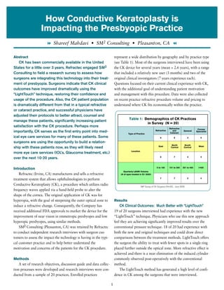
Abstract represent a wide distribution by geography and by practice type
CK has been commercially available in the United (see Table 1). Most of the surgeons interviewed have been using
States for a little over 3 years. Refractec engaged SM2 the CK device for several years (mean = 2.6 years), with a range
Consulting to field a research survey to assess how that included a relatively new user (3 months) and two of the
surgeons are integrating this technology into their treat- original clinical investigators (7 years experience each).
ment of presbyopia. Surgeons indicate that CK clinical Questions focused on their current clinical experience with CK,
outcomes have improved dramatically using the with the additional goal of understanding patient motivation
“LightTouch” technique, restoring their confidence and and management with this procedure. Data were also collected
usage of the procedure. Also, the CK patient population on recent practice refractive procedure volume and pricing to
is dramatically different from that in a typical refractive understand where CK fits economically within the practice.
or cataract practice, and successful physicians have
adjusted their protocols to better attract, counsel and
Table 1: Demographics of CK Practices
manage these patients, significantly increasing patient
in Survey (N = 20)
satisfaction with the CK procedure. Perhaps more
Refractive Cornea
Refractive
importantly, CK serves as the first entry point into med- and
Cataract
General Specialist
Type of Practice
ical eye care services for many of these patients. Some 6 6 3 5
surgeons are using the opportunity to build a relation-
North South
East West
ship with these patients now, as they will likely need Central Central
Location
more eye care services (IOL’s, Glaucoma treatment, etc.)
6 7 4 3
over the next 10-20 years.
0 to 150 151 to 300 301 to 450 >450
Introduction
Quarterly LASIK Volume
Refractec (Irvine, CA) manufactures and sells a refractive (# of eyes treated in Q1 2005)
treatment system that allows ophthalmologists to perform 7 7 2 4
Conductive Keratoplasty (CK), a procedure which utilizes radio SM2 Survey of CK Surgeons (N=20), June 2005
frequency waves applied via a hand-held probe to alter the
shape of the cornea. The original application of CK was for
hyperopia, with the goal of steepening the outer optical zone to Results
induce a refractive change. Consequently, the Company has CK Clinical Outcomes: Much Better with “LightTouch”
received additional FDA approvals to market the device for the 19 of 20 surgeons interviewed had experience with the new
improvement of near vision in emmetropic presbyopes and low “LightTouch” technique. Physicians who use this new approach
hyperopic presbyopes, using similar methods. feel they are achieving significantly improved results over the
SM2 Consulting (Pleasanton, CA) was retained by Refractec conventional pressure technique. 18 of 20 had experience with
to conduct independent research interviews with surgeon cus- both the new and original techniques and could draw direct
tomers to assess the impact the technology is having in the typi- comparisons between the treatment methods. LightTouch offers
cal customer practice and to help better understand the the surgeon the ability to treat with fewer spots in a single ring
motivation and concerns of the patients for the CK procedure. placed further outside the optical zone. More refractive effect is
achieved and there is a near elimination of the induced cylinder
Methods commonly observed post-operatively with the conventional
A set of research objectives, discussion guide and data collec- method.
tion processes were developed and research interviews were con- The LightTouch method has generated a high level of confi-
ducted from a sample of 20 practices. Enrolled practices dence in CK among the surgeons that were interviewed.
1
2. (cataract and/or refractive implants). See Figure 2.
Figure 1: As a surgeon, how would you rate your
CK Candidates: A Different Audience
confidence in the CK procedure?
The patient wanting CK is not likely to be found in the
7
SM2 Survey of CK Surgeons (N=20), June 2005
waiting room of a typical ophthalmic surgeon’s practice. They
6 fit somewhere between the typical LASIK patient (younger,
5 myopic, haven’t enjoyed unaided vision for years) and the typi-
# of Practices
Mean Rating = cal IOL patient (older, having other aging and health issues).
4
8.6 Many patients that choose CK have never had a relationship
3
with an eye care professional and only now are in need of help
2 with their vision. Blessed with good distance vision all their
1 lives, they reach their late 40s and find themselves increasingly
0 frustrated and even angry at their inability to perform near and
1 2 3 4 5 6 7 8 9 10 intermediate visual tasks. The frustration can be classified in
Confidence Rating (1 = lowest, 10 = highest) terms of the physical and emotional context of getting older.
When paired with the view that there is nothing redeeming
As shown in Figure 1, the average confidence was 8.6 (on a 1 to about wearing reading glasses, the CK candidate comes in say-
10 scale, 10 being highest) and no surgeon rated their confi- ing, “I feel old and I look old.” All surgeons reported the aver-
dence below a 7. Although not directly asked, surgeons indi- age CK patient age is from 48 to 52 years of age, which can be
cated that the reason for their high confidence was directly among the most active and financially productive years of a
attributable to the LightTouch method. Had they been asked person’s life. The motivation for CK is simple: reduce depend-
this question a year ago, confidence levels would have been ency on reading glasses.
rated much lower. Similar to research conducted about motivation for LASIK,
Many surgeons have noticed a difference in the immediacy the CK patient also wants to improve performance. In the con-
of the effect of CK, with LightTouch providing a stronger text of presbyopia, performance means being able to read the
“WOW! Factor” based on the statements of patients who have cell phone, price tags, restaurant menu, and newspaper without
had the new procedure. Although LightTouch has been in use a the need for reading glasses.
little more than one year, surgeons indicate that the incidence of Surgeons also reported that many of their CK patients would
retreatment is already significantly less than what they experi- never have had LASIK, either because they didn’t need it for dis-
enced with the original technique. tance correction or due to perceived risk. In contrast to using
The leading indication for CK use is for primary treatment of lasers and keratomes, CK allows surgeons to offer a less invasive
plano presbyopes (100%), followed by 11 of 20 surgeons (55%) approach which serves as a “stepping stone” before more dra-
who use CK for post-LASIK refinement. Thirty percent of those matic procedures will be required (or desired) later on in life.
interviewed will also treat mild hyperopes bilaterally, and 25% CK can be compared to other less invasive self-improvement
indicate they are using it for refinement of IOL implant results options available at midlife: Botox before a facelift, teeth
whitening before veneers, and collagen injection before knee
replacement.
Figure 2: Percentage of Practices Using CK by
Indication (Multiple responses allowed) This low level of invasiveness resonates very deeply with
someone who has had “perfect eyes” for 50 years. The more
Post-IOL SM2 Survey of CK Surgeons (N=20), June 2005 conservative and mature profile of this age group is a good fit
25%
Refinement
with a procedure that gives back capability they previously
Mild Hyperopes
30% enjoyed.
(Bilateral CK)
Post-LASIK 55% CK Surgeons: Setting Expectations
Refinement
How the surgeon sets expectations with patients is perhaps
Plano
Presbyopes 100% the single most critical factor in achieving success with CK.
These patients have emotional needs that differ greatly from that
0% 20% 40 % 60% 80% 100 %
of the typical LASIK or cataract patient. Sometimes described as
2
3. “high maintenance,” these patients are often dealing with vision CK Patients: How do you find them?
problems for the first time in their lives and are unaccustomed
to needing any help to function. Our interviews found that the The ideal CK candidate is one who says, “I’ve had great
process begins with the surgeon and how he views (and ulti- vision my whole life. I’m only 50 – I’m not old. I’m in the
mately presents) his expectations of the procedure. prime of my life and have the money to spend on things that
Surgeons who do best with this procedure are able to sepa- can help me. I want to feel better, look younger and perform
rate CK from LASIK in their minds. Objectively, they feel it is at the top of my game.”
not as “good” a procedure as LASIK; with a deficiency similar Because they’ve had good vision, many ideal CK patients
to how hyperopic LASIK is viewed when compared to myopic are not native to the practice. Nor are they being referred in
LASIK. That is, CK does not enjoy the same level of predictabil- great numbers by optometrists who co-manage, presumably
ity or stability. However, they recognize this procedure serves a because they have not had a strong need for primary eye
different purpose for a different audience. These surgeons are care services in their youth and younger adulthood. As with
able to view CK in its own right as a solution that fills the gap LASIK referrals patterns, word-of-mouth seems to be the
between LASIK and IOLs. While imperfect, it is a solution that dominant means of acquiring patients. This is especially true
is much better than what all of eye care has been doing for years now with the LightTouch method, with a greater initial
to this audience, namely turning them away and sending them “WOW! Factor” that is helping spur increased referrals.
to drug stores for reading glasses. “You need to make me and my friends aware of what you
can do to help me get rid of these reading glasses, which I
CK and the Patient Evaluation find truly annoying.”
There are 3 critical steps in evaluating a patient for CK, and Advertising and news stories are essential sources of CK
surgeons were adamant that they cannot be skipped in the patients, and 16 of the 20 surgeons interviewed conduct
process. external marketing; they spend an average of $12,650 per
First, surgeons employ a wide variety of analogies to help month total on their advertising (range $2,000 to $45,000).
educate the CK patient, generally focusing around giving back This equates to anywhere from 5% – 12% of their monthly
capability that has been lost. With experience, these surgeons refractive revenue (one exception is a surgeon who markets
have determined what words and phrases best describe the pro- and performs CK as his only refractive procedure; marketing
cedure and set proper expectations. “The procedure works well expenditures are at 30% of revenue).
but you will continue to age,” is a statement that captures this Most surgeons believe that “cross promotion” of proce-
sentiment. Surgeons believe it is essential that patients under- dures in their marketing has been effective. In other words,
stand that they will be given a “reprieve” until their accom- advertising for CK generates more LASIK business and
modative needs increase beyond what can be successfully vice-versa. Further analysis was done by looking at the
managed via CK. reported percentage of the marketing and advertising
Second, surgeons ask detailed and probing questions about message devoted to CK (as opposed to LASIK, IOLs, or
profession and hobbies. They describe this process as a form of a refractive surgery in general). All but one of the surgeons
psychological profile or personality test, looking for indicators who advertise had some portion of their advertising dedi-
that a patient might not be happy with anything less than 100% cated to CK, with a mean of 22% (range 5% to 100%).
crisp vision for the distance. Pilots, engineers, attorneys, and those Comparing advertising dollars invested towards CK to the
who are fanatical golf or tennis players probably won’t tolerate amount of revenue generated by CK yielded a mean ratio of
what they may have to give up at distance to achieve better 5.7 to 1. Because the spread of this ratio in this interview
unaided vision for near and intermediate. These simply are not sample is so large (from 1.2 to 13.3), we believe it is more
good candidates. However, patients with less demanding visual meaningful to express the average by using the median
needs who express high frustration with reading glasses (example: (3.8) rather than the mean (5.7) value. Using the median,
“these just aren’t for me”) are ideal candidates for CK. for every dollar spent on CK messaging, an average of
Third, surgeons perform one or more tests to determine $3.80 was generated in CK revenue.
compatibility with a mono- or blended- vision procedure. All “Reach out and find me today and help me improve my
surgeons employed a loose lens test followed by a contact lens vision. I will stay with you for the rest of my life.”
trial (if doubt persists) for one to two days. The critical decision
3
4. point here is whether or not the patient can “psycho adapt” to doing between 20 and 49 eyes. The mean among all surgeons
the reduction in binocularity, the key factor in predicting the was 32.1 eyes for the quarter and the median (half of surgeons
patient’s likelihood for success with this change in their vision. above, half below) was 18.6 eyes for the quarter. The range
Following the CK procedure, successful surgeons employ a includes one surgeon who did not perform CK in Q1 2005 and
different protocol for post-op management than is required for another who did 120 CK procedures during the same time
LASIK. The initial “WOW! Factor” experienced on the first day period. There was no correlation between high-volume LASIK
for near tasks is often followed by a temporary worsening of and high volume CK; both the highest and lowest volume CK
distance vision that lasts from 1 to 2 weeks and gradually surgeons interviewed were the highest-volume LASIK surgeons
improves. Thus, the postoperative Day 1 visit is often viewed as in this sample. Importantly, surgeons also indicated that their
a wasted effort as long as the patient is not experiencing some volumes are increasing at a rapid rate due to the improved
atypical symptom. CK’s safety profile affords this luxury, in con- results from the LightTouch technique.
trast to the post-op requirement for LASIK. However, what CK Using these figures, doing just several eyes per month cost
post-op management does require is a form of “psychological justifies the CK device and all but one of the surgeons inter-
hand holding” that is not generally present with the typically viewed met this criteria.
younger LASIK patient. Fortunately, the lack of instant gratifica-
tion is well-matched to the more tolerant, more mature patient Figure 3: # of CK Eyes Treated Per Quarter (Q1 2005)
that needs CK.
7
CK As An Investment SM2 Survey of CK Surgeons (N=20), June 2005
As with all technology, CK also needs to be measured on its 6 Mean = 32.1 eyes
Median = 18.6 eyes
ability to provide a return on investment for the surgeon. What
# of Practices
5
is unique about CK is that there is a three-tier rationale that
4
needs to be considered; we will describe these one at a time as
3
follows and how they support investment into this technology.
1) Positive ROI - Ability to do enough procedure volume to 2
pay for the device 1
At approximately $60,000, the up-front investment for a CK
0
device is nominal when compared with a laser (either excimer or 0 <10 10 to 19 20 to 49 50 to 75 120
femtosecond), yet has a disposable component of $175 or more # of CK Eyes
per eye that is roughly equivalent to that charged for use of a
laser. Additionally, the time required to perform the procedure is
minimal and does not require a special room or dedicated tech- 2) Part of the refractive toolkit - Surgeons indicated that CK
nicians. Some surgeons find it quick enough that they can add it was also effective at building other sources of revenue; LASIK
to an existing appointment or squeeze it in, eliminating the need procedure volume benefits from offering CK. This makes sense
to burden the schedule with CK-specific appointments. A con- intuitively when viewed from the perspective of the eye care
servative approach would factor in one-half of a full time equiv- consumer. The consumer knows he is bothered by having to
alent employee to assist with the additional hand holding wear glasses and wants to get rid of them, yet has no clue as to
required with CK patients. which vision solution will best meet his needs. News stories or
The average collected fee per CK treatment among these sur- advertising about any form of visual performance benefit is
geons was $1,637 (range: $1,250 to $2,000) and it is typically likely to generate interest from these consumers. Inquiries about
performed in only one eye. Surgeons who perform a bilateral CK can often lead to a LASIK procedure and vice-versa. it is the
treatment (for low hyperopes) tend to charge a discounted fee surgeon’s job to properly educate and steer patients to the right
for the second eye resulting in $2,500 collected OU. solution. As a result, all twenty surgeons interviewed believe
An analysis of the distribution of the number of CK eyes that CK is an essential tool and would purchase the system
treated during Q1 2005 in this sample follows a normal distri- again if given the choice today.
bution (see Figure 3), with the largest group of surgeons (35%)
4
5. 3) Building the patient base - Strategic significance for both surgeons and their patients. The other half (8) see CK
future eye care services remaining an important “niche player” much as it is today, with
The third rationale, described by several surgeons as key to its role limited to a step between the full refractive correction
their long-term strategy, was the use of CK as a way to begin a offered by LASIK for pre-presbyopes and by an IOL for presby-
relationship with patients that they would not otherwise see for opes both with and without a cataractous lens.
another 10 to 15 years. Leveraging the less invasive and less The remaining 20% of surgeons interviewed (4 of 20) feel
expensive aspects of the procedure (relative to an IOL), CK that CK will become obsolete once presbyopic LASIK or a
allows the surgeon to form a bond with a patient and begin a corneal inlay becomes available. They are hopeful that a differ-
dialog that will continue for many years down the road when ent solution will provide better predictability with less risk than
more invasive and expensive eye care services are needed or an intraocular implant. Admittedly, however, they do not have
required. As one physician noted, the reasoning is simple: enough data to evaluate whether or not a multifocal approach
“If I don’t begin this relationship now, that patient will never via ablation or inlay will be good enough in real world clinical
know I exist and will just as likely choose another surgeon when practice. Only time will tell. While these surgeons are expecting
it comes time for a cataract or refractive IOL.” advancements beyond CK, this is still an important tool in their
Thus, CK serves as an entry point for the patient into med- refractive practice today.
ical eye care, and for the surgeon to develop a long-term rela-
tionship with that patient. Summary
This survey yielded several key findings that should prove
CK’s Role In The Future helpful to surgeons who are considering adopting the technology
Refractive surgery is a rapidly advancing field, and the future as well as current CK users who are looking to expand their use
is filled with other developments, many of which are geared and success with this device:
towards alleviating presbyopia. These include accommodating,
multifocal and phakic IOLs, corneal inlays and onlays, and pres- Not unlike other eye care technologies such as phacoemulsi-
byopic LASIK. Even with these potentially more advanced devel- fication or excimer laser ablation, Conductive Keratoplasty has
opments on the horizon, 80% of surgeons interviewed (16 of undergone an evolution in its approach that has refined the
20) believe that CK will still be important over the next 3-5 technique and improved outcomes.
years (see Figure 4). This majority is impressed with CK’s safety
profile and what it offers to patients who are risk averse. Half The patient that is ideal for CK is different than other
of this group (8) believe CK will grow into a “big player” in the patients currently seen by the ophthalmologist; it will take time
refractive field. They view market adoption as only a matter of to make them aware of, interested in and educated about CK.
time, allowing for growing awareness and acceptance of CK by This was also true of laser vision correction in its early days.
Surgeons need to approach CK differently than LASIK.
Figure 4: Where do you believe CK will From their own mindset through counseling patients pre- and
be in 3 to 5 years? post-operatively, the expectations for CK and the management
of those expectations are indeed different.
Niche Player The low acquisition cost of the CK device makes it easy to
40%
cost justify, with breakeven procedure volumes that are a frac-
tion of what is required to breakeven with LASIK. CK has an
additive effect to existing LASIK procedure volume and can be
Obsolete
Big Player 20% used as a tool to build the future growth of the practice from
40% advanced IOLs.
Even with more advanced technology on the horizon, CK is
destined to have a role within the ophthalmic practice. This is
SM2 Survey of CK Surgeons (N=20), June 2005
5