Holmes adie syndrome
•Download as PPTX, PDF•
3 likes•1,325 views

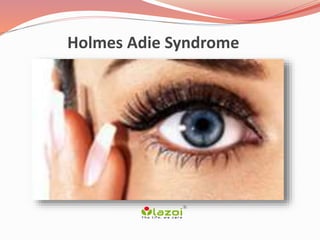
Holmes adie syndrome is a neurological disorder that affects the pupil of the eye and the autonomic nervous system.
Report
Share
Report
Share

Recommended
Nystagmus
This document provides an overview of nystagmus, including its definition, terminology, types, causes, and characteristics. Nystagmus is defined as a repetitive, to-and-fro movement of the eyes. It is classified as congenital or acquired, and includes types such as infantile nystagmus, spasmus nutans, end point nystagmus, vestibular nystagmus, optokinetic nystagmus, downbeat nystagmus, and nystagmus associated with strabismus. The document discusses the mechanisms, features, and treatments of different forms of nystagmus.
Approach to vision loss
1. The document provides an overview of approaches to evaluating vision loss, including determining whether it is monocular or binocular and transient or persistent.
2. Examinations should include visual acuity, color vision, visual fields, pupils, and examination of the eyes and optic discs.
3. Causes of transient and acute vision loss are discussed, including optic neuropathies, ischemic events, migraine, and PRES.
4. Progressive vision loss may be due to compressive lesions, glaucoma, or retinal disorders.
Horners syndrome
Horner's syndrome results from interruption of the sympathetic nerve supply to the eye, causing the classic triad of ptosis, miosis, and anhidrosis. It can occur from lesions anywhere along the three-neuron sympathetic pathway from the brainstem to the eye. Testing includes evaluating pupil response to light and pharmacologic tests like cocaine and apraclonidine to localize the lesion and guide further workup and treatment of the underlying cause when possible. The goal is to identify potentially serious underlying conditions causing Horner's syndrome.
Pupil
This document discusses the pupil in health and disease. It begins by describing the normal anatomy and function of the pupil, including its size, location, shape, and role in regulating light entry. It then covers various pupil reflexes and abnormalities such as anisocoria, mydriasis, miosis, light-near dissociation, Argyll Robertson pupils, and disorders of the third cranial nerve and sympathetic pathway. Causes, signs, and diagnostic tests for various pupil abnormalities are provided.
Approach to vision loss
This document discusses approaches to vision loss. It defines various patterns of vision loss including transient monocular, persistent monocular, and binocular vision loss. It describes causes of transient monocular vision loss such as amaurosis fugax which can be due to circulatory, ocular, or neurologic factors. Key factors in evaluating vision loss include whether it is monocular or binocular, the pattern and degree of loss, tempo of onset, and associated symptoms. Common causes of sudden monocular vision loss discussed include central retinal artery occlusion and optic neuritis.
Nystagmus
This document provides information about nystagmus, including its definition, mechanisms, causes, types, and clinical features. Some key points:
- Nystagmus is a periodic rhythmic oscillation of the eyes, characterized by a slow drift in one direction followed by a fast corrective movement. It can be caused by disturbances of vision, eye movements, or the vestibulo-ocular reflex.
- Types of nystagmus include jerk nystagmus, pendular nystagmus, see-saw nystagmus, convergence-retraction nystagmus, and various forms of congenital or acquired nystagmus.
- Causes can include lesions of the
Optic atrophy (b)
This document provides an overview of optic atrophy, including:
1. It defines optic atrophy as degeneration of the optic nerve due to damage to the visual pathways from the retina to the lateral geniculate body.
2. It classifies optic atrophy based on whether damage originates in the retina or more centrally, and by cause. Primary optic atrophy occurs without swelling, while secondary involves prior swelling.
3. Causes of primary optic atrophy include optic neuritis, compression, hereditary conditions, toxins, trauma, and multiple sclerosis. Secondary optic atrophy follows conditions like papilledema.
4. Treatment focuses on the underlying cause, with vitamins sometimes used
Chronic progressive external ophthalmoplegia
Chronic progressive external ophthalmoplegia (CPEO) is a descriptive term for a heterogeneous group of disorders characterized by chronic, progressive, bilateral, and usually symmetric ocular motility deficit and ptosis, without pain, proptosis and pupil involvement. Commonly a syndrome of Mitochondrial Cytopathy.
Optic neuritis
Optic neuritis is inflammation of the optic nerve that commonly presents as sudden, unilateral vision loss. It affects young females predominantly and is often the initial presentation of multiple sclerosis. Treatment with intravenous steroids followed by oral steroids accelerates visual recovery but provides no long-term benefit over oral steroids alone. Early treatment of multiple sclerosis after an initial demyelinating event such as optic neuritis can reduce future relapse rates and disability progression.
Ocular signs in medicine/ neurology
This document discusses ocular signs in neurology. It outlines how to systematically examine the eyes including evaluating pupil size and reaction, ocular motor function, visual acuity and fields. It describes various neuro-ophthalmic disorders like Horner syndrome, third nerve palsy, and internuclear ophthalmoplegia that can be identified on examination. It also covers types of nystagmus including congenital, sensory deprivation, and motor imbalance nystagmus and discusses evaluating nystagmus to determine its cause.
The patient with diplopia
Diplopia, or double vision, can be caused by ocular misalignment or optical abnormalities. The document discusses various types of diplopia including monocular and binocular diplopia. It describes how to evaluate a patient with diplopia through history, physical exam, and tests to determine the underlying cause which may be supranuclear, nuclear, internuclear, infranuclear, restrictive or orbital issues. Key examination findings that help localize the source of diplopia are discussed.
Gaze palsy
This document summarizes various types of gaze palsies, including their causes and clinical presentations. Supranuclear gaze palsies can result from lesions in areas that control eye movements, like the brainstem or cerebral cortex. Clinical exams help localize lesions and differentiate organic from functional disorders. Specific syndromes discussed include Parinaud's syndrome, progressive supranuclear palsy, internuclear ophthalmoplegia, and one-and-a-half syndrome. The document provides details on symptoms, locations of lesions, and distinguishing features of different supranuclear gaze palsy conditions.
Horner Syndrome
This document discusses Horner syndrome, providing details on its features, diagnosis, and evaluation. Some key points:
- Horner syndrome is caused by disruption of the sympathetic pathway to the eye, resulting in ptosis, miosis, and other signs.
- Physical exam looks for subtle signs like ptosis, miosis, and dilation lag. Pharmacologic tests can help diagnose and localize the lesion.
- Underlying causes can be preganglionic or postganglionic, and identifying the cause is important as it may indicate serious conditions like tumors or carotid dissection.
- Evaluation involves detailed history, physical exam, and may include imaging studies or pharmacologic testing to diagnose and localize
Pupil
The pupil is an opening in the iris that controls the amount of light entering the eye. It constricts and dilates under autonomic nervous system influence. The normal pupil diameter ranges from 2.5-4 mm in daylight and 1.3-10 mm in extremes. Unequal pupil sizes is called anisocoria. The pupil constricts in response to light and near vision. Abnormal pupils can be congenital, traumatic, inflammatory, or neurological in origin. Tests like cocaine, hydroxyamphetamine, and pilocarpine help localize lesions causing abnormalities like Horner's syndrome or Adie's tonic pupil.
Oculomotor nerve palsy
This document discusses the anatomy and function of the oculomotor nerve (cranial nerve 3). It describes the nucleus and course of the nerve, causes of lesions, clinical features of total oculomotor nerve palsy, treatment options which may include surgery or monitoring, and differential diagnoses. History, examination, and investigations are outlined to evaluate patients presenting with oculomotor nerve palsies.
Ocular myasthenia gravis
This document provides an overview of ocular myasthenia gravis (OMG), including pathophysiology, clinical presentation, diagnostic testing, and management. Key points include: OMG is caused by antibodies impairing acetylcholine receptors, causing muscle weakness that worsens with use. Common symptoms are fluctuating ptosis and diplopia. Diagnosis involves history, exam findings like fatigable ptosis, and may include tests like ice pack, Tensilon/neostigmine, EMG. Treatment focuses on pyridostigmine and immunosuppression with prednisone and azathioprine. Thymectomy may be considered for generalized cases under age 50.
Diplopia by yugandhar tummala
This document discusses diplopia (double vision), including its definition, causes, evaluation, and management. It begins by defining diplopia as seeing double due to misalignment of the eyes. The document then discusses the anatomy involved, types of diplopia (monocular vs binocular), approaches to evaluation, common causes like myasthenia gravis and various cranial nerve palsies, methods of examination, and treatment options which can include patching therapy, addressing underlying causes, eye exercises, and in some cases surgery.
Papilledema vs papillitis with notes timothy zagada
Papilledema and papillitis are two conditions that cause swelling of the optic disc. Papilledema is caused by increased intracranial pressure and results in bilateral swelling, while papillitis is caused by inflammation of the optic nerve and results in unilateral swelling. Key differences include papilledema causing an enlarged blind spot while papillitis causes vision loss, and papilledema showing engorged veins while papillitis can show hemorrhages. Treatment of papilledema involves treating the underlying cause of increased pressure, while papillitis is typically treated with corticosteroids.
Sixth nerve palsy
This document provides an overview of sixth nerve palsy, including:
- The sixth cranial nerve innervates the lateral rectus muscle to enable eye abduction. Sixth nerve palsy results in limited ability to turn the eye outward.
- Causes of sixth nerve palsy include idiopathic, vascular issues like hypertension and diabetes, trauma, and tumors. Symptoms include esotropia and diplopia.
- Diagnosis involves assessing eye movement limitations and diplopia. Treatment options include occlusion to control diplopia, botulinum toxin injection, or strabismus surgery if no spontaneous recovery occurs. Prognosis is generally good, with many cases recovering spontaneously in
3rd, 4th, & 6th cranial nerve palsy
The document describes the anatomy and causes of various cranial nerve palsies. It discusses the third, fourth, sixth, and trochlear nerves. For each nerve, it outlines the nuclear location, anatomical course, common causes of palsy for adults and children, associated signs and symptoms, and important diagnostic considerations. Evaluation may include medical history, examination of eye movements and pupil function, and neuroimaging in certain cases to identify potentially compressive lesions.
SIXTH CRANIAL NERVE PALSY- Diagnosis and management
Cover the fixing eye
Examiner: Note movement of uncovered eye
- Esotropia in primary position
- Exotropia on looking towards affected side
- No movement on looking towards normal side
- Quantifies the deviation
- Helps in diagnosis and management
Past pointing test
- Patient asked to point with finger to target
- Deviation of finger from target indicates diplopia
- Helps in localizing the diplopia
Diplopia charting
- Patient asked to draw position of second image
- Helps in localizing and quantifying diplopia
Worth 4 dot test
- Assesses binocular single vision
- Loss of fusion indicates diplop
Supranuclear pathways and lesions
This document discusses supranuclear pathways and lesions that can affect eye movements. It begins with the fundamentals of extraocular movements and anatomy of cortical and brainstem centers that control eye movements. It then covers the basic types of eye movements like saccades, smooth pursuit, vestibular-ocular reflex, and vergence movements. It provides a step-wise approach to evaluating eye movement disorders and localizing lesions based on the type of eye movement affected. Supranuclear lesions can cause bilateral eye movement involvement, while specific brainstem lesions impact horizontal or vertical eye movements or specific eye movement types like saccades or vestibular-ocular reflex.
Clinical approach to optic neuritis
Discussion of clinical approach to typical (demyelnating) and atypical optic neuritis (immune/inflammatory/infectious) optic neuritis with evidence-based review.
Target: Ophthalmologists/Neurologists
Ptosis
This document discusses ptosis, or drooping of the eyelid. It begins by describing the functional anatomy of the levator palpebrae superioris muscle and other muscles involved in eyelid elevation. It then defines ptosis and classifies it as congenital or acquired, with the acquired category further divided into neurogenic, myogenic, aponeurotic, mechanical, and neurotoxic causes. Several types of congenital ptosis are described in detail, including blepharophimosis syndrome and Marcus Gunn jaw-winking syndrome. Evaluation of ptosis involves assessing history, appearance of eyelids at rest, levator function testing, and identifying associated signs. Investigations may include imaging or blood tests depending on
Evaluation of a patient with diplopia
This document provides an overview of evaluating a patient presenting with diplopia (double vision). It discusses taking a thorough history and performing a physical exam to determine if the diplopia is monocular or binocular. A variety of tests are described to localize the cause and characterize the deviation, such as which muscles are affected and how the diplopia changes with different gazes or head positions. Causes can be supranuclear, nuclear, internuclear, infranuclear or myogenic/restrictive. Imaging may be needed to identify structural lesions.
Abducent nerve
The abducens nerve originates in the pons and contains motor neurons that innervate the lateral rectus muscle, allowing for eye movement outward. It exits the brainstem and travels through the cavernous sinus before entering the orbit through the superior orbital fissure to innervate the lateral rectus. Pathologies of the abducens nerve can occur at the nuclear, central, cisternal, petrous, cavernous, or orbital segments and result in lateral rectus palsy and limitations in abduction of the eye. Common causes include pontine hemorrhage, MS, meningiomas, and traumatic injuries.
Normal fundus
This document provides information on key anatomical structures of the fundus including:
1) It describes several structures that can be seen on examination of the fundus including the optic disc, macula, retinal blood vessels, and peripheral retina.
2) It explains features of the optic disc including that it is insensitive, round or oval in shape, and contains the lamina cribrosa through which ganglion cell axons exit the eye.
3) The macula is described as being located temporally from the optic disc, containing the highest concentration of cones, and responsible for photopic vision and color vision.
Orbital Apex Syndrome
This document provides information on orbital apex syndrome (OAS) and related conditions. It begins with an overview of applied anatomy of the superior orbital fissure and orbital apex. It then discusses the classification of OAS, cavernous sinus syndrome, and superior orbital fissure syndrome. The clinical presentation, etiology, and management of these conditions is summarized. Common causes include tumors, infections, inflammation, and vascular abnormalities. The document provides details on specific pathologies, treatments, and outcomes.
mydriasis, nystagmus, papilledema.pptx
Mydriasis refers to the dilation of the pupil. It can occur normally in low light environments but prolonged or inappropriate mydriasis may indicate an underlying cause like injury, drugs, or conditions like Adie Syndrome. Treatment depends on the cause but may include medications. Mydriasis is a risk factor for angle closure glaucoma in individuals with narrow eye angles.
Nystagmus causes uncontrollable eye movements and can be congenital or acquired later in life due to conditions like MS, stroke or ear problems. Symptoms include blurred vision and dizziness. It is usually managed with glasses, contacts or rarely surgery but the underlying cause also needs to be addressed.
Papilledema refers to swelling of
Blindness and its management
The document discusses visually handicapped and visually challenged individuals. It defines different types of blindness from complete blindness to mild or moderate vision impairment. The leading causes of vision loss globally are uncorrected refractive errors and cataracts. Some key causes of blindness include glaucoma, macular degeneration, diabetic retinopathy, and certain eye diseases. Symptoms may include cloudy vision, inability to see shapes, or poor night vision. Blindness is diagnosed through eye exams and can sometimes be treated through glasses, surgery, medication or learning adaptive skills depending on the cause and severity of vision loss.
More Related Content
What's hot
Optic neuritis
Optic neuritis is inflammation of the optic nerve that commonly presents as sudden, unilateral vision loss. It affects young females predominantly and is often the initial presentation of multiple sclerosis. Treatment with intravenous steroids followed by oral steroids accelerates visual recovery but provides no long-term benefit over oral steroids alone. Early treatment of multiple sclerosis after an initial demyelinating event such as optic neuritis can reduce future relapse rates and disability progression.
Ocular signs in medicine/ neurology
This document discusses ocular signs in neurology. It outlines how to systematically examine the eyes including evaluating pupil size and reaction, ocular motor function, visual acuity and fields. It describes various neuro-ophthalmic disorders like Horner syndrome, third nerve palsy, and internuclear ophthalmoplegia that can be identified on examination. It also covers types of nystagmus including congenital, sensory deprivation, and motor imbalance nystagmus and discusses evaluating nystagmus to determine its cause.
The patient with diplopia
Diplopia, or double vision, can be caused by ocular misalignment or optical abnormalities. The document discusses various types of diplopia including monocular and binocular diplopia. It describes how to evaluate a patient with diplopia through history, physical exam, and tests to determine the underlying cause which may be supranuclear, nuclear, internuclear, infranuclear, restrictive or orbital issues. Key examination findings that help localize the source of diplopia are discussed.
Gaze palsy
This document summarizes various types of gaze palsies, including their causes and clinical presentations. Supranuclear gaze palsies can result from lesions in areas that control eye movements, like the brainstem or cerebral cortex. Clinical exams help localize lesions and differentiate organic from functional disorders. Specific syndromes discussed include Parinaud's syndrome, progressive supranuclear palsy, internuclear ophthalmoplegia, and one-and-a-half syndrome. The document provides details on symptoms, locations of lesions, and distinguishing features of different supranuclear gaze palsy conditions.
Horner Syndrome
This document discusses Horner syndrome, providing details on its features, diagnosis, and evaluation. Some key points:
- Horner syndrome is caused by disruption of the sympathetic pathway to the eye, resulting in ptosis, miosis, and other signs.
- Physical exam looks for subtle signs like ptosis, miosis, and dilation lag. Pharmacologic tests can help diagnose and localize the lesion.
- Underlying causes can be preganglionic or postganglionic, and identifying the cause is important as it may indicate serious conditions like tumors or carotid dissection.
- Evaluation involves detailed history, physical exam, and may include imaging studies or pharmacologic testing to diagnose and localize
Pupil
The pupil is an opening in the iris that controls the amount of light entering the eye. It constricts and dilates under autonomic nervous system influence. The normal pupil diameter ranges from 2.5-4 mm in daylight and 1.3-10 mm in extremes. Unequal pupil sizes is called anisocoria. The pupil constricts in response to light and near vision. Abnormal pupils can be congenital, traumatic, inflammatory, or neurological in origin. Tests like cocaine, hydroxyamphetamine, and pilocarpine help localize lesions causing abnormalities like Horner's syndrome or Adie's tonic pupil.
Oculomotor nerve palsy
This document discusses the anatomy and function of the oculomotor nerve (cranial nerve 3). It describes the nucleus and course of the nerve, causes of lesions, clinical features of total oculomotor nerve palsy, treatment options which may include surgery or monitoring, and differential diagnoses. History, examination, and investigations are outlined to evaluate patients presenting with oculomotor nerve palsies.
Ocular myasthenia gravis
This document provides an overview of ocular myasthenia gravis (OMG), including pathophysiology, clinical presentation, diagnostic testing, and management. Key points include: OMG is caused by antibodies impairing acetylcholine receptors, causing muscle weakness that worsens with use. Common symptoms are fluctuating ptosis and diplopia. Diagnosis involves history, exam findings like fatigable ptosis, and may include tests like ice pack, Tensilon/neostigmine, EMG. Treatment focuses on pyridostigmine and immunosuppression with prednisone and azathioprine. Thymectomy may be considered for generalized cases under age 50.
Diplopia by yugandhar tummala
This document discusses diplopia (double vision), including its definition, causes, evaluation, and management. It begins by defining diplopia as seeing double due to misalignment of the eyes. The document then discusses the anatomy involved, types of diplopia (monocular vs binocular), approaches to evaluation, common causes like myasthenia gravis and various cranial nerve palsies, methods of examination, and treatment options which can include patching therapy, addressing underlying causes, eye exercises, and in some cases surgery.
Papilledema vs papillitis with notes timothy zagada
Papilledema and papillitis are two conditions that cause swelling of the optic disc. Papilledema is caused by increased intracranial pressure and results in bilateral swelling, while papillitis is caused by inflammation of the optic nerve and results in unilateral swelling. Key differences include papilledema causing an enlarged blind spot while papillitis causes vision loss, and papilledema showing engorged veins while papillitis can show hemorrhages. Treatment of papilledema involves treating the underlying cause of increased pressure, while papillitis is typically treated with corticosteroids.
Sixth nerve palsy
This document provides an overview of sixth nerve palsy, including:
- The sixth cranial nerve innervates the lateral rectus muscle to enable eye abduction. Sixth nerve palsy results in limited ability to turn the eye outward.
- Causes of sixth nerve palsy include idiopathic, vascular issues like hypertension and diabetes, trauma, and tumors. Symptoms include esotropia and diplopia.
- Diagnosis involves assessing eye movement limitations and diplopia. Treatment options include occlusion to control diplopia, botulinum toxin injection, or strabismus surgery if no spontaneous recovery occurs. Prognosis is generally good, with many cases recovering spontaneously in
3rd, 4th, & 6th cranial nerve palsy
The document describes the anatomy and causes of various cranial nerve palsies. It discusses the third, fourth, sixth, and trochlear nerves. For each nerve, it outlines the nuclear location, anatomical course, common causes of palsy for adults and children, associated signs and symptoms, and important diagnostic considerations. Evaluation may include medical history, examination of eye movements and pupil function, and neuroimaging in certain cases to identify potentially compressive lesions.
SIXTH CRANIAL NERVE PALSY- Diagnosis and management
Cover the fixing eye
Examiner: Note movement of uncovered eye
- Esotropia in primary position
- Exotropia on looking towards affected side
- No movement on looking towards normal side
- Quantifies the deviation
- Helps in diagnosis and management
Past pointing test
- Patient asked to point with finger to target
- Deviation of finger from target indicates diplopia
- Helps in localizing the diplopia
Diplopia charting
- Patient asked to draw position of second image
- Helps in localizing and quantifying diplopia
Worth 4 dot test
- Assesses binocular single vision
- Loss of fusion indicates diplop
Supranuclear pathways and lesions
This document discusses supranuclear pathways and lesions that can affect eye movements. It begins with the fundamentals of extraocular movements and anatomy of cortical and brainstem centers that control eye movements. It then covers the basic types of eye movements like saccades, smooth pursuit, vestibular-ocular reflex, and vergence movements. It provides a step-wise approach to evaluating eye movement disorders and localizing lesions based on the type of eye movement affected. Supranuclear lesions can cause bilateral eye movement involvement, while specific brainstem lesions impact horizontal or vertical eye movements or specific eye movement types like saccades or vestibular-ocular reflex.
Clinical approach to optic neuritis
Discussion of clinical approach to typical (demyelnating) and atypical optic neuritis (immune/inflammatory/infectious) optic neuritis with evidence-based review.
Target: Ophthalmologists/Neurologists
Ptosis
This document discusses ptosis, or drooping of the eyelid. It begins by describing the functional anatomy of the levator palpebrae superioris muscle and other muscles involved in eyelid elevation. It then defines ptosis and classifies it as congenital or acquired, with the acquired category further divided into neurogenic, myogenic, aponeurotic, mechanical, and neurotoxic causes. Several types of congenital ptosis are described in detail, including blepharophimosis syndrome and Marcus Gunn jaw-winking syndrome. Evaluation of ptosis involves assessing history, appearance of eyelids at rest, levator function testing, and identifying associated signs. Investigations may include imaging or blood tests depending on
Evaluation of a patient with diplopia
This document provides an overview of evaluating a patient presenting with diplopia (double vision). It discusses taking a thorough history and performing a physical exam to determine if the diplopia is monocular or binocular. A variety of tests are described to localize the cause and characterize the deviation, such as which muscles are affected and how the diplopia changes with different gazes or head positions. Causes can be supranuclear, nuclear, internuclear, infranuclear or myogenic/restrictive. Imaging may be needed to identify structural lesions.
Abducent nerve
The abducens nerve originates in the pons and contains motor neurons that innervate the lateral rectus muscle, allowing for eye movement outward. It exits the brainstem and travels through the cavernous sinus before entering the orbit through the superior orbital fissure to innervate the lateral rectus. Pathologies of the abducens nerve can occur at the nuclear, central, cisternal, petrous, cavernous, or orbital segments and result in lateral rectus palsy and limitations in abduction of the eye. Common causes include pontine hemorrhage, MS, meningiomas, and traumatic injuries.
Normal fundus
This document provides information on key anatomical structures of the fundus including:
1) It describes several structures that can be seen on examination of the fundus including the optic disc, macula, retinal blood vessels, and peripheral retina.
2) It explains features of the optic disc including that it is insensitive, round or oval in shape, and contains the lamina cribrosa through which ganglion cell axons exit the eye.
3) The macula is described as being located temporally from the optic disc, containing the highest concentration of cones, and responsible for photopic vision and color vision.
Orbital Apex Syndrome
This document provides information on orbital apex syndrome (OAS) and related conditions. It begins with an overview of applied anatomy of the superior orbital fissure and orbital apex. It then discusses the classification of OAS, cavernous sinus syndrome, and superior orbital fissure syndrome. The clinical presentation, etiology, and management of these conditions is summarized. Common causes include tumors, infections, inflammation, and vascular abnormalities. The document provides details on specific pathologies, treatments, and outcomes.
What's hot (20)
Papilledema vs papillitis with notes timothy zagada
Papilledema vs papillitis with notes timothy zagada
SIXTH CRANIAL NERVE PALSY- Diagnosis and management
SIXTH CRANIAL NERVE PALSY- Diagnosis and management
Similar to Holmes adie syndrome
mydriasis, nystagmus, papilledema.pptx
Mydriasis refers to the dilation of the pupil. It can occur normally in low light environments but prolonged or inappropriate mydriasis may indicate an underlying cause like injury, drugs, or conditions like Adie Syndrome. Treatment depends on the cause but may include medications. Mydriasis is a risk factor for angle closure glaucoma in individuals with narrow eye angles.
Nystagmus causes uncontrollable eye movements and can be congenital or acquired later in life due to conditions like MS, stroke or ear problems. Symptoms include blurred vision and dizziness. It is usually managed with glasses, contacts or rarely surgery but the underlying cause also needs to be addressed.
Papilledema refers to swelling of
Blindness and its management
The document discusses visually handicapped and visually challenged individuals. It defines different types of blindness from complete blindness to mild or moderate vision impairment. The leading causes of vision loss globally are uncorrected refractive errors and cataracts. Some key causes of blindness include glaucoma, macular degeneration, diabetic retinopathy, and certain eye diseases. Symptoms may include cloudy vision, inability to see shapes, or poor night vision. Blindness is diagnosed through eye exams and can sometimes be treated through glasses, surgery, medication or learning adaptive skills depending on the cause and severity of vision loss.
Macular Degeneration : causes, symptoms and treatment
Macular degeneration is an eye disease and is the most common type of macular damage in adults. Because the disease develops as a person ages, it is often known as age-related macular degeneration (AMD).
GLAUCOMA PRESENTATION.pptx
Glaucoma is a group of eye conditions that damage the optic nerve. The optic nerve sends visual information from your eye to your brain and is vital for good vision. Damage to the optic nerve is often related to high pressure in your eye. But glaucoma can happen even with normal eye pressure.
Glaucoma can occur at any age but is more common in older adults. It is one of the leading causes of blindness for people over the age of 60.
Many forms of glaucoma have no warning signs. The effect is so gradual that you may not notice a change in vision until the condition is in its later stages.
It's important to have regular eye exams that include measurements of your eye pressure. If glaucoma is recognized early, vision loss can be slowed or prevented. If you have glaucoma, you'll need treatment or monitoring for the rest of your life.
Products & Services
Book: Mayo Clinic Guide to Better Vision
Symptoms
The symptoms of glaucoma depend on the type and stage of your condition.
Open-angle glaucoma
No symptoms in early stages
Gradually, patchy blind spots in your side vision. Side vision also is known as peripheral vision
In later stages, difficulty seeing things in your central vision
Acute angle-closure glaucoma
Severe headache
Severe eye pain
Nausea or vomiting
Blurred vision
Halos or colored rings around lights
Eye redness
Normal-tension glaucoma
No symptoms in early stages
Gradually, blurred vision
In later stages, loss of side vision
Glaucoma in children
A dull or cloudy eye (infants)
Increased blinking (infants)
Tears without crying (infants)
Blurred vision
Nearsightedness that gets worse
Headache
Pigmentary glaucoma
Halos around lights
Blurred vision with exercise
Gradual loss of side vision
When to see a doctor
If you experience symptoms that come on suddenly, you may have acute angle-closure glaucoma. Symptoms include severe headache and severe eye pain. You need treatment as soon as possible. Go to an emergency room or call an eye doctor's (ophthalmologist's) office immediately.Causes
Glaucoma develops when the optic nerve becomes damaged. As this nerve gradually deteriorates, blind spots develop in your vision. For reasons that doctors don't fully understand, this nerve damage is usually related to increased pressure in the eye.
Elevated eye pressure happens as the result of a buildup of fluid that flows throughout the inside of the eye. This fluid also is known as the aqueous humor. It usually drains through a tissue located at the angle where the iris and cornea meet. This tissue also is called the trabecular meshwork. The cornea is important to vision because it lets light into the eye. When the eye makes too much fluid or the drainage system doesn't work properly, eye pressure may increase.
Open-angle glaucoma
This is the most common form of glaucoma. The drainage angle formed by the iris and cornea remains open.
Glaucoma | Glaucoma Surgery, Glaucoma Eye Surgery Centre | Glaucoma Centre in...
Glaucoma | Glaucoma Surgery, Glaucoma Eye Surgery Centre | Glaucoma Centre in...Vinayak Netralaya | Eye Hospital In Indore
Glaucoma treatment In Indore. Glaucoma treatment at Vinayak Netralaya With Laser for the different type of Glaucoma. Glaucoma Clinic with Latest Equipment for diagnosis and treatment of Glaucoma.
Glaucoma is the name for a group of eye conditions in which optic nerve is damaged at the point where it leaves the eye. This nerve carries information from the light sensitive layer, the retina, to the brain where it is perceived as a picture.
In some people, the glaucoma damage is caused by raised eye pressure. Others may have an eye pressure within normal limits but damage occurs because there is weakness in the optic nerve.
Different types of Glaucoma
Open angle glaucomas (chronic glaucoma): It is most common. The eye is anatomically normal, but blockage or malfunction of the drainage channels slowly over many years causes elevated eye pressure. There is no pain but the field of vision gradually becomes impaired. We need to use chemical cleaner (eye drops) to open the drain or turn down the faucet. If this is insufficient, we can stake the drain (laser trabeculoplasty) & if that doesn’t work. We need to put in new plumbing (surgery / implants)
Angle closure glaucoma (Acute glaucoma): The trabecular meshwork is normal, but the iris is pushed against the meshwork & there is sudden and more complete blockage to the flow of aqueous. It means the drainage channels are covered by a stopper & we need to remove the stopper (laser iridotomy). This glaucoma can be quite painful & will cause permanent damage to sight if not treated promptly. Glued iol surgery in indore with best doctors at glaucoma treatment hospital in indore.
Secondary and developmental glaucoma: When a rise in eye pressure is cause by another eye condition it is called secondary glaucoma. Glaucoma in childhood is called developmental or congenital which is caused by malformation in the eye.
Risk factors
> Hypertension
> Diabetes
> People over the age of 45.
> People with family history of glaucoma.
> People with myopia are more prone to develop open angle glaucoma & those with hyperopia are more prone to develop angle closure.
Warning Signs of Glaucoma
> Trouble adjusting to dark rooms
> Difficulty focusing on near or distant objects
> Squinting or blinking due to sensitivity to light or glare
> Recurrent pain in or around eyes
> Double vision
> Dark spot at the center of viewing
> Lines and edges appear distorted or wavy
> Excess “watery eyes”
> Dry eyes with itching or burning
Surgical facility includes
> Trabeculectomy with anti-fibrotic agents (MMC)
> Trabeculotomy for congenital glaucoma
> GLAUCOMA VALVE IMPLANT/ GLAUCOMA
> DRAINAGE DEVICE for complicated cases
Causes and Symptoms of Glaucoma in Dogs
Dr. Joel Todd Leroy Prince is a veterinarian who has treated many cases of glaucoma in dogs. Glaucoma occurs when the eye produces more fluid than it can drain, leading to increased pressure inside the eye. If not treated by reducing the pressure, glaucoma will cause vision loss. Symptoms can include a red eye, cloudy cornea, abnormal pupil size or response to light, bulging or recessed eyeball, blinking, head rubbing, and discomfort. A veterinarian can test for glaucoma by measuring intraocular pressure.
Common eye disease
This document summarizes several common eye disorders and diseases: refractive errors like myopia and presbyopia can usually be corrected with glasses or contacts; age-related macular degeneration damages central vision and has wet and dry forms; cataracts are the leading cause of blindness and can be treated surgically; diabetic retinopathy damages blood vessels in the retina due to diabetes; glaucoma damages the optic nerve and can cause vision loss; amblyopia is reduced vision in one eye caused by conditions like strabismus where the eyes do not align properly.
Genetic Disorders Toy Poodle
Genetic Disorders in Toy Poodles can affect the musculoskeletal system, eyes, blood, nervous system, reproductive/urinary systems, and skin. Common issues include patellar luxation, retinal dysplasia, cataracts, corneal dystrophy, entropion, progressive retinal atrophy, glaucoma, thrombocytopenia, hydrocephalus, epilepsy, deafness, cryptorchidism, and congenital hypotrichosis. Breeders should test for these conditions and buyers should have the puppy examined by a veterinarian to check for any health issues.
Pupils
The pupil is an opening in the iris that controls the amount of light entering the eye. When light hits the retina, signals are sent through the optic nerve and oculomotor nerve to the iris sphincter muscle, causing it to contract and reduce pupil size in the pupillary light reflex. Various conditions can cause the pupil to constrict (miosis) or dilate (mydriasis) abnormally. Adie's tonic pupil is characterized by a dilated pupil due to damage to parasympathetic innervation. The Argyll Robertson pupil constricts with accommodation but not light, indicating neurosyphilis. Parinaud and Marcus Gunn syndromes involve impaired light reflexes due to lesions in the
Second lecture neuro ophthalmology
This document discusses various conditions that affect the pupil, including Adie's tonic pupil, Argyll Robertson pupils, and pituitary adenomas. Adie's tonic pupil is caused by damage to the ciliary ganglion and results in a dilated, poorly reactive pupil. Argyll Robertson pupils are caused by neurosyphilis and show a dissociation between the light and near reflexes. Pituitary adenomas are tumors of the pituitary gland that can compress the optic chiasm and cause visual field defects such as bitemporal hemianopia. MRI is useful for evaluating these conditions.
Abnormalities of pupil
Abnormalities of pupil
Written By - Kh. Md .Imrul Morshed
B.Optom
Institute of community Ophthalmology, Chittagong , Bangladesh.
Amblyopia, Diagnosis and Management
The document discusses amblyopia, including its definition, causes, classification, risk factors, diagnosis, critical period of visual development, and management through eliminating obstacles to vision, correcting refractive errors, and forcing use of the amblyopic eye through occlusion of the better eye or use of cycloplegic drugs. Amblyopia is a potentially reversible reduction in visual acuity that develops due to abnormal visual experience during the critical period of visual development from birth to around age 8.
Abnormalities of the Pupil copy copy copy.pptx
Dr. Daniel Emediong Walter gave a presentation on abnormalities of the pupil. He discussed normal pupil size and shape. Abnormal pupils included anisocoria (unequal pupils) and abnormally large or small pupils. Causes of an abnormally large pupil included trauma, nerve palsy, drugs, and Adie's pupil. Causes of an abnormally small pupil included drugs, inflammation, and Horner's syndrome. Other topics included Marcus-Gunn pupil, congenital abnormalities like aniridia and iris coloboma, acquired issues like pseudoexfoliation and tears, and neurological conditions such as Horner's syndrome, Adie's tonic pupil, and Argyll-Robertson
Glaucoma
Glaucoma is a disease of the optic nerve that can cause vision loss and blindness. It is usually associated with elevated pressure in the eye, though some cases occur with normal pressure. There are five main types of glaucoma: open-angle glaucoma, which has no symptoms and is the most common type; angle-closure glaucoma, which is an emergency; congenital glaucoma present at birth; secondary glaucoma caused by other eye conditions or medications; and normal tension glaucoma of unknown cause. Worldwide, glaucoma is the second leading cause of blindness.
Glaucoma
This document provides information about glaucoma, including:
- Glaucoma is a disease of the optic nerve that can cause vision loss and blindness if left untreated. It has no symptoms in its early stages.
- The two main types are open-angle glaucoma, which accounts for 95% of cases, and closed-angle glaucoma, which develops more rapidly.
- Risk factors include family history, age over 40 for African Americans and 60 for others, high eye pressure, thin corneas, and certain medical conditions.
- Regular eye exams including tonometry to measure pressure and examination of the optic nerve are important for early detection and treatment to prevent vision loss. Glau
Abnormalities of the Pupil.pptx
This presentation discusses various abnormalities of the pupil. It begins by describing the normal anatomy and function of the pupil. It then discusses several types of abnormal pupils including anisocoria, abnormally large pupils, abnormally small pupils, and Marcus-Gunn pupil. It also covers several diseases that can affect the pupils, including congenital abnormalities like aniridia, iris coloboma, and leukocoria, as well as acquired structural abnormalities and various neurological abnormalities such as Horner's syndrome, Adie's tonic pupil, and Argyll-Robertson's pupil. For each abnormality, it provides details on signs and symptoms, causes, and potential treatments.
access-ms002-0115
This document provides information about age-related macular degeneration (AMD) including what it is, its symptoms, diagnosis and treatment. AMD affects the macula, which is responsible for central vision and can cause vision loss. There are two types, dry AMD and wet AMD. Wet AMD can be treated with injections if caught early, while dry AMD currently has no treatment. Risk factors include age, family history, smoking, diet and sunlight exposure. Maintaining a healthy lifestyle and diet rich in nutrients like lutein can help protect vision.
Presentation13
Amblyopia, also known as "lazy eye", is a condition where the eye and brain are not working together, reducing vision in one eye. Common causes include an inability to focus light on the retina in one eye. Treatment may involve patching the good eye to strengthen the weak eye. Diplopia, or double vision, can be caused by problems with binocular or monocular vision. Mydriatic eye drops dilate the pupil to treat some eye inflammations, while miotic drops cause pupil constriction. Presbyopia is age-related farsightedness that causes blurred close vision and may be treated with glasses, contacts, or surgery.
Causes Of Blurry Vision And Dizziness
Blurred vision and dizziness can be caused by several medical conditions:
1) A stroke decreases blood flow to the brain, which can cause blurred vision and dizziness.
2) Antihistamines used to treat allergies can cause angle-closure glaucoma in some people, resulting in blurred vision, headaches, and halos around lights.
3) Conditions like presbyopia (age-related blurred close vision), cataracts (cloudy vision and night halos), and retinal tears (sudden blurred vision) can be diagnosed and treated by an eye specialist.
Pilocarpine use in glaucoma
Pilocarpine is an eye drop medication used to treat glaucoma by reducing the pressure inside the eye. It works by stimulating muscarinic receptors in the eye, causing the iris sphincter muscle and ciliary muscle to contract. This contraction allows fluid to drain from the eye more easily, thereby lowering intraocular pressure. Pilocarpine should not be used by patients with uveitis or pupillary block glaucoma, or those with an allergy to pilocarpine. Common side effects include blurred vision, eye pain, headaches, and nausea.
Similar to Holmes adie syndrome (20)
Macular Degeneration : causes, symptoms and treatment
Macular Degeneration : causes, symptoms and treatment
Glaucoma | Glaucoma Surgery, Glaucoma Eye Surgery Centre | Glaucoma Centre in...
Glaucoma | Glaucoma Surgery, Glaucoma Eye Surgery Centre | Glaucoma Centre in...
More from Lazoi Lifecare Private Limited
Periodontitis (advance gum disease)
Periodontitis is a serious gum infection that damages the soft tissue and destroys the bone that supports your teeth.
Amyotrophic lateral sclerosis (als)
Amyotrophic lateral sclerosis (als) is a neurological disease, which is categorized under a group of disorders called motor neuron diseases.
Ichthyosis vulgaris
Ichthyosis vulgaris is a genetic skin condition caused by a mutation in the filaggrin gene. It causes thick, dry scales to accumulate on the skin due to an inability to shed dead skin cells. It affects about 1 in 200 people and causes itchy, flaky skin that can crack and become infected. While there is no cure, treatment focuses on moisturizing the skin and using medications to slow skin cell production and relieve symptoms.
Fatty liver
Fatty liver, also known as steatosis, is a condition where fat builds up in the liver. It is common and reversible through lifestyle changes. Fatty liver may cause no symptoms and does not typically cause permanent damage. It is diagnosed through blood tests, ultrasound imaging showing white fatty areas of the liver, or liver biopsy. Risk factors include obesity, diabetes, excessive alcohol use, malnutrition, and certain medications. Treatment focuses on managing underlying conditions and making healthy lifestyle modifications to reduce fat in the liver. The two main types are alcoholic fatty liver disease caused by heavy drinking and non-alcoholic fatty liver disease which has other potential causes like obesity, genetics, or high cholesterol.
Pneumothorax
Pneumothorax, also referred to as lung collapse, is the result of unwanted air trapped around the lungs.
Primary ciliary dyskinesia (pcd)
Primary ciliary dyskinesia (pcd) is an autosomal recessive genetic condition in which the microscopic cells in the respiratory system called cilia do not function normally.
Periventricular leukomalacia (pvl)
Periventricular leukomalacia (pvl) is a form of brain damage that affects the white matter of brain, resulting in the cells in the white matter of brain either decaying or dying.
Polycystic ovarian syndrome (pcos)
Polycystic ovarian syndrome (pcos) is a state in which a woman's level of sex hormones estrogen and progesterone are out of balance.
Polycystic kidney disease
Polycystic kidney disease is a genetic disorder characterized by the growth of several cysts in the kidneys.
Peripheral vascular disease (pvd)
Peripheral vascular disease (pvd) is a slow blood circulation disorder that leads to blockage in blood vessels, outside your heart and brain.
Narcolepsy
Narcolepsy is a chronic neurological disorder which is caused by the brains' inability to regulate sleep cycles.
Menopause
Menopause is defined as occurring 12 months after your last menstrual period and marks the end of menstrual cycles.
Melasma
Melasma is a skin condition characterized by brown patches on the face and neck. It is associated with female hormones and sun exposure. Areas like the forehead, cheeks, and upper lip are most commonly affected. Melasma occurs more often in women, especially during pregnancy, and in those with brown skin tones. While not dangerous, it can cause self-consciousness due to changes in appearance.
Meniere's disease
Meniere's disease is a disorder of the inner ear that causes episodic vertigo (spinning sensation), tinnitus (ringing in the ear), hearing loss, and a feeling of fullness in the ear. It occurs when the fluid balance system in the inner ear is disrupted, causing the membranous labyrinth to balloon and allow fluid mixing between the endolymph and perilymph. The exact cause is unknown, but risk factors include head injuries, ear infections, allergies, smoking, and stress. Symptoms vary but include severe vertigo attacks that cause nausea and vomiting, as well as tinnitus. There is no cure, but treatment focuses on fluid reduction through diet, medication
Liver disease
The liver is located under the rib cage and is essential for digesting food, absorbing nutrients, eliminating toxins, and storing vitamins and minerals. Liver disease can be genetic, caused by viruses like hepatitis, or damage from excessive alcohol intake or obesity. Over time, liver damage leads to scarring called cirrhosis and can cause liver failure. Symptoms of liver disease include jaundice, abdominal pain, fatigue, and bruising easily. Treatment depends on the cause but may include lifestyle changes, medication, or transplantation for severe liver failure. Prevention involves limiting alcohol, vaccinations for hepatitis if at risk, taking medications properly, and maintaining a healthy weight.
Kluver bucy syndrome
Kluver bucy syndrome is a very rare cerebral neurological disorder associated with damage to both temporal lobes resulting in abnormalities in memory, social and sexual functioning and idiosyncratic behaviours.
Kyphosis (curved thoracic spine)
Kyphosis is also known by other names such as hunchback, scheuerman's disease, postural kyphosis, etc.
Inflammatory bowel disease (ibd)
Inflammatory bowel disease or IBD in short, is a problem that occurs in the gastrointestinal tract, causing the intestines to inflame.
Hypothyroidism
Hypothyroidism is a health condition that occurs when the thyroid gland produces extremely less amounts of thyroid hormone.
Hantavirus pulmonary syndrome
Hantavirus pulmonary syndrome is an infectious disease characterized by flu-like symptoms that can progress rapidly to potentially life-threatening breathing problems.
More from Lazoi Lifecare Private Limited (20)
Recently uploaded
Ketone bodies and metabolism-biochemistry
This slide consists of all the topics of ketone . This can be used for exam purpose for writing about Diabetic keto acidosis etc . Thank you
Local Advanced Lung Cancer: Artificial Intelligence, Synergetics, Complex Sys...
Overall life span (LS) was 1671.7±1721.6 days and cumulative 5YS reached 62.4%, 10 years – 50.4%, 20 years – 44.6%. 94 LCP lived more than 5 years without cancer (LS=2958.6±1723.6 days), 22 – more than 10 years (LS=5571±1841.8 days). 67 LCP died because of LC (LS=471.9±344 days). AT significantly improved 5YS (68% vs. 53.7%) (P=0.028 by log-rank test). Cox modeling displayed that 5YS of LCP significantly depended on: N0-N12, T3-4, blood cell circuit, cell ratio factors (ratio between cancer cells-CC and blood cells subpopulations), LC cell dynamics, recalcification time, heparin tolerance, prothrombin index, protein, AT, procedure type (P=0.000-0.031). Neural networks, genetic algorithm selection and bootstrap simulation revealed relationships between 5YS and N0-12 (rank=1), thrombocytes/CC (rank=2), segmented neutrophils/CC (3), eosinophils/CC (4), erythrocytes/CC (5), healthy cells/CC (6), lymphocytes/CC (7), stick neutrophils/CC (8), leucocytes/CC (9), monocytes/CC (10). Correct prediction of 5YS was 100% by neural networks computing (error=0.000; area under ROC curve=1.0).
TEST BANK For An Introduction to Brain and Behavior, 7th Edition by Bryan Kol...
TEST BANK For An Introduction to Brain and Behavior, 7th Edition by Bryan Kol...rightmanforbloodline
TEST BANK For An Introduction to Brain and Behavior, 7th Edition by Bryan Kolb, Ian Q. Whishaw, Verified Chapters 1 - 16, Complete Newest Versio
TEST BANK For An Introduction to Brain and Behavior, 7th Edition by Bryan Kolb, Ian Q. Whishaw, Verified Chapters 1 - 16, Complete Newest Version
TEST BANK For An Introduction to Brain and Behavior, 7th Edition by Bryan Kolb, Ian Q. Whishaw, Verified Chapters 1 - 16, Complete Newest VersionJournal Article Review on Rasamanikya
Rasamanikya is a excellent preparation in the field of Rasashastra, it is used in various Kushtha Roga, Shwasa, Vicharchika, Bhagandara, Vatarakta, and Phiranga Roga. In this article Preparation& Comparative analytical profile for both Formulationon i.e Rasamanikya prepared by Kushmanda swarasa & Churnodhaka Shodita Haratala. The study aims to provide insights into the comparative efficacy and analytical aspects of these formulations for enhanced therapeutic outcomes.
TEST BANK For Basic and Clinical Pharmacology, 14th Edition by Bertram G. Kat...
TEST BANK For Basic and Clinical Pharmacology, 14th Edition by Bertram G. Kat...rightmanforbloodline
TEST BANK For Basic and Clinical Pharmacology, 14th Edition by Bertram G. Katzung, Verified Chapters 1 - 66, Complete Newest Version.
TEST BANK For Basic and Clinical Pharmacology, 14th Edition by Bertram G. Katzung, Verified Chapters 1 - 66, Complete Newest Version.
TEST BANK For Basic and Clinical Pharmacology, 14th Edition by Bertram G. Katzung, Verified Chapters 1 - 66, Complete Newest Version.
TEST BANK For Basic and Clinical Pharmacology, 14th Edition by Bertram G. Katzung, Verified Chapters 1 - 66, Complete Newest Version.Role of Mukta Pishti in the Management of Hyperthyroidism
Muktapishti is a traditional Ayurvedic preparation made from Shoditha Mukta (Purified Pearl), is believed to help regulate thyroid function and reduce symptoms of hyperthyroidism due to its cooling and balancing properties. Clinical evidence on its efficacy remains limited, necessitating further research to validate its therapeutic benefits.
Histololgy of Female Reproductive System.pptx
Dive into an in-depth exploration of the histological structure of female reproductive system with this comprehensive lecture. Presented by Dr. Ayesha Irfan, Assistant Professor of Anatomy, this presentation covers the Gross anatomy and functional histology of the female reproductive organs. Ideal for students, educators, and anyone interested in medical science, this lecture provides clear explanations, detailed diagrams, and valuable insights into female reproductive system. Enhance your knowledge and understanding of this essential aspect of human biology.
share - Lions, tigers, AI and health misinformation, oh my!.pptx
• Pitfalls and pivots needed to use AI effectively in public health
• Evidence-based strategies to address health misinformation effectively
• Building trust with communities online and offline
• Equipping health professionals to address questions, concerns and health misinformation
• Assessing risk and mitigating harm from adverse health narratives in communities, health workforce and health system
Cell Therapy Expansion and Challenges in Autoimmune Disease
There is increasing confidence that cell therapies will soon play a role in the treatment of autoimmune disorders, but the extent of this impact remains to be seen. Early readouts on autologous CAR-Ts in lupus are encouraging, but manufacturing and cost limitations are likely to restrict access to highly refractory patients. Allogeneic CAR-Ts have the potential to broaden access to earlier lines of treatment due to their inherent cost benefits, however they will need to demonstrate comparable or improved efficacy to established modalities.
In addition to infrastructure and capacity constraints, CAR-Ts face a very different risk-benefit dynamic in autoimmune compared to oncology, highlighting the need for tolerable therapies with low adverse event risk. CAR-NK and Treg-based therapies are also being developed in certain autoimmune disorders and may demonstrate favorable safety profiles. Several novel non-cell therapies such as bispecific antibodies, nanobodies, and RNAi drugs, may also offer future alternative competitive solutions with variable value propositions.
Widespread adoption of cell therapies will not only require strong efficacy and safety data, but also adapted pricing and access strategies. At oncology-based price points, CAR-Ts are unlikely to achieve broad market access in autoimmune disorders, with eligible patient populations that are potentially orders of magnitude greater than the number of currently addressable cancer patients. Developers have made strides towards reducing cell therapy COGS while improving manufacturing efficiency, but payors will inevitably restrict access until more sustainable pricing is achieved.
Despite these headwinds, industry leaders and investors remain confident that cell therapies are poised to address significant unmet need in patients suffering from autoimmune disorders. However, the extent of this impact on the treatment landscape remains to be seen, as the industry rapidly approaches an inflection point.
Adhd Medication Shortage Uk - trinexpharmacy.com
The UK is currently facing a Adhd Medication Shortage Uk, which has left many patients and their families grappling with uncertainty and frustration. ADHD, or Attention Deficit Hyperactivity Disorder, is a chronic condition that requires consistent medication to manage effectively. This shortage has highlighted the critical role these medications play in the daily lives of those affected by ADHD. Contact : +1 (747) 209 – 3649 E-mail : sales@trinexpharmacy.com
REGULATION FOR COMBINATION PRODUCTS AND MEDICAL DEVICES.pptx
It includes regulation of combination products and medical devices. FDA and industry liaisons.
The Best Ayurvedic Antacid Tablets in India
Treat the symptoms of indigestion, heartburn and stomach reflux with the 10 Best Ayurvedic Antacid Tablets in India.
Top Effective Soaps for Fungal Skin Infections in India
Swisschem Dermacare has mentioned the List of The Best Antifungal Soap In India 2022. All of these soaps are trusted by various Dermatology Experts.
Recently uploaded (20)
Local Advanced Lung Cancer: Artificial Intelligence, Synergetics, Complex Sys...
Local Advanced Lung Cancer: Artificial Intelligence, Synergetics, Complex Sys...
TEST BANK For An Introduction to Brain and Behavior, 7th Edition by Bryan Kol...
TEST BANK For An Introduction to Brain and Behavior, 7th Edition by Bryan Kol...
TEST BANK For Basic and Clinical Pharmacology, 14th Edition by Bertram G. Kat...
TEST BANK For Basic and Clinical Pharmacology, 14th Edition by Bertram G. Kat...
CHEMOTHERAPY_RDP_CHAPTER 6_Anti Malarial Drugs.pdf
CHEMOTHERAPY_RDP_CHAPTER 6_Anti Malarial Drugs.pdf
Role of Mukta Pishti in the Management of Hyperthyroidism
Role of Mukta Pishti in the Management of Hyperthyroidism
share - Lions, tigers, AI and health misinformation, oh my!.pptx
share - Lions, tigers, AI and health misinformation, oh my!.pptx
Cell Therapy Expansion and Challenges in Autoimmune Disease
Cell Therapy Expansion and Challenges in Autoimmune Disease
Vestibulocochlear Nerve by Dr. Rabia Inam Gandapore.pptx
Vestibulocochlear Nerve by Dr. Rabia Inam Gandapore.pptx
REGULATION FOR COMBINATION PRODUCTS AND MEDICAL DEVICES.pptx
REGULATION FOR COMBINATION PRODUCTS AND MEDICAL DEVICES.pptx
Top Effective Soaps for Fungal Skin Infections in India
Top Effective Soaps for Fungal Skin Infections in India
Holmes adie syndrome
- 2. Holmes Adie Syndrome Holmes-Adie Syndrome is a neurological disorder that affects the pupil of the eye and the autonomic nervous system. In Holmes-Adie Syndrome, the pupil of the affected eye is larger than the unaffected eye. The affected pupil constricts slowly when exposed to bright light. Patients with this disease also experience loss of deep tendon reflexes and excessive sweating. The symptoms of Holmes-Adie Syndrome may appear alone or along with other neurological disorders. Predominantly females between the ages of 20 and 40 seem to be affected by this eye disease.
- 3. Causes of Holmes-Adie Syndrome Holmes-Adie Syndrome cannot be classified as an inherited disease barring few cases. The disease is caused by a viral or a bacterial infection. The infection causes damage to the postganglionic fibers of the eye. Some eye experts believe that Holmes-Adie Syndrome is an autoimmune disorder. The immune system of the body makes antibodies that in turn affect those specific optic nerves. Arteritis, alcoholism, and diabetes can also cause the disease.
- 4. Symptoms People with Holmes-Adie Syndrome develop various distinct symptoms. The pupil of the affected eye first appears larger than the normal eye and reacts abnormally to light. At first, the pupil reacts slowly during close tasks such as reading because the eye begins to lose its close- range focusing power. Occasionally, the iris becomes de- pigmented, losing most or its entire colour. Blurred vision, especially at close range, is another common symptom of Holmes-Adie Syndrome, as well as excessive sweating.
- 5. Diagnosis of Holmes-Adie Syndrome Your ophthalmologist will ask questions to determine when your pupil size difference or blurry vision became noticeable. Your eye doctor will most likely to conduct an eye examination. This will include shining a very bright light into your eyes to test your pupil reactions. He/she may instil special diagnostic eye drops to assess the location in the nerve pathway that problems may be occurring.
- 6. Treatments Prescription of reading glasses is one of the options for the ophthalmologists. Reading glasses help in reducing the effects of the weakened vision of the affected eye. Pilocarpine drops must be applied at least 3 times daily or as prescribed by the eye doctor to the affected eye to constrict the dilated pupil. The ultimate treatment of excessive sweating is Thoracic Sympathectomy. Here, the involved nerve that causes the excessive sweating is severed. Holmes-Adie Syndrome is not a life-threatening disease. It does not lead to permanent disability. However, the loss of deep tendon reflexes seems to remain permanent. Sadly, Holmes-Adie Syndrome may progress with time. For most of the patients Pilocarpine drops and reading glasses seem to be enough for tackling the disease.
- 7. Logon to www.lazoi.com Like us on Facebook https://www.facebook.com/LazoiTheLife Follow us on Twitter https://www.twitter.com/lazoithelife Follow us on Pinterest https://www.in.pinterest.com/lazoithelife