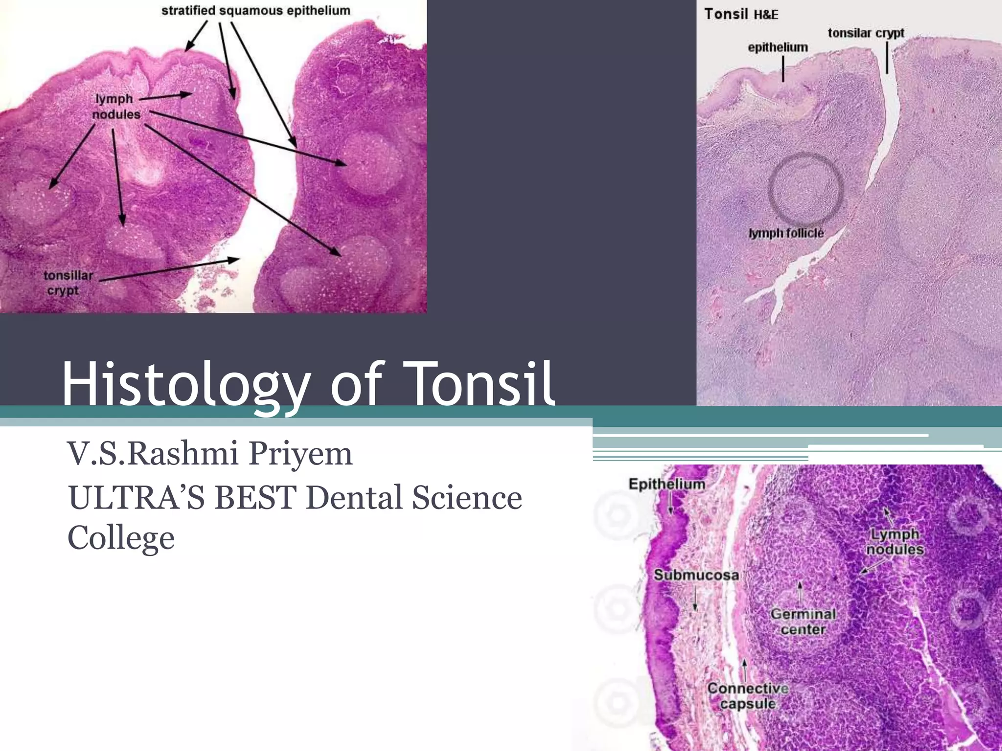
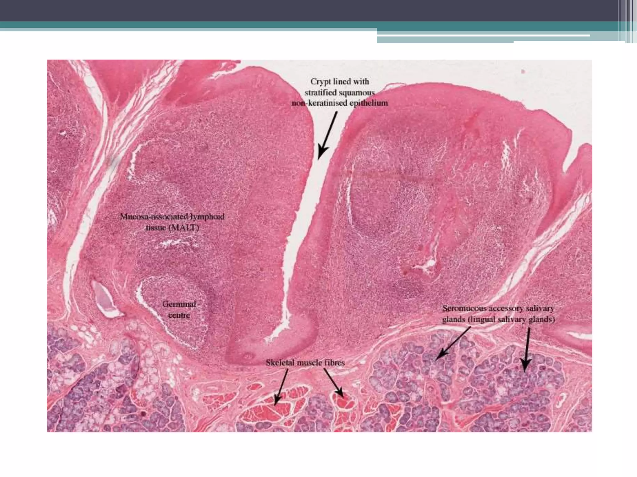
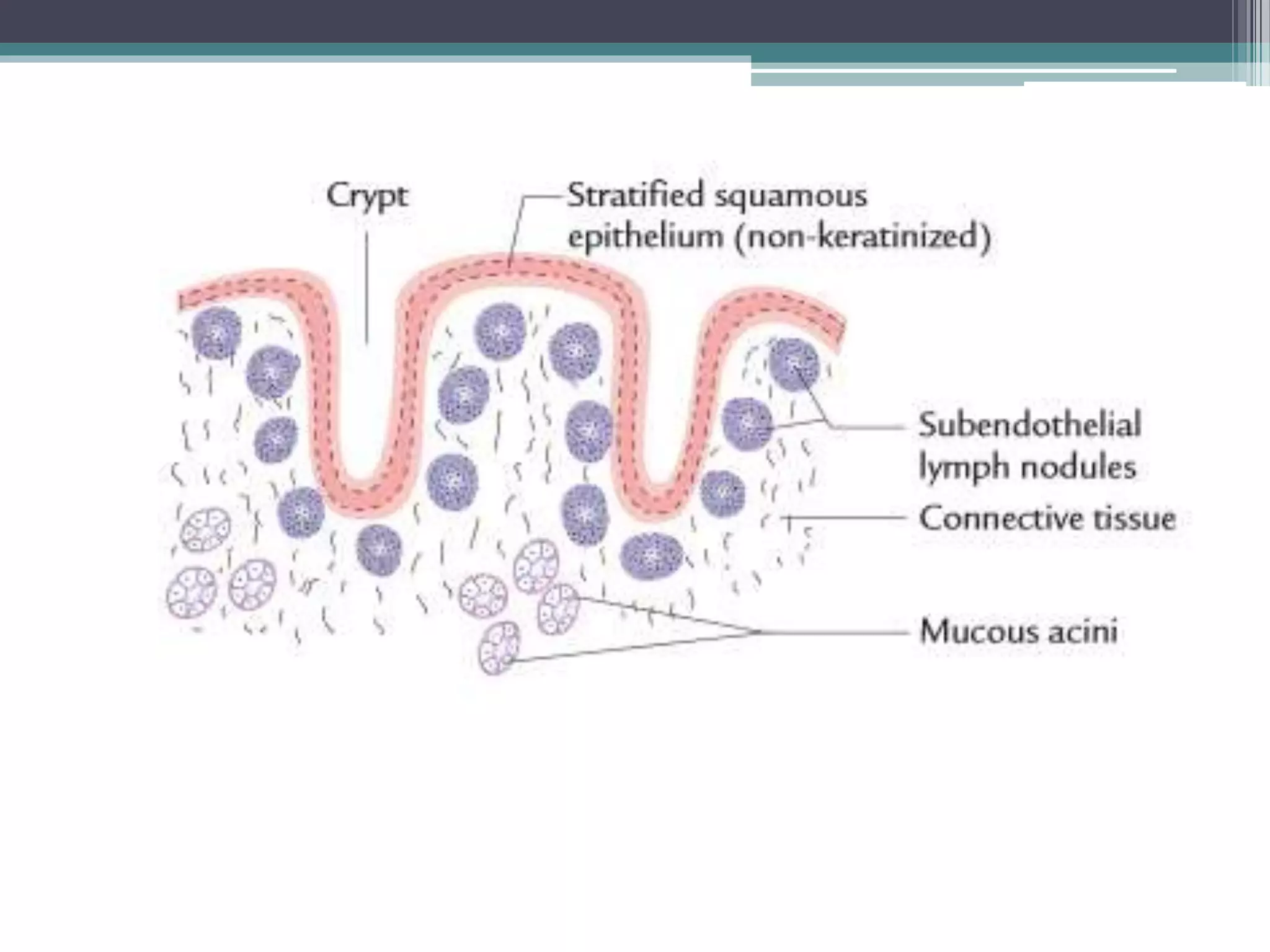
Histology of Tonsil
V.S.Rashmi Priyem
ULTRA’S BEST Dental Science
College
Histologically,
1. Its Oral Surface is lined by Stratified Squamous
Non-keratinized Epithelium, which Dips into
Underlying Tissues to Form Crypts
2. Presence of Lymphatic Nodules on the Sides of
the Crypts, Many with Prominent:
a. Germinal Centres
b. Mantle Zones (Corona)
3. Presence of Mucous Glands
4. Incomplete Connective Tissue Capsule
Function
• Immune Function at the Entrance of the
Oropharynx
• 1. Form the Protective Mucosal Lining
• 2. Increase Surface Area for Contact between the
Oropharyngeal Content and the Immune Cells
• 3. Separate Tonsils from underlying Connective
Tissue and Wall Them off in the Event of
Infection
Palatine Tonsil
Pharyngeal Tonsil
Lingual Tonsil
Location
• Either Side of the Oropharynx between
Palatopharyngeal and Palatoglossal Arches
• 1. Pharyngeal Surface of the Tonsil
• 2. Extend into Tonsillar Parenchyma
• 3. Between the Tonsil and underlying Connective
Tissue
• 4. Throughout Tonsillar Parenchyma
Palatoglossal and Palatopharyngeal
Arches
Clinical Significance
• Tonsillitis:
• Inflammation of the Tonsils as the result of
Bacterial or Viral Infection.
• Red, swollen palatine tonsils with Purulent
Exudates (Pus) are easily observed when the
patient opens the mouth and the tongue is
depressed.
• Patients present with sore throat, pain, fever, and
dysphagia.
• In severe cases, the infection may extend to involve
the pharynx, larynx, and auditory tube.

Histology of Tonsil

  • 1.
    Histology of Tonsil V.S.RashmiPriyem ULTRA’S BEST Dental Science College
  • 3.
    Histologically, 1. Its OralSurface is lined by Stratified Squamous Non-keratinized Epithelium, which Dips into Underlying Tissues to Form Crypts 2. Presence of Lymphatic Nodules on the Sides of the Crypts, Many with Prominent: a. Germinal Centres b. Mantle Zones (Corona) 3. Presence of Mucous Glands 4. Incomplete Connective Tissue Capsule
  • 5.
    Function • Immune Functionat the Entrance of the Oropharynx • 1. Form the Protective Mucosal Lining • 2. Increase Surface Area for Contact between the Oropharyngeal Content and the Immune Cells • 3. Separate Tonsils from underlying Connective Tissue and Wall Them off in the Event of Infection
  • 6.
  • 7.
  • 8.
  • 9.
    Location • Either Sideof the Oropharynx between Palatopharyngeal and Palatoglossal Arches • 1. Pharyngeal Surface of the Tonsil • 2. Extend into Tonsillar Parenchyma • 3. Between the Tonsil and underlying Connective Tissue • 4. Throughout Tonsillar Parenchyma
  • 10.
  • 12.
    Clinical Significance • Tonsillitis: •Inflammation of the Tonsils as the result of Bacterial or Viral Infection. • Red, swollen palatine tonsils with Purulent Exudates (Pus) are easily observed when the patient opens the mouth and the tongue is depressed. • Patients present with sore throat, pain, fever, and dysphagia. • In severe cases, the infection may extend to involve the pharynx, larynx, and auditory tube.