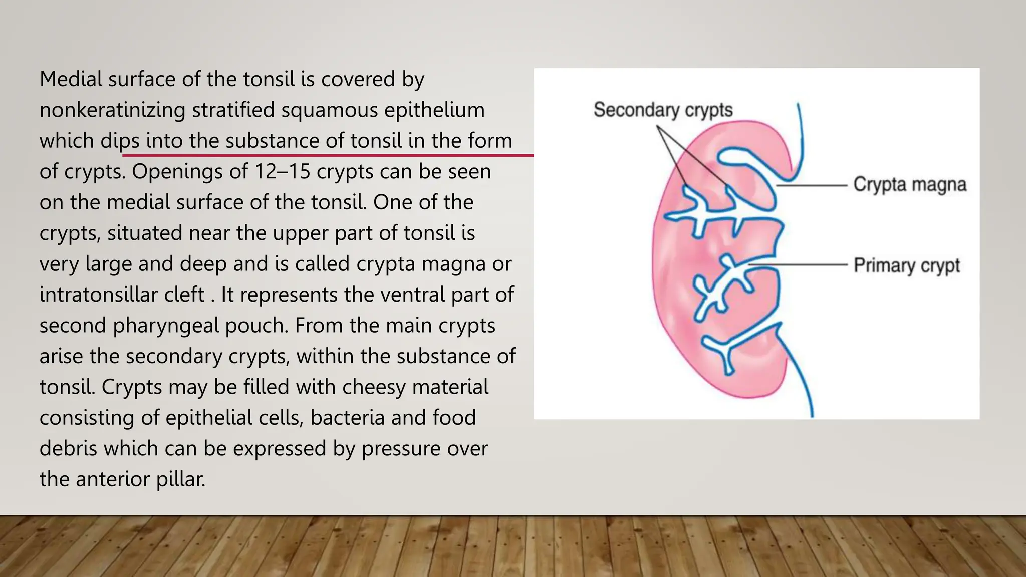
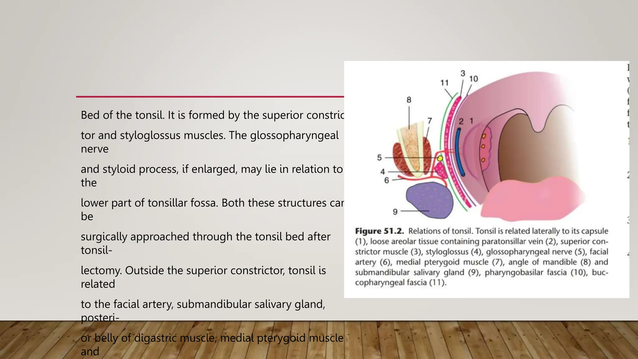
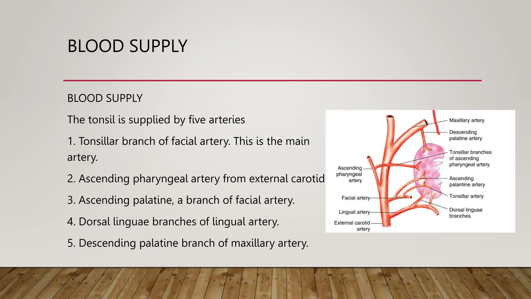
ANATOMY OF
TONSILS
SANSKRITI KUMAR
210201064
APPLIED ANATOMY
Each tonsil is an ovoid mass of lymphoid tissue situated in the lateral wall of oro-
pharynx between the anterior and posterior pillars. Actual size of the tonsil is
bigger than the one that appears from its surface as parts of tonsil extend upwards
into the soft palate, downwards into the base of tongue and anteriorly into
palatoglossal arch. A tonsil presents two surfaces—a medial and a lateral, and two
poles and upper and a lower.
Medial surface of the tonsil is covered by
nonkeratinizing stratified squamous epithelium
which dips into the substance of tonsil in the form
of crypts. Openings of 12–15 crypts can be seen
on the medial surface of the tonsil. One of the
crypts, situated near the upper part of tonsil is
very large and deep and is called crypta magna or
intratonsillar cleft . It represents the ventral part of
second pharyngeal pouch. From the main crypts
arise the secondary crypts, within the substance of
tonsil. Crypts may be filled with cheesy material
consisting of epithelial cells, bacteria and food
debris which can be expressed by pressure over
the anterior pillar.
Lateral surface of the tonsil presents a well-defined fibrous capsule. Between the
capsule and the bed of tonsil is the loose areolar tissue which makes it easy to dissect
the tonsil in the plane during tonsillectomy. It is also the site for collection of pus in
peritonsillar abscess. Some fibres of palatoglossus and palatopharyngeus muscles are
attached to the capsule of the tonsil.
Upper pole of the tonsil extends into soft palate. Its medial surface is covered by a
semilunar fold, extending between anterior and posterior pillars and enclosing a
potential space called supratonsillar fossa.
Lower pole of the tonsil is attached to the tongue. A triangular fold of mucous
membrane extends from anterior pillar to the anteroinferior part of tonsil and encloses
a space called anterior tonsillar space. The tonsil is separated from the tongue by a
sulcus called tonsillolingual sulcuswhich may be the seat of carcinoma.
Bed of the tonsil. It is formed by the superior constric-
tor and styloglossus muscles. The glossopharyngeal
nerve
and styloid process, if enlarged, may lie in relation to
the
lower part of tonsillar fossa. Both these structures can
be
surgically approached through the tonsil bed after
tonsil-
lectomy. Outside the superior constrictor, tonsil is
related
to the facial artery, submandibular salivary gland,
posteri-
or belly of digastric muscle, medial pterygoid muscle
and
BLOOD SUPPLY
BLOOD SUPPLY
The tonsil is supplied by five arteries
1. Tonsillar branch of facial artery. This is the main
artery.
2. Ascending pharyngeal artery from external carotid.
3. Ascending palatine, a branch of facial artery.
4. Dorsal linguae branches of lingual artery.
5. Descending palatine branch of maxillary artery.
VENOUS DRAINAGE
Veins from the tonsil drain into paratonsillar vein which joins the common facial
vein and the
LYMPHATIC DRAINAGE
Lymphatics from the tonsils pierce the superior constructor and drain into upper
deep cervical nodes particularly the jugulodigastric node situated below the angle
of mandible .
NERVE SUPPLY
Lesser palatine branches of sphenopalatine ganaglion ( CN V) and the
glossopharyngeal nerve provide sensory nerve supply.

Anatomy and histology of the palatine tonsils.pptx

  • 1.
  • 2.
    APPLIED ANATOMY Each tonsilis an ovoid mass of lymphoid tissue situated in the lateral wall of oro- pharynx between the anterior and posterior pillars. Actual size of the tonsil is bigger than the one that appears from its surface as parts of tonsil extend upwards into the soft palate, downwards into the base of tongue and anteriorly into palatoglossal arch. A tonsil presents two surfaces—a medial and a lateral, and two poles and upper and a lower.
  • 3.
    Medial surface ofthe tonsil is covered by nonkeratinizing stratified squamous epithelium which dips into the substance of tonsil in the form of crypts. Openings of 12–15 crypts can be seen on the medial surface of the tonsil. One of the crypts, situated near the upper part of tonsil is very large and deep and is called crypta magna or intratonsillar cleft . It represents the ventral part of second pharyngeal pouch. From the main crypts arise the secondary crypts, within the substance of tonsil. Crypts may be filled with cheesy material consisting of epithelial cells, bacteria and food debris which can be expressed by pressure over the anterior pillar.
  • 4.
    Lateral surface ofthe tonsil presents a well-defined fibrous capsule. Between the capsule and the bed of tonsil is the loose areolar tissue which makes it easy to dissect the tonsil in the plane during tonsillectomy. It is also the site for collection of pus in peritonsillar abscess. Some fibres of palatoglossus and palatopharyngeus muscles are attached to the capsule of the tonsil. Upper pole of the tonsil extends into soft palate. Its medial surface is covered by a semilunar fold, extending between anterior and posterior pillars and enclosing a potential space called supratonsillar fossa. Lower pole of the tonsil is attached to the tongue. A triangular fold of mucous membrane extends from anterior pillar to the anteroinferior part of tonsil and encloses a space called anterior tonsillar space. The tonsil is separated from the tongue by a sulcus called tonsillolingual sulcuswhich may be the seat of carcinoma.
  • 5.
    Bed of thetonsil. It is formed by the superior constric- tor and styloglossus muscles. The glossopharyngeal nerve and styloid process, if enlarged, may lie in relation to the lower part of tonsillar fossa. Both these structures can be surgically approached through the tonsil bed after tonsil- lectomy. Outside the superior constrictor, tonsil is related to the facial artery, submandibular salivary gland, posteri- or belly of digastric muscle, medial pterygoid muscle and
  • 6.
    BLOOD SUPPLY BLOOD SUPPLY Thetonsil is supplied by five arteries 1. Tonsillar branch of facial artery. This is the main artery. 2. Ascending pharyngeal artery from external carotid. 3. Ascending palatine, a branch of facial artery. 4. Dorsal linguae branches of lingual artery. 5. Descending palatine branch of maxillary artery.
  • 7.
    VENOUS DRAINAGE Veins fromthe tonsil drain into paratonsillar vein which joins the common facial vein and the
  • 8.
    LYMPHATIC DRAINAGE Lymphatics fromthe tonsils pierce the superior constructor and drain into upper deep cervical nodes particularly the jugulodigastric node situated below the angle of mandible .
  • 9.
    NERVE SUPPLY Lesser palatinebranches of sphenopalatine ganaglion ( CN V) and the glossopharyngeal nerve provide sensory nerve supply.