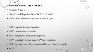
HEPATITIS A ,D, E & G
INTRODUCTION
• VIRAL HEPATITIS- is a primary infection of the liver caused by heterogeneous
group of ‘hepatitis viruses’.
• hepatitis viruses cause inflammation of liver producing identical histopathologic
lesions similar to illness such as fever, nausea, vomiting and jaundice.
CLASSIFICATION
1. INFECTIVE OR INFECTIOUS HEPATITIS
- occurs sporadically or as epidemics.
- affects mainly children and young adults.
- transmitted through faecal-oral route.
2. HOMOLOGOUS SERUM JAUNDICE/SERUM HEPATITIS/TRANSFUSION
HEPATITIS
-transmitted mainly by inoculation or blood transmission.
-associated with human or homologous antisera used for prophylaxis or therapy
TYPES OF HEPATITIS VIRUSES
• Hepatitis A and B
• Non-A non-B hepatitis (NANB): C, D, E and G.
• All are RNA viruses except type B a DNA type.
• HAV: causes infectious hepatitis.
• HBV: causes serum hepatitis.
• HCV: causes post-transfusion hepatitis.
• HDV: is defective virus, need HBV for replication.
• HEV: agent of enterically transmitted Non-A, Non-B hepatitis.
• HGV.
HEPATITIS A VIRUS -HAV
• belongs to the family PICORNAVIRIDAE.
• GENUS: Hepatovirus.
MORPHOLOGY
• Size: 27-32nm
• Shape: spherical particles with icosahedral symmetry-with
linear ssRNA.
• Has one serotype
• Does not cross-react with other hepatitis viruses.
• Genotypes: about seven are known.
Electron micrograph of 27-nm
hepatitis A virus aggregated
with antibody
RESISTANCE
• HAV is resistant to disinfectants.
-It is stable to acid, heat (60°C for 1 hour) and 20% ether
-preservation: in dried state for 1 month and stored for years at -20°C.
-It is destroyed by autoclaving, boiling in water for 5 minutes, by hot air oven, by
UV rays and by treatment with formalin or chlorine (10-15 ppm for 30 minutes).
MODE OF TRANSMISSION
• Is by faecal-oral route.
• Rarely transmitted by sexual (homosexuals through oro-genital contact)
• And parental routes (infected blood products or needle pricks).
Epidemiology
• Hosts: humans.
• Age: children and adolescents (5-14 years), majority is subclinical (80-95%) who
excrete viruses in faeces for longer time.
• Adults: more icteric (75-90%) than children-higher mortality rate
EPIDEM. CONTI…
Risk factors
• Poor personal hygiene and overcrowding.
-Mostly in children of age 10 years (90%) in developing countries.
-Adults have protective antibodies and are immune to HAV.
-Outbreaks: common in summer camps, day-care centers, families and institutions,
neonatal ICUs and among military troops.
• Recurrent epidemics: common due to faecal contamination of a single source, e.g.
Drinking water, milk or food such as raw vegetables, salad, frozen strawberries, green
onions and shell fish.
• Seasonal incidence: usually in rainfall and early winter.
• Virus excretion: in faeces may be 2 weeks before 2 weeks after appearance of jaundice.
CLINICAL MANIFESTATION
• Incubation period: 15 to 45 days (mean 30)
• Onset is subacute.
Characteristics of infection
-Pre-icteric phase: mainly GIT symptoms such as nausea and vomiting
-Icteric phase or jaundice: dark urine, yellowish sclera and mucus membrane.
• Complete recovery: occurs in 98% cases.
• No chronic or carrier cases.
COMPLICATIONS OF HAV
• Fulminant hepatitis -with severe necrosis of hepatocytes.
• Relapsing hepatitis- develops weeks to months after apparent recovery from
acute hepatitis.
• Cholestatic hepatitis: with protracted cholestatic jaundice and pruritus.
LABORATORY DIAGNOSIS OF HAV
• Specimen: Stool and serum.
• Direct demonstration: virus can be visualized by immunoelectron microscopy (IEM) in fecal
extracts during late incubation period and in the preicteric phase.
• Serology: diagnosis is usually by antibody demonstration.
• Anti HAV antibody detection by ELISA.
-Appearance of IgM during acute phase
- Liver enzymes elevated after 2 weeks- rise in IgM abs
-IgM disappears within 3-6 months
-IgG antibodies appear after a week after IgM appears, it persists for years.
Interpretation
• Anti-HAV IgM positive: indicates acute infection with HAV.
• Anti-HAV IgG antibody detection-in absence of IgM: indicates past infection or recovery.
• Detection of HAV particles
-HAV appears in stool from -2 to +2 weeks of jaundice.
-HAV is also detected in liver, bile and blood by immunoelectron microscopy.
• HAV antigen detection
-ELISA is used to detect HAV antigen from stool sample from -2 to +2 weeks of
jaundice.
Isolation
• HAV is difficult to grow in cell line.
• Non-specific findings: elevated liver enzymes and serum bilirubin level.
TREATMENT
• No specific antiviral drug available.
Prevention
• Improving hygiene
-Hand hygiene before and after use of toilet.
- Sanitary disposal of infected faecal material b (disinfection with 0.5%
hypochlorite).
-Purification of drinking water by effective filtration and adequate chlorination with
1 mg/l chlorine.
-Use of boiled water during outbreaks.
VACCINES
• Formaldehyde inactivated vaccine:
-Prepared from human fetal lung fibroblast cell lines such as MRC5 and WI 38.
-Given to children after 12 months of age.
-Single dose given intramuscularly
-booster dose: given at 6-12 months gap.
• Live attenuated vaccine
-Is a single dose given subcutaneously
- Uses H2 and L-A-1 strains of HAV
- Prepaid in human diploid cell line
• Human immunoglobulin (HAV-Ig)
-Used for post-exposure prophylaxis of intimate contacts of persons with HAV or to
travellers.
Dosage: 0.02ml/kg- gives protection for 1-2 months.
HEPATITIS D VIRUS
• Is a defective virus
• Cannot replicate by itself- depends on HBV for survival.
Morphology
• Resembles viroids
• Small size: 35nm
• Consists of circular, negative-sense ssRNA.
• Protein coat made of single protein-hepatitis d antigen (HDAg).
• Surrounded by envelope protein derived from HBsAg from HBV-hence called
defective virus.
TRANSMISSION
Parenteral route, sexual and vertical routes.
Co-infection: occurs when person is exposed to serum containing both HDV and HBV.
Super-infection: occurs when a chronic carrier of HBV is exposed to serum with HDV.
-Results in disease 30-50 days later with two phases
Acute phase- HDV replicates actively with high transaminase levels with suppression of
HBV.
Chronic phase- HDV replication decreases, HBV replication increases, transaminase
levels fluctuate, disease progresses to cirrhosis and hepatocellular carcinoma (HCC).
- Mortality rate higher (> 20%).
LABORATORY DIAGNOSIS
A. In co-infection: IgM against both HDAg and HBcAg are elevated
-IgM anti-HDV appears late and is short-lived.
B. In super-infection: HBV appears already as carrier, IgG anti-HBc is detected
-Anti-HDV would be IgM type initially
-In chronic state: mixture of IgM and igg persists for months or longer.
C. Anti HBc antibody differentiates between co-infection and super-infection.
-IgM anti HBc + IgM anti-HDV: indicates co-infection.
-IgG anti HBc + mixture of IgM and IgG anti-HDV: indicates super-infection.
• HDV RNA is detected in blood and liver before and early days of acute phase of both
infections.
Epidemiology
• Globally- 15 million people are infected with HDV- where 5% of 350 million are infected
with HBV.
• Infection occurs world wide.
Treatment
-IFN-a
Prevention
HBV vaccination and prophylaxis measures.
HEPATITIS E VIRUS (HEV)
• Causes an enterically transmitted hepatitis- in young adults.
Morphology
• Resembles caliciviruses.
• Size: small (30-32 nm), non-enveloped with icosahedral symmetry.
• Contains positive-sense, ssRNA and a specific antigen (HEV-Ag).
Genotypes
• HEV has a single serotype, five genotypes.
• Genotypes: -four are detected in humans.
- Type 1 and 2 appear to be more virulent.
- Type 3 and 4 are more attenuated and account for subclinical infections.
CLINICAL MANIFESTATION
• Incubation period: 14-60 days.
• Mostly present as self-limiting acute hepatitis lasting for several weeks.
• Fulminant hepatitis- occurs in 1-2% of cases; except for pregnant women at
higher risk (20%).
• No chronic infection or carrier state.
EPIDEMIOLOGY
• HEV is a zoonotic disease- affects monkeys, cats, pigs and dogs.
• Transmission: faecal-oral route via sewage contamination of drinking water or
food.
• Epidemics: most common in India, Asia, Africa and Central America.
• Age: young adults (20-40 years).
LABORATORY DIAGNOSIS
• Specimens: stool, serum.
• HEV RNA is detected by reverse transcriptase PCR.
• HEV virions- detected by electron microscopy.
• Serum antibody detection- by ELISA.
IgM anti-HEV: appears in serum same time with appearance of liver enzymes-
indicates acute infection.
 Igg anti-HEV: replace IgM in 2-4 weeks- indicates recovery or past infection.
TREATMENT
• No specific antiviral drugs available.
Prevention
• Same prevention measures like for HAV.
HEPATITIS G VIRUS (HGV)
• Also referred as GB virus C.
• Related to hepatitis c.
• Family: flaviviridae, genus: Pegivirus.
• Transmission: contaminated blood or blood products via sexual contact.
• Does not cause hepatitis- replicates in bone marrow and spleen.
• Classified into 6 genotypes.
• Commonly co-infects people with HIV.
THANK YOU

Hepatitis A, D, E & G

  • 1.
  • 2.
    INTRODUCTION • VIRAL HEPATITIS-is a primary infection of the liver caused by heterogeneous group of ‘hepatitis viruses’. • hepatitis viruses cause inflammation of liver producing identical histopathologic lesions similar to illness such as fever, nausea, vomiting and jaundice.
  • 3.
    CLASSIFICATION 1. INFECTIVE ORINFECTIOUS HEPATITIS - occurs sporadically or as epidemics. - affects mainly children and young adults. - transmitted through faecal-oral route. 2. HOMOLOGOUS SERUM JAUNDICE/SERUM HEPATITIS/TRANSFUSION HEPATITIS -transmitted mainly by inoculation or blood transmission. -associated with human or homologous antisera used for prophylaxis or therapy
  • 4.
    TYPES OF HEPATITISVIRUSES • Hepatitis A and B • Non-A non-B hepatitis (NANB): C, D, E and G. • All are RNA viruses except type B a DNA type. • HAV: causes infectious hepatitis. • HBV: causes serum hepatitis. • HCV: causes post-transfusion hepatitis. • HDV: is defective virus, need HBV for replication. • HEV: agent of enterically transmitted Non-A, Non-B hepatitis. • HGV.
  • 5.
    HEPATITIS A VIRUS-HAV • belongs to the family PICORNAVIRIDAE. • GENUS: Hepatovirus. MORPHOLOGY • Size: 27-32nm • Shape: spherical particles with icosahedral symmetry-with linear ssRNA. • Has one serotype • Does not cross-react with other hepatitis viruses. • Genotypes: about seven are known.
  • 6.
    Electron micrograph of27-nm hepatitis A virus aggregated with antibody
  • 7.
    RESISTANCE • HAV isresistant to disinfectants. -It is stable to acid, heat (60°C for 1 hour) and 20% ether -preservation: in dried state for 1 month and stored for years at -20°C. -It is destroyed by autoclaving, boiling in water for 5 minutes, by hot air oven, by UV rays and by treatment with formalin or chlorine (10-15 ppm for 30 minutes).
  • 8.
    MODE OF TRANSMISSION •Is by faecal-oral route. • Rarely transmitted by sexual (homosexuals through oro-genital contact) • And parental routes (infected blood products or needle pricks). Epidemiology • Hosts: humans. • Age: children and adolescents (5-14 years), majority is subclinical (80-95%) who excrete viruses in faeces for longer time. • Adults: more icteric (75-90%) than children-higher mortality rate
  • 9.
    EPIDEM. CONTI… Risk factors •Poor personal hygiene and overcrowding. -Mostly in children of age 10 years (90%) in developing countries. -Adults have protective antibodies and are immune to HAV. -Outbreaks: common in summer camps, day-care centers, families and institutions, neonatal ICUs and among military troops. • Recurrent epidemics: common due to faecal contamination of a single source, e.g. Drinking water, milk or food such as raw vegetables, salad, frozen strawberries, green onions and shell fish. • Seasonal incidence: usually in rainfall and early winter. • Virus excretion: in faeces may be 2 weeks before 2 weeks after appearance of jaundice.
  • 10.
    CLINICAL MANIFESTATION • Incubationperiod: 15 to 45 days (mean 30) • Onset is subacute. Characteristics of infection -Pre-icteric phase: mainly GIT symptoms such as nausea and vomiting -Icteric phase or jaundice: dark urine, yellowish sclera and mucus membrane. • Complete recovery: occurs in 98% cases. • No chronic or carrier cases.
  • 11.
    COMPLICATIONS OF HAV •Fulminant hepatitis -with severe necrosis of hepatocytes. • Relapsing hepatitis- develops weeks to months after apparent recovery from acute hepatitis. • Cholestatic hepatitis: with protracted cholestatic jaundice and pruritus.
  • 12.
    LABORATORY DIAGNOSIS OFHAV • Specimen: Stool and serum. • Direct demonstration: virus can be visualized by immunoelectron microscopy (IEM) in fecal extracts during late incubation period and in the preicteric phase. • Serology: diagnosis is usually by antibody demonstration.
  • 13.
    • Anti HAVantibody detection by ELISA. -Appearance of IgM during acute phase - Liver enzymes elevated after 2 weeks- rise in IgM abs -IgM disappears within 3-6 months -IgG antibodies appear after a week after IgM appears, it persists for years. Interpretation • Anti-HAV IgM positive: indicates acute infection with HAV. • Anti-HAV IgG antibody detection-in absence of IgM: indicates past infection or recovery.
  • 14.
    • Detection ofHAV particles -HAV appears in stool from -2 to +2 weeks of jaundice. -HAV is also detected in liver, bile and blood by immunoelectron microscopy. • HAV antigen detection -ELISA is used to detect HAV antigen from stool sample from -2 to +2 weeks of jaundice. Isolation • HAV is difficult to grow in cell line. • Non-specific findings: elevated liver enzymes and serum bilirubin level.
  • 15.
    TREATMENT • No specificantiviral drug available. Prevention • Improving hygiene -Hand hygiene before and after use of toilet. - Sanitary disposal of infected faecal material b (disinfection with 0.5% hypochlorite). -Purification of drinking water by effective filtration and adequate chlorination with 1 mg/l chlorine. -Use of boiled water during outbreaks.
  • 16.
    VACCINES • Formaldehyde inactivatedvaccine: -Prepared from human fetal lung fibroblast cell lines such as MRC5 and WI 38. -Given to children after 12 months of age. -Single dose given intramuscularly -booster dose: given at 6-12 months gap. • Live attenuated vaccine -Is a single dose given subcutaneously - Uses H2 and L-A-1 strains of HAV - Prepaid in human diploid cell line
  • 17.
    • Human immunoglobulin(HAV-Ig) -Used for post-exposure prophylaxis of intimate contacts of persons with HAV or to travellers. Dosage: 0.02ml/kg- gives protection for 1-2 months.
  • 18.
    HEPATITIS D VIRUS •Is a defective virus • Cannot replicate by itself- depends on HBV for survival. Morphology • Resembles viroids • Small size: 35nm • Consists of circular, negative-sense ssRNA. • Protein coat made of single protein-hepatitis d antigen (HDAg). • Surrounded by envelope protein derived from HBsAg from HBV-hence called defective virus.
  • 19.
    TRANSMISSION Parenteral route, sexualand vertical routes. Co-infection: occurs when person is exposed to serum containing both HDV and HBV. Super-infection: occurs when a chronic carrier of HBV is exposed to serum with HDV. -Results in disease 30-50 days later with two phases Acute phase- HDV replicates actively with high transaminase levels with suppression of HBV. Chronic phase- HDV replication decreases, HBV replication increases, transaminase levels fluctuate, disease progresses to cirrhosis and hepatocellular carcinoma (HCC). - Mortality rate higher (> 20%).
  • 20.
    LABORATORY DIAGNOSIS A. Inco-infection: IgM against both HDAg and HBcAg are elevated -IgM anti-HDV appears late and is short-lived. B. In super-infection: HBV appears already as carrier, IgG anti-HBc is detected -Anti-HDV would be IgM type initially -In chronic state: mixture of IgM and igg persists for months or longer. C. Anti HBc antibody differentiates between co-infection and super-infection. -IgM anti HBc + IgM anti-HDV: indicates co-infection. -IgG anti HBc + mixture of IgM and IgG anti-HDV: indicates super-infection.
  • 21.
    • HDV RNAis detected in blood and liver before and early days of acute phase of both infections. Epidemiology • Globally- 15 million people are infected with HDV- where 5% of 350 million are infected with HBV. • Infection occurs world wide. Treatment -IFN-a Prevention HBV vaccination and prophylaxis measures.
  • 22.
    HEPATITIS E VIRUS(HEV) • Causes an enterically transmitted hepatitis- in young adults. Morphology • Resembles caliciviruses. • Size: small (30-32 nm), non-enveloped with icosahedral symmetry. • Contains positive-sense, ssRNA and a specific antigen (HEV-Ag). Genotypes • HEV has a single serotype, five genotypes. • Genotypes: -four are detected in humans. - Type 1 and 2 appear to be more virulent. - Type 3 and 4 are more attenuated and account for subclinical infections.
  • 23.
    CLINICAL MANIFESTATION • Incubationperiod: 14-60 days. • Mostly present as self-limiting acute hepatitis lasting for several weeks. • Fulminant hepatitis- occurs in 1-2% of cases; except for pregnant women at higher risk (20%). • No chronic infection or carrier state.
  • 24.
    EPIDEMIOLOGY • HEV isa zoonotic disease- affects monkeys, cats, pigs and dogs. • Transmission: faecal-oral route via sewage contamination of drinking water or food. • Epidemics: most common in India, Asia, Africa and Central America. • Age: young adults (20-40 years).
  • 25.
    LABORATORY DIAGNOSIS • Specimens:stool, serum. • HEV RNA is detected by reverse transcriptase PCR. • HEV virions- detected by electron microscopy. • Serum antibody detection- by ELISA. IgM anti-HEV: appears in serum same time with appearance of liver enzymes- indicates acute infection.  Igg anti-HEV: replace IgM in 2-4 weeks- indicates recovery or past infection.
  • 26.
    TREATMENT • No specificantiviral drugs available. Prevention • Same prevention measures like for HAV.
  • 27.
    HEPATITIS G VIRUS(HGV) • Also referred as GB virus C. • Related to hepatitis c. • Family: flaviviridae, genus: Pegivirus. • Transmission: contaminated blood or blood products via sexual contact. • Does not cause hepatitis- replicates in bone marrow and spleen. • Classified into 6 genotypes. • Commonly co-infects people with HIV.
  • 28.