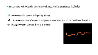
SPIROCHETES II
BY MARY MWINGA
BORRELIA
Borreliae are large, motile, refractile spirochetes.
Have 3-10 irregular and wide open coils.
Size: 5-30 µm long and 0.2-0.6 µm wide.
They stain readily with dyes and are Gram negative.
It is a normal mouth commensal.
Pathogenic under conditions such as malnutrition or viral infections.
Important pathogenic borreliae of medical importance includes:
-B. recurrentis: cause relapsing fever.
-B. vicentii: causes Vincent’s angina in association with fusiform bacilli.
-B. burgdorferi- causes Lyme disease.
LEPTOSPIRA
Belongs to the genus Leptospira.
They are actively motile with endoflagellum, delicate spirochetes.
Possess numerous closely wound spirals with hooked ends.
They do not stain readily.
They may be observed by dark ground illumination.
History:
first described by Weil (1886) –caused leptospiral jaundice in humans.
Stimson (1907) observed slender spirochetes in silver-stained sections
of kidneys from a fatal case of jaundice.
Saprophytic leptospires were isolated from water, sewage and other
sources.
Classification: Leptospira species
a)L interrogans:-causes leptospirosis or Weil’s disease.
-Pathogenic for humans.
b) L. biflexa –saprophyte (non-pathogenic).
LEPTOSPIRA INTERROGANS
Morphology
They are spiral bacteria.
Size: 5-20µm long x 0.1µm thick.
They have numerous closely set coils wand hooked ends.
Actively motile, posses a single endoflagellum attached to the pole.
Stain poorly with aniline dyes but observable by fluorescent antibody
and silver impregnation techniques.
Best observed by dark ground, phase contrast or electron microscopy
due to narrow diameters.
They rotate readily about their long axis and bending or flexing sharply.
Figure 1: Leptospira- dark ground
illumination
CULTURE CHARACTERISTICS
They are aerobic and microaerophilic.
They grow in media enriched with rabbit serum.
Liquid and semi-solid media such as Korthof’s, Stuarts's and
Fletcher’s media are used.
Semisynthetic media: such as Ellinghausen, McCullough, Johnson,
Harris (EMJH) media.
In semi-solid media: grow a few millimetres below the surface.
Incubation at 25-30°C and pH 7.2-7.5.
Generation time in media is 12-16 hours, in inoculated animals 4-8
hours.
Leptospires can also be grown on chorioallantoic membrane (CAM) of
chick embryos.
Resistance:
heat labile: killed at 50°C in 10 minutes and at 60°C in 10 seconds.
Sensitive to acid, they are destroyed by gastric juice and bile.
Readily destroyed by chlorine and other antiseptics and disinfectants.
survive for days in moist conditions at pH 6.8-8.
PATHOGENICITY
Leptospiral infections are asymptomatic.
Diseases results when infection is transmitted from one animal to another.
Leptospires have endotoxin-like component- cause fever, inflammation and
necrosis of tissues.
Haemolysin- lysis RBCs results in anaemia.
cytotoxicity factor- cause muscle spasm and dyspnoea.
Diseases
Leptospirosis:
is a zoonotic disease in rodents caused by L. interrogans.
Transmission in Humans:
Indirect contact: though water, soil or moist surface
contaminated by urine of carrier animals
Direct contact:-Through cuts or abrasions on skin or
 -Through intact mucosa of the mouth, nose or
conjunctiva.
Incubation period: 10 days (range from 2 – 26 days).
Risk factors: -lower socioeconomic status
-rainfall and floods
-occupational exposure to urine. e.g. rice fields, farmers.
Epidemiology
It is distributed worldwide.
High in areas highly populated such as Brazil, India and Thailand.
Weil’s disease:
involves hepatorenal damage associated with mild and undifferentiated
pyrexia.
in severe cases: onset is acute, with rigor, vomiting, headache and
intense injection of the eyes.
fever is irregular and subsides within 10days.
Other diseases:
Aseptic meningitis.
Renal failure.
Jaundice.
LABORATORY DIAGNOSIS
Specimens: CFS, blood and urine.
1. MICROSCOPY:
Wet films: observed in blood and urine under phase contrast or dark ground
microscope.
staining: they are stained by silver impregnation stains such as Fontana stain and
modified Steiner technique.
Appearance: L. interrogans is 6-12µm long and 0.1µm wide.
-tightly and regularly coiled with characteristic hooked ends(resemble an umbrella
handle).
-highly motile; exhibit spinning ad translational movements.
Disadvantage of Microscopy:
Less sensitive
Requires technical expertise
Serum proteins and fibrin strands in blood may resemble similar to
Leptospires.
2. CULTURE
Conditions: Leptospira is an obligate aerobe, slow growing
Cultures inoculated at 30°C for 4-6 weeks at pH 7.2 – 7.5.
Media: it is highly fastidious and requires enriched media.
On EMJH medium: it is composed of albumin fatty acid supplement
added to the basal media containing 0.1% agar.
-Growth: dense ring of organism under the surface of the medium
(Dinger’s ring).
Korthof’s and Fletcher’ semi-solid media can also be used.
3. ANIMAL INOCULATION
Blood or urine from patients is inoculated intraperitoneally into young
guinea pigs.
Peritoneal fluid is examined for Leptospires by dark ground
illumination.
4. SEROLOGICAL TESTS
a) Antibody detection
IgM antibodies: appear early within one week of illness.
IgG antibodies: appear later than IgM after few weeks.
Antibody tests
Screening tests- they are genus specific.
 includes complement fixation test, hemagglutination test, enzyme-
linked immunosorbent assay (ELISA), sensitised erythrocyte lysis
(SEL), agglutination test and indirect immunofluorescence.
Serotype Specific tests
Microscopic and macroscopic agglutination test (MAT) detects
antibodies against serovars of L. interrogans.
it is a gold standard method for diagnosis of leptospirosis.
b) Cross agglutination and absorption test (CAAT)
Detects the relatedness between the strains.
5. MOLECULAR METHODS
Polymerase chain reaction (PCR).
TREATMENT
Mild leptospirosis: oral doxycycline (100mg twice a day for 7days) or amoxicillin.
Severe leptospirosis: penicillin (1.5 million units a day for 7 days) or tetracycline,
erythromycin, ceftriaxone or cefotaxime.
PROPHYLAXIS
Chemoprophylaxis with doxycycline.
Rodent control.
Disinfection of water.
Wearing protective clothing.
Health education.

Spirochetes ii

  • 1.
  • 2.
    BORRELIA Borreliae are large,motile, refractile spirochetes. Have 3-10 irregular and wide open coils. Size: 5-30 µm long and 0.2-0.6 µm wide. They stain readily with dyes and are Gram negative. It is a normal mouth commensal. Pathogenic under conditions such as malnutrition or viral infections.
  • 3.
    Important pathogenic borreliaeof medical importance includes: -B. recurrentis: cause relapsing fever. -B. vicentii: causes Vincent’s angina in association with fusiform bacilli. -B. burgdorferi- causes Lyme disease.
  • 4.
    LEPTOSPIRA Belongs to thegenus Leptospira. They are actively motile with endoflagellum, delicate spirochetes. Possess numerous closely wound spirals with hooked ends. They do not stain readily. They may be observed by dark ground illumination.
  • 5.
    History: first described byWeil (1886) –caused leptospiral jaundice in humans. Stimson (1907) observed slender spirochetes in silver-stained sections of kidneys from a fatal case of jaundice. Saprophytic leptospires were isolated from water, sewage and other sources. Classification: Leptospira species a)L interrogans:-causes leptospirosis or Weil’s disease. -Pathogenic for humans. b) L. biflexa –saprophyte (non-pathogenic).
  • 6.
    LEPTOSPIRA INTERROGANS Morphology They arespiral bacteria. Size: 5-20µm long x 0.1µm thick. They have numerous closely set coils wand hooked ends. Actively motile, posses a single endoflagellum attached to the pole. Stain poorly with aniline dyes but observable by fluorescent antibody and silver impregnation techniques. Best observed by dark ground, phase contrast or electron microscopy due to narrow diameters. They rotate readily about their long axis and bending or flexing sharply.
  • 7.
    Figure 1: Leptospira-dark ground illumination
  • 8.
    CULTURE CHARACTERISTICS They areaerobic and microaerophilic. They grow in media enriched with rabbit serum. Liquid and semi-solid media such as Korthof’s, Stuarts's and Fletcher’s media are used. Semisynthetic media: such as Ellinghausen, McCullough, Johnson, Harris (EMJH) media. In semi-solid media: grow a few millimetres below the surface. Incubation at 25-30°C and pH 7.2-7.5. Generation time in media is 12-16 hours, in inoculated animals 4-8 hours.
  • 9.
    Leptospires can alsobe grown on chorioallantoic membrane (CAM) of chick embryos. Resistance: heat labile: killed at 50°C in 10 minutes and at 60°C in 10 seconds. Sensitive to acid, they are destroyed by gastric juice and bile. Readily destroyed by chlorine and other antiseptics and disinfectants. survive for days in moist conditions at pH 6.8-8.
  • 10.
    PATHOGENICITY Leptospiral infections areasymptomatic. Diseases results when infection is transmitted from one animal to another. Leptospires have endotoxin-like component- cause fever, inflammation and necrosis of tissues. Haemolysin- lysis RBCs results in anaemia. cytotoxicity factor- cause muscle spasm and dyspnoea.
  • 11.
    Diseases Leptospirosis: is a zoonoticdisease in rodents caused by L. interrogans. Transmission in Humans: Indirect contact: though water, soil or moist surface contaminated by urine of carrier animals Direct contact:-Through cuts or abrasions on skin or  -Through intact mucosa of the mouth, nose or conjunctiva.
  • 12.
    Incubation period: 10days (range from 2 – 26 days). Risk factors: -lower socioeconomic status -rainfall and floods -occupational exposure to urine. e.g. rice fields, farmers. Epidemiology It is distributed worldwide. High in areas highly populated such as Brazil, India and Thailand.
  • 13.
    Weil’s disease: involves hepatorenaldamage associated with mild and undifferentiated pyrexia. in severe cases: onset is acute, with rigor, vomiting, headache and intense injection of the eyes. fever is irregular and subsides within 10days. Other diseases: Aseptic meningitis. Renal failure. Jaundice.
  • 14.
    LABORATORY DIAGNOSIS Specimens: CFS,blood and urine. 1. MICROSCOPY: Wet films: observed in blood and urine under phase contrast or dark ground microscope. staining: they are stained by silver impregnation stains such as Fontana stain and modified Steiner technique. Appearance: L. interrogans is 6-12µm long and 0.1µm wide. -tightly and regularly coiled with characteristic hooked ends(resemble an umbrella handle). -highly motile; exhibit spinning ad translational movements.
  • 15.
    Disadvantage of Microscopy: Lesssensitive Requires technical expertise Serum proteins and fibrin strands in blood may resemble similar to Leptospires.
  • 16.
    2. CULTURE Conditions: Leptospirais an obligate aerobe, slow growing Cultures inoculated at 30°C for 4-6 weeks at pH 7.2 – 7.5. Media: it is highly fastidious and requires enriched media. On EMJH medium: it is composed of albumin fatty acid supplement added to the basal media containing 0.1% agar. -Growth: dense ring of organism under the surface of the medium (Dinger’s ring). Korthof’s and Fletcher’ semi-solid media can also be used.
  • 17.
    3. ANIMAL INOCULATION Bloodor urine from patients is inoculated intraperitoneally into young guinea pigs. Peritoneal fluid is examined for Leptospires by dark ground illumination. 4. SEROLOGICAL TESTS a) Antibody detection IgM antibodies: appear early within one week of illness. IgG antibodies: appear later than IgM after few weeks.
  • 18.
    Antibody tests Screening tests-they are genus specific.  includes complement fixation test, hemagglutination test, enzyme- linked immunosorbent assay (ELISA), sensitised erythrocyte lysis (SEL), agglutination test and indirect immunofluorescence. Serotype Specific tests Microscopic and macroscopic agglutination test (MAT) detects antibodies against serovars of L. interrogans. it is a gold standard method for diagnosis of leptospirosis.
  • 19.
    b) Cross agglutinationand absorption test (CAAT) Detects the relatedness between the strains. 5. MOLECULAR METHODS Polymerase chain reaction (PCR). TREATMENT Mild leptospirosis: oral doxycycline (100mg twice a day for 7days) or amoxicillin. Severe leptospirosis: penicillin (1.5 million units a day for 7 days) or tetracycline, erythromycin, ceftriaxone or cefotaxime.
  • 20.
    PROPHYLAXIS Chemoprophylaxis with doxycycline. Rodentcontrol. Disinfection of water. Wearing protective clothing. Health education.