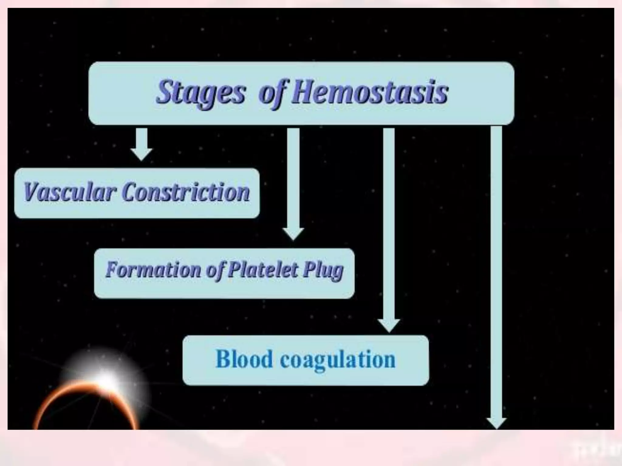
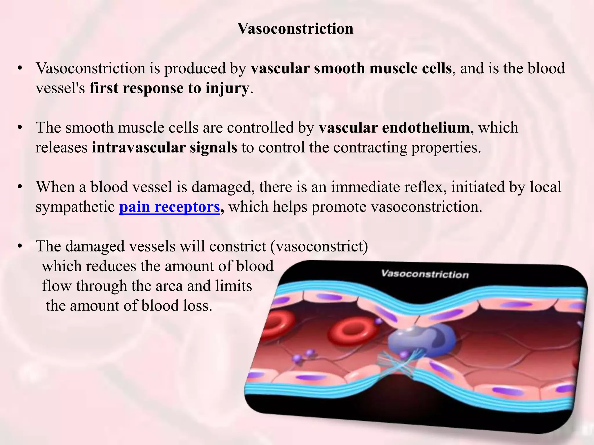
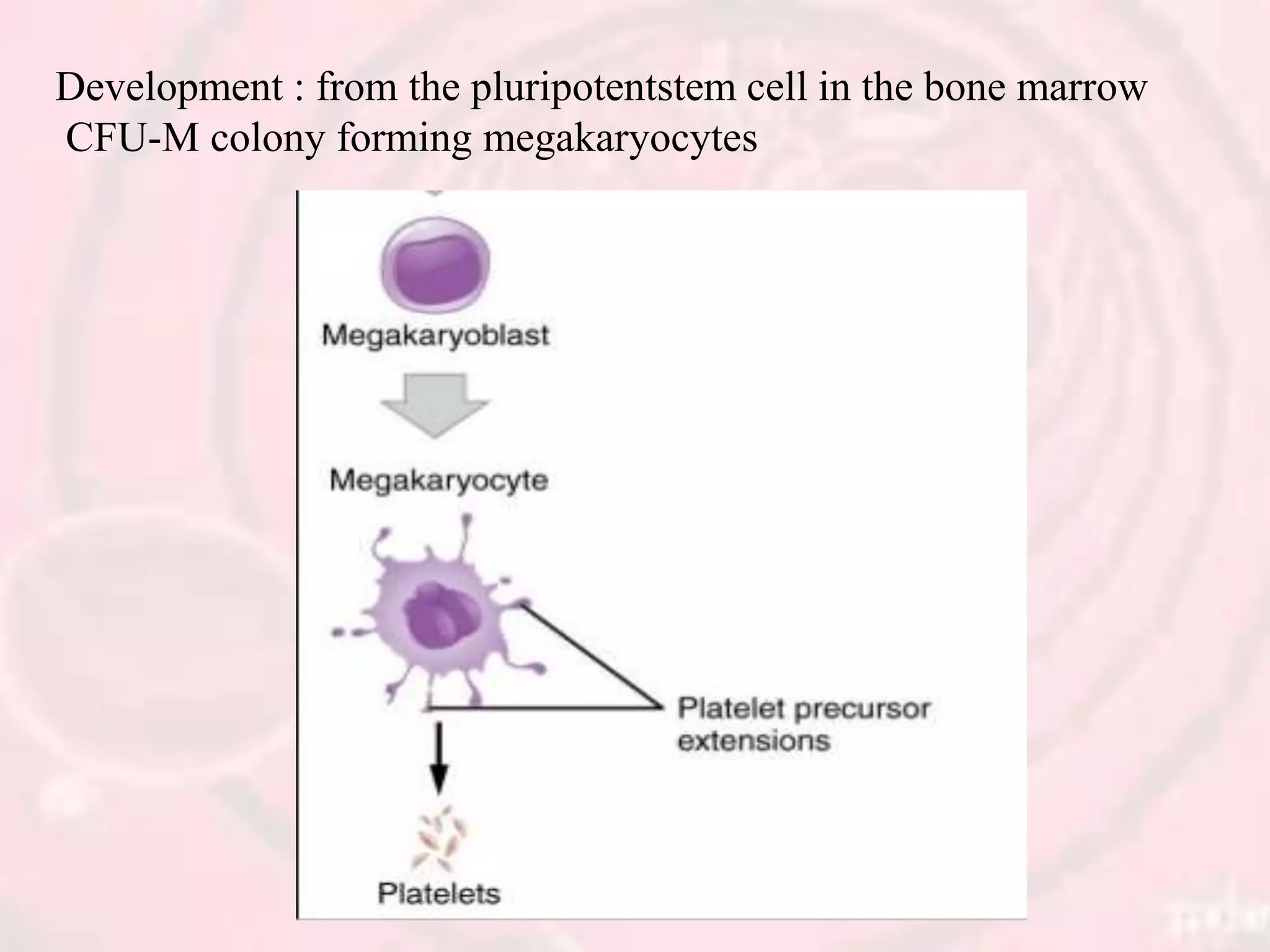
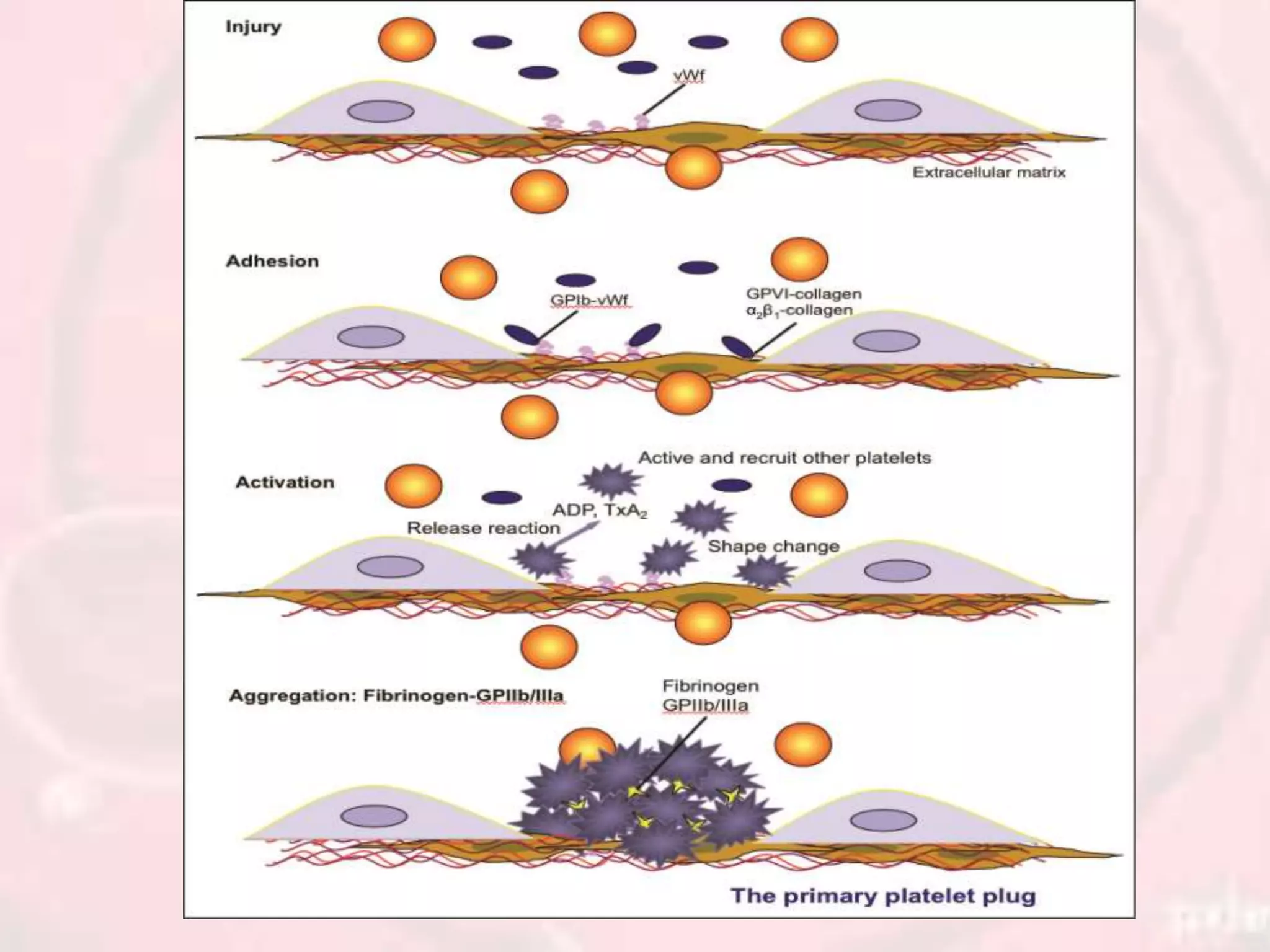
Vasoconstriction, the platelet cell membrane, the formation of a platelet plug, and the significance of the platelet mechanism for sealing vascular holes. PHARMACOLOGICAL AGENTS, INTERACTION BETWEEN THE INTRINSIC AND EXTRINSIC PATHWAYS, BLOOD CLOT, AND THE MECHANISM OF BLOOD COAGULATION
description about RBC membrane and its structural peculiarities,how it differs from other cells of our body. How this specialized cell manage homeostasis and function in a well defined manner. This presentation will also help in understanding various RBC storage lesions ,an important aspect of blood banking.
This document provides information on the various components that can be derived from human plasma. It begins by explaining that plasma is the straw-colored liquid portion of blood that suspends the blood cells and contains water, salts, enzymes, antibodies and proteins. The document then describes different plasma derivatives like fresh frozen plasma, cryoprecipitate, coagulation factors, albumin and fibrin glue/sealants. It provides details on the preparation, composition, storage, indications and administration for each plasma component.
Hemostasis is the process by which the body stops bleeding from damaged blood vessels through primary and secondary hemostasis. Primary hemostasis involves platelet adhesion and aggregation to form a platelet plug. Secondary hemostasis activates the coagulation cascade of clotting factors to form a fibrin clot over the platelet plug. A balance between coagulation and fibrinolysis maintains normal blood fluidity. Disorders can cause bleeding from inadequate clotting or thrombosis from excessive clotting.
Hemostasis is achieved through vascular constriction, platelet plug formation, and blood clotting. When a vessel is severed, platelets adhere to collagen in the damaged vessel, activate, and secrete factors that promote vasoconstriction and recruit additional platelets to form a platelet plug. Simultaneously, the coagulation cascade is initiated through intrinsic and extrinsic pathways, culminating in the formation of thrombin which converts fibrinogen to fibrin to form a blood clot.
This document discusses hemostasis, which is the biological process that controls bleeding at the site of injured blood vessels. It summarizes the key components and steps of hemostasis, including:
1. Vasoconstriction of injured blood vessels to reduce blood flow and platelet plug formation via adhesion and aggregation of platelets at the injury site.
2. Activation of the coagulation cascade through intrinsic and extrinsic pathways leading to thrombin generation and fibrin clot formation to strengthen the platelet plug.
3. Fibrinolysis by the plasminogen system which acts to dissolve clots when healing is complete to restore blood flow.
This document discusses reticulocytes, which are immature red blood cells that spend 1-3 days maturing in the bone marrow before circulating in the bloodstream. Key points include:
- Reticulocytes have residual RNA that allows them to be stained and counted to assess bone marrow activity.
- An elevated reticulocyte count indicates the bone marrow is responding to anemic stress by increasing red blood cell production.
- The reticulocyte count is used to evaluate bone marrow function and health in conditions like anemia, blood loss, or following chemotherapy/radiation.
Hemostasis and thrombosis involve the regulation of blood clotting. Normal hemostasis maintains blood fluidity but allows clotting at sites of injury. Thrombosis is pathological clotting in uninjured or minimally injured vessels. It involves platelet adhesion and activation, coagulation cascade activation, and fibrin clot formation. Counter-regulatory mechanisms normally limit clotting to the injury site. Abnormalities in blood components, vessel walls, or flow can cause hypercoagulability and thrombosis.
Apheresis is a medical technology in which blood is withdrawn from a donor or patient, separated into components, and at least one component is retained while the remainder is returned to the circulation. It is used to collect blood components like platelets, plasma, and stem cells for transfusion or therapeutic purposes. Apheresis can be performed manually or using automated machines that utilize centrifugation or filtration to separate components. It has various applications including collection of platelets, plasma exchange to remove antibodies or toxins, and stem cell collection for transplantation. Complications are usually minor but may include hypocalcemia, hypotension, and allergic reactions.
Red blood cell (RBC) disorders are conditions that affect red blood cells, the cells of blood that carry oxygen from the lungs to all parts of the body.
White blood cells - morphology, functions and variationsJilsha Cecil
White blood cells (WBCs), also known as leukocytes, are nucleated cells that perform defense functions in the body. There are 5 main types of WBCs - neutrophils, eosinophils, basophils, lymphocytes, and monocytes. Each type has distinct morphological features and functions. Neutrophils are the most abundant WBC and form the first line of defense via phagocytosis. Eosinophils and basophils are involved in allergic responses. Lymphocytes mediate humoral and cell-mediated immunity. Monocytes differentiate into macrophages and also phagocytose pathogens. WBC counts can become elevated or decreased in various physiological and pathological conditions.
This document discusses coagulation factors and blood clotting. It defines coagulation as the process where blood loses fluidity and forms a jelly-like clot. Thirteen coagulation factors are involved in a cascade of reactions to form a clot. The cascade involves the formation of prothrombin activator through the intrinsic and extrinsic pathways, followed by the conversion of prothrombin to thrombin and fibrinogen to fibrin. Deficiencies in specific factors can cause bleeding disorders like hemophilia. The clot then undergoes retraction and may be broken down through fibrinolysis.
Secondary Haemostasis involves the formation of fibrin clots via the blood coagulation cascade. This involves complex sequential reactions between coagulation factors. The coagulation factors are classified based on their physical properties like molecular weight or functional properties like being substrates, cofactors, or enzymes. Vitamin K plays an important role by allowing coagulation factors to bind to phospholipid surfaces. The coagulation cascade is tightly regulated by naturally occurring inhibitors like antithrombin III and protein C to prevent excessive clotting.
The document discusses haematopoiesis, or blood cell formation. It defines the process and sites where it occurs, including the bone marrow, thymus, lymph nodes, spleen and liver. It describes the progression of haematopoiesis from embryonic stages through postnatal development when the bone marrow becomes the primary site. The functions of different haematopoietic organs are outlined. Stem cells differentiate into committed progenitor cells that give rise to mature blood cells through the influence of various growth factors and cytokines.
Here's important & condensed ppt slides about hemostasis and its orchestrated steps and cogulation cascade, roles of endothelium,platelets and Coagulation protiens....!
Total leukocyte count is determined by counting white blood cells in a hemocytometer. Leukocytosis is an increase above 10,000 cells/mm3 while leukopenia is a decrease below 4,000 cells/mm3. Leukocytosis can be caused by infections, inflammation, or conditions like leukemia. Leukopenia is seen in some viral infections, bone marrow suppression, anemia, or drug/radiation effects. A differential count identifies percentages of neutrophils, lymphocytes, monocytes, eosinophils, and basophils from a blood smear to help diagnose conditions.
The fibrinolytic system prevents excessive fibrin deposition and regulates clot dissolution to maintain a balance between coagulation and fibrinolysis. Key components include plasminogen and its activators, such as tissue plasminogen activator and urokinase, as well as inhibitors that regulate the system. Fibrinolysis is initiated when fibrin is formed and binds plasminogen and activators, localizing plasmin generation. Plasmin then degrades fibrin into degradation products, preventing inappropriate clotting. tight regulation by activator inhibitors and antiplasmins ensures fibrin deposition is removed in a controlled manner without causing bleeding.
The document discusses the process of coagulation and fibrinolysis. It describes the three major systems involved - the vessel wall, platelets, and the coagulation cascade. The coagulation cascade involves multiple coagulation factors and pathways. Fibrinolysis is the breakdown of clots by plasmin. The document also discusses inhibitors and regulators of coagulation, including the roles of vitamin K, thrombomodulin, and tissue factor pathway inhibitor.
Reticulocytes are immature red blood cells that contain RNA and cytoplasmic remnants from earlier stages of development. A reticulocyte count provides information about bone marrow response and red blood cell production. There are four stages of reticulocyte maturation defined by their morphological appearance after staining. A reticulocyte count can be performed manually using supravital staining or automatically using flow cytometry to measure RNA levels. An increased reticulocyte count indicates bone marrow response to anemia while a decreased count suggests impaired red blood cell production.
1. Hyperemia and congestion refer to localized increases in blood volume within dilated vessels and are associated with edema.
2. Edema occurs when hydrostatic pressure is increased or oncotic pressure is decreased, overwhelming the lymphatic system and causing fluid accumulation in tissues.
3. Common causes of edema include increased venous pressure from heart failure, decreased plasma proteins, lymphatic obstruction, sodium retention, and inflammation.
This is the powerpoint for the students, faculties as well as any person who study medical and any life sciences subjects , the hemostasis portion is very comprehensively covered by diagrams and descriptions from standard books. Go through this, all the best.
This document discusses the structure, composition, functions, formation and lifespan of platelets. It provides details on platelets, including that they are the smallest blood cells, lack a nucleus, and function in hemostasis through adhesion, aggregation and clot formation. Platelets are formed from megakaryocytes in the bone marrow, have a lifespan of 8-12 days, and are destroyed by macrophages in the spleen.
This document summarizes various platelet disorders including their causes, characteristics, diagnosis and treatment. It discusses decreased platelet production from bone marrow issues as well as increased platelet destruction from immune or non-immune causes. Specific disorders covered include idiopathic thrombocytopenic purpura, hemolytic uremic syndrome, thrombotic thrombocytopenic purpura, sequestration, Kasabach-Merritt syndrome, and others. Diagnostic tests and treatment approaches are provided for each condition.
This document provides information on platelets (thrombocytes), including their morphology, ultrastructure, functional organization, metabolism, and functions in hemostasis and thrombosis. It discusses qualitative and quantitative platelet disorders and their causes. Platelets are cytoplasmic fragments of megakaryocytes that circulate in the blood. They have a discoid shape and contain granules. Platelets play an essential role in hemostasis, thrombosis, inflammation, and wound healing through adhesion, aggregation, and secretion of various factors. Qualitative platelet disorders can be hereditary or acquired and involve defects in platelet structure, number, or function.
The document provides instructions for drawing blood from donors. It states that donors must be healthy and comfortable, and the skin where blood will be drawn must be thoroughly cleaned. It also notes that blood is drawn into bottles or bags containing an anticoagulant solution to prevent clotting, and several safety and quality control procedures are followed before and after drawing blood.
This document summarizes the process of hemostasis, including platelet activation and aggregation to form a loose plug, coagulation involving prothrombin and fibrinogen to stabilize the clot, and fibrinolysis by plasmin to dissolve the clot. It discusses the roles of platelets, coagulation factors, fibrinogen and fibrin in clot formation. It also mentions how anticoagulants prevent coagulation to treat cardiovascular diseases and how fibrinolytics dissolve clots to limit tissue damage.
This document discusses proteins and nucleic acids in tissues and various methods for demonstrating them. It covers:
1. Protein composition and types including structural, enzymes, and complement proteins.
2. Methods for demonstrating proteins including immunohistochemistry, histochemical staining of amino acids or enzymes, and physical properties.
3. Nucleic acid composition, structure of DNA and RNA, and their functions in storage of genetic information and protein coding.
4. Techniques for demonstrating DNA including Feulgen reaction, fluorescent staining, and in situ hybridization. Methods for RNA include methyl green-pyronin staining and enzymatic digestion.
This document summarizes blood coagulation and hemostasis. It discusses platelets, the platelet plug formation process, and the intrinsic and extrinsic pathways of blood coagulation. Disorders of coagulation covered include hemophilia A/B/C, von Willebrand disease, and purpura. Tests of hemostatic function like bleeding time, clotting time, and prothrombin time are also outlined. Anticoagulants like heparin and warfarin are briefly described.
Platelets are disk-like cell fragments that originate from megakaryocytes and play a key role in hemostasis. When a blood vessel is damaged, platelets adhere to the site of injury and aggregate to form a platelet plug. This triggers the coagulation cascade, resulting in a fibrin clot that further strengthens the platelet plug. The clot then contracts through actin and myosin in the platelets. Over time, plasmin dissolves the clot through fibrinolysis, restoring blood flow in the vessel. A balance of pro-coagulant and anticoagulant factors normally maintains blood in a fluid state.
This document summarizes the process of hemostasis (blood clotting and dissolution). It discusses the key events in primary hemostasis involving platelet plug formation and secondary hemostasis involving the coagulation cascade and fibrin clot formation. It describes the clotting factors, coagulation pathways (extrinsic and intrinsic), and natural anticoagulants like heparin. Applied aspects such as causes of bleeding disorders and thromboembolic conditions are also covered at a high level.
The document discusses hemostasis, the process by which bleeding is prevented after vascular injury. It describes the four main mechanisms: vasoconstriction, formation of a platelet plug, coagulation pathways, and fibrinolytic phase. Specifically, it details the roles of platelets, calcium ions, coagulation factors, fibrinogen and thrombin in forming a blood clot via the intrinsic and extrinsic coagulation pathways. It also discusses anticoagulants that prevent unnecessary clotting and plasmin which lyses blood clots.
This document discusses hemostasis, which is the biological process that controls bleeding at the site of injured blood vessels. It begins with an overview of hemostasis and its importance in maintaining blood fluidity and integrity of blood vessels. It then covers the components involved, including blood vessels, platelets, plasma coagulation factors, and the fibrinolytic system. The key stages and mechanisms of hemostasis are explained, including primary hemostasis mediated by platelets and secondary hemostasis involving the coagulation cascade and thrombin production. Inhibitors that regulate coagulation are also summarized.
Hemostasis and coagulation of blood For M.Sc & Basic Medical Students by Pand...Pandian M
Blood coagulation
Mechanism of coagulation
STAGES OF HEMOSTASIS
Coagulation of blood
Factors involved in blood clotting
Enzyme cascade theory
Mechanisms for formation of prothrombin activator
Fibrinolysis
Anticlotting mechanism in the body
Applied physiology
This document provides information about platelets, including their structure, formation, and role in hemostasis (blood clotting).
Platelets are cell fragments without a nucleus that play a key role in hemostasis. They have a cell membrane, microtubules, and cytoplasm containing granules and proteins. Platelets are formed from fragmentation of large megakaryocytes in the bone marrow.
During hemostasis, platelets initially adhere to damaged blood vessel walls, become activated, and aggregate to form a platelet plug. This is followed by a blood coagulation cascade that converts prothrombin to thrombin, leading to conversion of fibrinogen to fibrin and formation of a fibrin clot. Overall, the
This is the power point that explains about the blood and blood cells. Power point describes about the mechanism of coagulation and defense cells of our circulatory system.
This document summarizes information about blood coagulation, blood groups, and related topics. It discusses hemostasis, the three stages of coagulation (formation of prothrombin activator, conversion of prothrombin to thrombin, and conversion of fibrinogen to fibrin). It also describes blood clotting factors, tests for blood clotting like prothrombin time, and bleeding disorders like hemophilia. Additionally, it covers blood grouping systems like ABO and Rh, and provides guidance on dental management of patients with bleeding disorders.
Hemostasis is the process by which bleeding is stopped. It occurs via mechanical, chemical, and thermal means. Mechanical hemostasis involves direct pressure, gauze packing, and suturing or ligating cut blood vessels. Chemical hemostasis occurs via platelet plug formation and blood coagulation, while thermal hemostasis involves vasoconstriction to reduce blood flow to the site of injury. Together, these processes form a clot to seal the damaged vessel until tissue repair can take place.
Hemostasis is normal physiological mechanism by which blood in fluid state in vascular system normally and prevention of bleeding by Hemostasis by complex interactions of blood vessels wall, plasma proteins and platelets.
Platelets play a key role in hemostasis through adhesion, activation, and aggregation at the site of vascular injury to form a platelet plug. The coagulation cascade then forms a blood clot through a series of coagulation factor activations. This process is regulated by anti-coagulation mechanisms including thrombomodulin and the fibrinolytic system. Abnormalities can cause excessive bleeding from issues like thrombocytopenia or vitamin K deficiency, or excessive clotting from conditions like deep vein thrombosis. Laboratory tests evaluate platelet count and function as well as coagulation factor levels.
The document discusses the control of coagulation through various proteins and pathways that regulate clot formation and dissolution. Coagulation begins with tissue factor activating the coagulation cascade to form a clot. This is regulated by tissue factor pathway inhibitor and proteins that inhibit specific coagulation factors, such as protein C inhibiting factors V and VIII. Clot dissolution is mediated by plasmin and inhibited by alpha2-antiplasmin. Together these pathways maintain a balance between clot formation and removal. Deficiencies can lead to bleeding disorders or thrombosis.
PC of Blood and Blood forming agents.pdfRAMDAS BHAT
This document provides an overview of drugs acting on blood and blood forming agents. It discusses coagulants that promote coagulation like calcium salts and vitamin K. It also discusses anticoagulants that prevent coagulation, including heparin, low molecular weight heparins, direct thrombin inhibitors, factor Xa inhibitors, and vitamin K antagonists like warfarin. The document provides details on the mechanisms of coagulation, platelet function, fibrinolysis, and conditions requiring treatment with coagulants or anticoagulants.
This document discusses platelets and the process of hemostasis. It notes that platelets form from megakaryocytes in the bone marrow, do not have nuclei, and play an important role in clotting. When platelets come into contact with collagen in damaged blood vessels, they swell, adhere to collagen, and secrete substances that activate nearby platelets, forming a platelet plug to stop bleeding. Blood coagulation further involves the conversion of prothrombin to thrombin and fibrinogen to fibrin fibers that trap platelets and blood cells to form a clot.
The document discusses normal coagulation, coagulopathies, and hemorrhage. It begins by introducing coagulation as a defense mechanism to maintain circulatory system integrity during vascular injury. Coagulation involves thrombin generation, fibrin clot formation, and fibrin clot dissolution through a balance of procoagulant, anticoagulant, and fibrinolytic factors. The mechanisms of hemostasis include vasoconstriction, platelet activation and aggregation, coagulation, and fibrinolysis. The major components and processes of coagulation are then described, including vitamin K-dependent proteins, fibrinolysis proteins, the roles of endothelium and platelets, the coagulation cascade, and tests to monitor blood coagulation.
The seminar presentation covered hemostasis and approaches to bleeding disorders in pediatrics. It discussed the pathophysiology, clinical features, laboratory findings and management of idiopathic thrombocytopenic purpura, Von Willebrand's disease, and hemophilia. It provided an overview of hemostasis and the coagulation cascade, approaches to evaluating a child with bleeding, and specifics on selected bleeding disorders. The presentation included descriptions of laboratory tests used to evaluate coagulation factors and identify bleeding disorders.
Coagulants and anticoagulants are used to control bleeding and clotting. Coagulants promote clotting while anticoagulants prevent clotting. Coagulants include thrombin and thromboplastin, which can be applied locally to control oozing of blood from small vessels. Transfusional coagulants include specific clotting factors administered to replace deficient factors. Nontransfusional coagulants include vitamin K, which is necessary for the production of several clotting factors in the liver.
Hemostasis is the biological process that controls bleeding at the site of injured blood vessels. It involves three key steps:
1) Platelet adhesion and activation forms a platelet plug to block blood loss.
2) Coagulation factors in the bloodstream form a fibrin clot over the platelet plug via the intrinsic and extrinsic pathways.
3) The fibrin clot is eventually dissolved by the fibrinolytic system to restore blood flow without risk of hemorrhage. Precise regulation of hemostasis maintains blood fluidity while enabling rapid clot formation in response to vessel injury.
TRANSPLANTATION IMMUNOLOGY- MLR, HLA TYPING.pptxBharath S R
PURPOSE OF HLA TYPING, CONDITIONS THAT REQUIRING TRANSPLANTATION, THE PROCESS OF HLA TYPING, HLA TYPING IMPORTANT ROLE, SEROLOGICAL TEST, Microlymphocytotoxic test, MIXED LYMPHOCYTE REACTION, Molecular HLA typing, PCR BASED METHODS/ THREE CATEGORIES, STEPS OF MOLECULAR CLONING, Sequence specific priming, Hybridization with sequence specific oligonucleotide probes (SSOP), SEQUENCE BASED HLA TYPING, CLINICAL SIGNIFICANCE OF HLA TYPING,
Genetic markers, Classical markers, DNA markers, MICROSATELLITES, AFLP, SNP: Single Nucleotide Polymorphism, QTL: Quantitative Trait Locus, Activities of marker-assisted breeding, Marker-based breeding and conventional breeding Perspectives,The application of molecular technologies to plant breeding is still facing the following drawbacks and/or challenges
Vermiculture and Vermicomposting ppt/ slideshareBharath S R
1. VERMICULTURE
Vermis = worm, cultura = growth
Composting done with the help of earthworms is known as vermicomposting
The habitat, habits, nutrition,
reproduction of different
earthworms differ, hence the
need to select the proper species
for the given need
2. IMPORTANT ASPECTS
Habits & habitat
Found everywhere expect sandy soil and soil deficient in humus
Found in upper layers of slightly damp soil
One acre of land = 50,000 earthworms on an average
3. Nutrition
Feed on dead organic matter
Secretions of the intestinal tract release the plant nutrients in simple forms in the form of castings
Castings contain 5-11 times of available N, P and K
4. Organic wastages that is paper waste, garden waste, industrial waste contribute negative impact on the environment.
Vermicomposting method is fully utilized to manage the wastes towards a more sustainable approach.
The worms that are used in the vermicomposting are Eisenia foetida and Eudrillus euginae.
With the help of earthworms, organic waste is subjected to decomposition and compost is formed and it is good manure for growth of plant with a permissible NPK values.
5. PRINCIPLES OF COMPOSTING PROCESSES
The organic material present in the municipal wastes can be converted to stable from either aerobically or anaerobically.
In case of aerobic decomposition micro-organisms oxidized organic compounds to CO2, NO2 .& NO3.
Here carbon is used as source of energy and nitrogen is recycled. This is an exothermic reaction, hence temperature rises.
In case of anaerobic decomposition organic compounds while metabolizing nutrients breakdown by the process of reduction where CH4 & CO2 are released with small amount of energy.
This is an endothermic reaction where temperature doesn’t raise much.
6. Advantages of Vermicomposting:
• Least expensive method.
• It works relatively low temperature which is helpful.
• To destroy pathogens.
• It improves the pH of the soil. Earthworm is having characteristics of changing acidic or alkaline soil to neutral soil. Waste land can also be converted to fertile land.
• Vermicomposting increases the soil texture, soil aeration, fertility and soil moisture and reduces in the water requirement in long run.
• The optimal carbon/nitrogen (C/N) ratio is available in vermicomposting, which determines the quality of compost.
7.
TEST BANK For Community Health Nursing A Canadian Perspective, 5th Edition by...Donc Test
TEST BANK For Community Health Nursing A Canadian Perspective, 5th Edition by Stamler, Verified Chapters 1 - 33, Complete Newest Version Community Health Nursing A Canadian Perspective, 5th Edition by Stamler, Verified Chapters 1 - 33, Complete Newest Version Community Health Nursing A Canadian Perspective, 5th Edition by Stamler Community Health Nursing A Canadian Perspective, 5th Edition TEST BANK by Stamler Test Bank For Community Health Nursing A Canadian Perspective, 5th Edition Pdf Chapters Download Test Bank For Community Health Nursing A Canadian Perspective, 5th Edition Pdf Download Stuvia Test Bank For Community Health Nursing A Canadian Perspective, 5th Edition Study Guide Test Bank For Community Health Nursing A Canadian Perspective, 5th Edition Ebook Download Stuvia Test Bank For Community Health Nursing A Canadian Perspective, 5th Edition Questions and Answers Quizlet Test Bank For Community Health Nursing A Canadian Perspective, 5th Edition Studocu Test Bank For Community Health Nursing A Canadian Perspective, 5th Edition Quizlet Test Bank For Community Health Nursing A Canadian Perspective, 5th Edition Stuvia Community Health Nursing A Canadian Perspective, 5th Edition Pdf Chapters Download Community Health Nursing A Canadian Perspective, 5th Edition Pdf Download Course Hero Community Health Nursing A Canadian Perspective, 5th Edition Answers Quizlet Community Health Nursing A Canadian Perspective, 5th Edition Ebook Download Course hero Community Health Nursing A Canadian Perspective, 5th Edition Questions and Answers Community Health Nursing A Canadian Perspective, 5th Edition Studocu Community Health Nursing A Canadian Perspective, 5th Edition Quizlet Community Health Nursing A Canadian Perspective, 5th Edition Stuvia Community Health Nursing A Canadian Perspective, 5th Edition Test Bank Pdf Chapters Download Community Health Nursing A Canadian Perspective, 5th Edition Test Bank Pdf Download Stuvia Community Health Nursing A Canadian Perspective, 5th Edition Test Bank Study Guide Questions and Answers Community Health Nursing A Canadian Perspective, 5th Edition Test Bank Ebook Download Stuvia Community Health Nursing A Canadian Perspective, 5th Edition Test Bank Questions Quizlet Community Health Nursing A Canadian Perspective, 5th Edition Test Bank Studocu Community Health Nursing A Canadian Perspective, 5th Edition Test Bank Quizlet Community Health Nursing A Canadian Perspective, 5th Edition Test Bank Stuvia
ABDOMINAL TRAUMA in pediatrics part one.drhasanrajab
Abdominal trauma in pediatrics refers to injuries or damage to the abdominal organs in children. It can occur due to various causes such as falls, motor vehicle accidents, sports-related injuries, and physical abuse. Children are more vulnerable to abdominal trauma due to their unique anatomical and physiological characteristics. Signs and symptoms include abdominal pain, tenderness, distension, vomiting, and signs of shock. Diagnosis involves physical examination, imaging studies, and laboratory tests. Management depends on the severity and may involve conservative treatment or surgical intervention. Prevention is crucial in reducing the incidence of abdominal trauma in children.
These lecture slides, by Dr Sidra Arshad, offer a quick overview of the physiological basis of a normal electrocardiogram.
Learning objectives:
1. Define an electrocardiogram (ECG) and electrocardiography
2. Describe how dipoles generated by the heart produce the waveforms of the ECG
3. Describe the components of a normal electrocardiogram of a typical bipolar lead (limb II)
4. Differentiate between intervals and segments
5. Enlist some common indications for obtaining an ECG
6. Describe the flow of current around the heart during the cardiac cycle
7. Discuss the placement and polarity of the leads of electrocardiograph
8. Describe the normal electrocardiograms recorded from the limb leads and explain the physiological basis of the different records that are obtained
9. Define mean electrical vector (axis) of the heart and give the normal range
10. Define the mean QRS vector
11. Describe the axes of leads (hexagonal reference system)
12. Comprehend the vectorial analysis of the normal ECG
13. Determine the mean electrical axis of the ventricular QRS and appreciate the mean axis deviation
14. Explain the concepts of current of injury, J point, and their significance
Study Resources:
1. Chapter 11, Guyton and Hall Textbook of Medical Physiology, 14th edition
2. Chapter 9, Human Physiology - From Cells to Systems, Lauralee Sherwood, 9th edition
3. Chapter 29, Ganong’s Review of Medical Physiology, 26th edition
4. Electrocardiogram, StatPearls - https://www.ncbi.nlm.nih.gov/books/NBK549803/
5. ECG in Medical Practice by ABM Abdullah, 4th edition
6. Chapter 3, Cardiology Explained, https://www.ncbi.nlm.nih.gov/books/NBK2214/
7. ECG Basics, http://www.nataliescasebook.com/tag/e-c-g-basics
NVBDCP.pptx Nation vector borne disease control programSapna Thakur
NVBDCP was launched in 2003-2004 . Vector-Borne Disease: Disease that results from an infection transmitted to humans and other animals by blood-feeding arthropods, such as mosquitoes, ticks, and fleas. Examples of vector-borne diseases include Dengue fever, West Nile Virus, Lyme disease, and malaria.
Does Over-Masturbation Contribute to Chronic Prostatitis.pptxwalterHu5
In some case, your chronic prostatitis may be related to over-masturbation. Generally, natural medicine Diuretic and Anti-inflammatory Pill can help mee get a cure.
Promoting Wellbeing - Applied Social Psychology - Psychology SuperNotesPsychoTech Services
A proprietary approach developed by bringing together the best of learning theories from Psychology, design principles from the world of visualization, and pedagogical methods from over a decade of training experience, that enables you to: Learn better, faster!
Integrating Ayurveda into Parkinson’s Management: A Holistic ApproachAyurveda ForAll
Explore the benefits of combining Ayurveda with conventional Parkinson's treatments. Learn how a holistic approach can manage symptoms, enhance well-being, and balance body energies. Discover the steps to safely integrate Ayurvedic practices into your Parkinson’s care plan, including expert guidance on diet, herbal remedies, and lifestyle modifications.
8 Surprising Reasons To Meditate 40 Minutes A Day That Can Change Your Life.pptxHolistified Wellness
We’re talking about Vedic Meditation, a form of meditation that has been around for at least 5,000 years. Back then, the people who lived in the Indus Valley, now known as India and Pakistan, practised meditation as a fundamental part of daily life. This knowledge that has given us yoga and Ayurveda, was known as Veda, hence the name Vedic. And though there are some written records, the practice has been passed down verbally from generation to generation.
3. Hemostasis:
The stoppage of bleeding or hemorrhage. Also, the stoppage of
blood flow through a blood vessel or organ of the body.
Hemostasis is the arrest of bleeding, whether it be by normal
vasoconstriction (the vessel walls closing temporarily), by an
abnormal obstruction (such as a plaque) or by coagulation or
surgical means (such as ligation).
The term comes from the Greek roots heme, blood + stasis, halt =
halt of the blood.
4. Events in Hemostasis
Hemostasis means prevention of blood loss, Hemostasis achieved several mechanism
1) Vasoconstriction
2) Formation of platelet plug
3) Blood coagulation, or formation of a fibrin clot
4) Growth of fibrous tissue in the clot
5. Vasoconstriction
• Vasoconstriction is produced by vascular smooth muscle cells, and is the blood
vessel's first response to injury.
• The smooth muscle cells are controlled by vascular endothelium, which
releases intravascular signals to control the contracting properties.
• When a blood vessel is damaged, there is an immediate reflex, initiated by local
sympathetic pain receptors, which helps promote vasoconstriction.
• The damaged vessels will constrict (vasoconstrict)
which reduces the amount of blood
flow through the area and limits
the amount of blood loss.
6. • Collagen is exposed at the site of injury, the collagen promotes platelets to adhere to
the injury site.
• Platelets release cytoplasmic granules which contain Serotonin, ADP and thromboxane
A2, all of which increase the effect of vasoconstriction.
• The spasm response becomes more effective as the amount of damage is increased.
• Vascular spasm is much more effective in smaller blood vessels.
7. Development : from the pluripotentstem cell in the bone marrow
CFU-M colony forming megakaryocytes
10. CELL MEMBRANE OF PLATELET
It is 6nm thick and contains lipids (phosholipids,
cholestrol and glycolipids), Carbohydrates (glyocalyx),
proteins and glycoproteins
Out of all glycoprotein and phospholipids are
functionally important
11. Glycoproteins
Prevents the adherence of platelets to normal
endothelium
Accelerates the adherence of platelets to collagen and
damaged endothelium in rupture blood vessels
Forms a receptor for ADP and thrombin
12. CYTOPLASM
The cytoplasm of the platelet include:
• Golgi apparatus
• ER
• Mitochondria
• Microvessels
• Microtubule
• Microfilaments
• Granules
Cytoplasm also contains
• Proteins
• Enzymes
• Hormones
• Chemical substances
13. PROTEINS
The major proteins are contractile proteins which are responsible
for the contraction of platelets:
• Actin
• Myosin
• Thrombosthenin
CHEMICAL SUBSTANCES
• Calcium ions
• Mg- ions
• Adenosine triphosphate (ATP)
• Adenosine diphosphate (ADP)
14. FUNCTION OF PLATELETS
Its surface has glycoprotein coat that adhere it to injured
endothelial cells.. Preventing bleeding
Actin, myosin& thrombostheninn that are contractile proteins..
Cause clot retraction
Secretes growth factor that promotes growth&multiplication of
vascular endothelial cells, vascular smooth cells& fibrblasts…
repair damaged vascular wall
Its membrane has phopholipids that activate intrinsic system of
blood clotting
15. LIFE SPAN OF PLATELETS
Platelets are eliminated from the circulation mainly by
the tissue macrophage system in the spleen. Its half time
is 8 to 12 days
17. PLATELET ADHESION
-{vWF} von willebrand factor – immobilizes under endothelial damage
-Collagen (hemodynamic changes)
• They have AI in its structure ; while platelet have GPIb-IX-V complex
receptor this complex will got GPIb alpha I subunit and this subunit can
bind with AI subunit of vWF domain
• In the collagen platelet interaction collagen displays alpha 1 CB3 peptide
• Platelet which have a cell surface receptor of alpha 2 beta 1 integral
• During endothelial damage alpha 2 beta integral 1 receives alpha 1 CB3
peptide of collagen and mediate them, that platelet enhnance the damaging
site
• And also GPVI proteins
interacts with collagen
18. PLATELET ACTIVATION
• Glycoprotein VI or GP-6; it is receptor for collagen , it mediates
collagen induced released of several factors
• ADP releases from dense granules, vWF from alpha granules
and also secretion of thromboxane -2
• ADP mediates in two pathways
Purinogenic signalling (receptors)
• P2Y12 receptor and P2Y1 receptor
• P2Y12 is belongs to Gi- Class of GPCR ; P2Y1 is belongs of
Gq class of GPCR
20. • ADP molecules binds the P2Y12 receptor , it cause activation of
GPCR by GDP to GTP
• Gi have a cAMP synthesis
• Activation of alpha subunit, inhibit the adenyl cylase which
inhibit the cAMP pathway
• cAMP being the seconary messenger which enter and
inactivates
Which Accumulate the calcium inside platelets off inactivating
• Calcium effective pump platelet activation
21. When ADP binds with P2Y1 it cause activation of Gq of GPCR
by GDP to GTP
PLC activation and it activates….
IP3 pathway
Intracellular release of calcium also cause platelet activation
PLC activation also have drives the granule secretion from
platelet
Its seecretion includes serotonin/PDGF, TXA-2, ADP/PAF
Serotonin and TXA-2 is mainly for vasoconstriction
TXA-2 and ADP is for platelet activation
22. PLATELET AGGREGATION
Platelet aggregation by ADY; which leads to Aformentioned confirmational
changes in platelet receptors
GPII b / III a receptors on platelets
It’s a conformational change upon platelet activation
TXA-2 secreted by activated platelet
Stimulates other platelets via, paracrinesignalling which lead to platelet aggregation
GPII b / III a receptors binds fibrinogen
Platelets stick together
Leads to platelet aggregation
23.
24. Importance of the Platelet Mechanism for Closing Vascular Holes.
This mechanism is extremely important for closing minute ruptures in very small
blood vessels that occur many thousands of times daily.
Indeed, multiple small holes through the endothelial cells themselves are often
closed by platelets actually fusing with the endothelial cells to form additional
endothelial cell membrane.
Literally thousands of small hemorrhagic areas develop each day under the skin
and throughout the internal tissues of a person who has few blood platelets.
This phenomenon does not occur in persons with normal numbers of platelets.
25. BLOOD COAGULATION IN THE RUPTURED VESSEL
The clot begins to develop in 15 to 20 seconds -been minor
1 to 2 minutes –severe
Activator substances from platelets, and from blood proteins
adhering to the traumatized vascular wall initiate the clotting
process.
Platelets also play an important role in this clot retraction
26.
27. MECHANISM OF BLOOD COAGULATION
GENERAL MECHANISM
Clotting takes place in three essential steps:
1. In response to rupture of the vessel or damage to the blood itself, a complex
cascade of chemical reactions occurs in the blood involving more than a
dozen blood coagulation factors. The net result is formation of a complex of
activated substances collectively called prothrombin activator.
2. The prothrombin activator catalyzes conversion of prothrombin into
thrombin.
3. The thrombin acts as an enzyme to convert fibrinogen into fibrin fibers that
enmesh platelets, blood cells, and plasma to form the clot.
28. CONVERSION OF PROTHROMBIN TO THROMBIN
First, prothrombin activator is formed as a result of rupture of a blood vessel or as a
result of damage to special substances in the blood.
Second, the prothrombin activator, in the presence of sufficient amounts of ionic calcium
(Ca++), causes conversion of prothrombin to thrombin
Third, the thrombin causes polymerization of fibrinogen molecules into fibrin fibers
within another 10 to 15 seconds.
Thus, the rate-limiting factor in causing blood coagulation is usually the formation of
prothrombin activator and not the subsequent reactions beyond that point, because these
terminal steps normally occur rapidly to form the clot.
Platelets also play an important role in the conversion of prothrombin to thrombin
because much of the prothrombin first attaches to prothrombin receptors on the
platelets already bound to the damaged tissue.
29. Prothrombin and Thrombin.
Prothrombin is a plasma protein, an α2-globulin, having a molecular weight of 68,700.
It is present in normal plasma in a concentration of about 15 mg/dl.
It is an unstable protein that can split easily into smaller compounds, one of which is
thrombin, which has a molecular weight of 33,700, almost exactly one half that of
prothrombin.
Prothrombin is formed continually by the liver, and it is continually being used
throughout the body for blood clotting.
If the liver fails to produce prothrombin, in a day or so prothrombin concentration in
the plasma falls too low to provide normal blood coagulation.
Vitamin K is required by the liver for normal activation of prothrombin, as well as a
few other clotting factors.
Either lack of vitamin K or the presence of liver disease that prevents normal
prothrombin formation can decrease the prothrombin to such a low level that a
bleeding tendency results.
30. CONVERSION OF FIBRINOGEN TO FIBRIN—FORMATION OF THE CLOT
Fibrinogen Formed in the Liver Is Essential for Clot Formation.
Action of Thrombin on Fibrinogen to Form Fibrin.
Thrombin is a protein enzyme with weak proteolytic capabilities.
It acts on fibrinogen to remove four low-molecular-weight peptides from each molecule
of fibrinogen, forming one molecule of fibrin monomer that has the automatic capability
to polymerize with other fibrin monomer molecules to form fibrin fibers.
Therefore, many fibrin monomer molecules polymerize within seconds into long fibrin
fibers that constitute the reticulum of the blood clot.
The fibrin monomer molecules are held together by weak non-covalent hydrogen
bonding, and the newly forming fibers are not cross-linked with one another
Therefore, the resultant clot is weak and can be broken apart with ease.
However, another process occurs during the next few minutes that greatly strengthens
the fibrin reticulum. This process involves a substance called fibrin-stabilizing factor
31. BLOOD CLOT
The clot is a meshwork
Fibrin fibers also adherse to damaged surfaces of blood vessels
CLOT RETRACTION- SERUM
Contraction causes expression of fluid from clot-serum
Platelet contractile proteins contributes greatly to the clot
retraction by activting Platelet Thrombosthenin
They also compress fibrin meshwork into smaller mass
As the clot contracts, the edges are further pulled together,
contributing ultimate state of Hemostasis
32. INITIATION OF COAGULATION
FORMTION OF PROTHROMBIN ACTIVATOR
Three mechanism are set into play by
Trauma to the vascular wall and the adjacent tissues
Contract of the blood with damaged endothelial cells
They formed in two ways
• Extrinsic pathway for initiating blood clotting
• Intrinsic pathway for initiating blood clotting
36. ROLE OF CALCIUM IONS IN BOTH PATHWAYS
• Except for the first steps in the intrinsic pathway Ca2+
ions for the promotion/ acceleration of all the blood-
clotting reactions
• In the living body Ca2+ ion concenrtion seldom falls
low enough to significantly affect the kinetics
37. INTERACTION B/W THE INTRINSIC AND EXTRINSIC
PATHWAYS
- SUMMARY OF INITIATION
LYSIS OF BLOOD CLOTS
Plasminogen or profibrinolysin when activated forms
plasmin(fibrinolysin)
Plasmin is a proteolytic enzyme
that digests fibrin fibers protein
coagulants such as fibrinogen,
Factor V, Factor VIII, prothrobin
and Factor XII
40. THERMAL ENERGY METHOD
• Heat (cautery)
• Electo cautery: it is the use of high frequency alternating current for
cutting, coagulation, dissication or fulgurating tissue in both open
and laproscopic procedure
• Monopolar electro surgery
• Bipolar electro surgery
• Bipolar electro surgery vessel sealing technology
• Argon enhanced coagulation technology
• Ultrasonic device
• Lasers