Downloaded 996 times


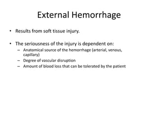

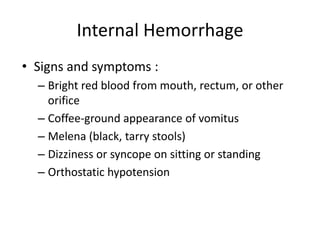


This document discusses hemorrhage, or abnormal blood loss. It describes external hemorrhage from soft tissue injuries and internal hemorrhage that can result from trauma or medical illnesses in body cavities like the chest, abdomen, pelvis or retroperitoneum. Signs of internal hemorrhage include blood from orifices or vomit. The body's response to hemorrhage is hemostasis to stop bleeding. Stages of hemorrhage are described based on percentage of circulating blood volume lost. Assessment of hemorrhage includes mental status, vital signs and interventions to control bleeding, provide oxygen and treat for shock.