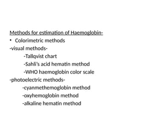
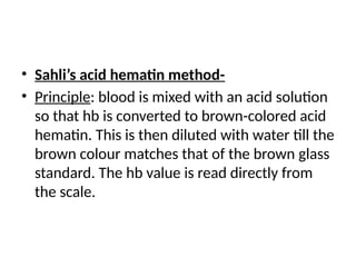
Haemoglobin Estimation
Methods for estimation of Haemoglobin-
• Colorimetric methods
-visual methods-
-Tallqvist chart
-Sahli’s acid hematin method
-WHO haemoglobin color scale
-photoelectric methods-
-cyanmethemoglobin method
-oxyhemoglobin method
-alkaline hematin method
• Gasometric method
• Chemical method
• Specific gravity method
• Sahli’s acid hematin method-
• Principle: blood is mixed with an acid solution
so that hb is converted to brown-colored acid
hematin. This is then diluted with water till the
brown colour matches that of the brown glass
standard. The hb value is read directly from
the scale.
Sahli’s acid hemati method
Equipments-
• Sahli’s hemoglobinometer
• Sahli’s pipette or hb pipette
• Small glass rod
• Dropping pipette
Reagents-
• N/10 HCl
• Distilled water
Specimen-EDTA-anticoagulated venous blood or blood
obtained by skin puncture
method
• Place N/10 HCl into Sahli’s graduated tube up to the
mark of 2 grams.
• Take blood sample in Sahli’s pipette exactly up to 20µl
mark.
• Add blood sample to the acid solution, mix with a glass
stirrer, & allow to stand for 10 mins
• Add distilled water drop by drop till the color of the
solution matches that of the brown glass standard
• Take the reading of the lower meniscus from the
graduated tube in grams
Disadvantages of Sahli’s method:
• About 95% color of acid hematin is attained at the end of 10
mins. For max color devp, much longer time(1 hr) is required.
• Perfect matching with the brown glass standard is not possible
• Carboxyhb, methhb, & sulfhb are not converted to acid
hematin. HbF cannot be estimated.
• Devp of color is slow & acid hematin solution is not stable.
• Source of light will influence the visual comparison of colors
• Personal error in matching brown glass standard with test
solution is 10%
• Color of the brown glass standard fades with time.
Cyanmethemoglobin
(hemiglobincynide)method
• Method of choice bcoz- 1) all forms of hb are
converted to cyanmethemoglobin(except sulfhb)&
2)a stable and reliable standard is available.
• Principle-potassium ferricyanide converts hb to
methhb. The methhemoglobin then further reacts
with pot.cyanide to form a stable methemoglobin
complex. The intensity of the complex found is
directly proportional to the amt of hb present in
the sample.
Drabkins solution(ph7.0-7.4)- contents
• Potassium ferricyanide- 200mg
• Potassium cyanide-50mg
• Potassium dihydrogen phosphate- 140mg
• Non-ionic detergent -1ml
• Distilled water to make-1000ml
• Cyanmethemoglobin standard solution with
known hb value.
Specimen- EDTA-anticoagulated blood or blood obtained from skin puncture.
Method-
• In a test tube take 5ml of Drabkin’s solution & to it add 20µl of blood.
Stopper the tube, mix by inverting several times, and allow to stand
for atleast 5 mins. This time is adequate for conversion of hb to
cyanmethb.
• Transfer the test sample to a cuvette. Read the absorbance in
spectrophotometer at 540 nm or in a photoelectric colorimeter using
a yellow-green filter.
• Also take the absorbance of the standard solutn. absorbance should
be read against reagent blank(drabkin’s solution).
• Hb value is derived from the formula-
• Hb in gm/dl = absorbance of test
sample/absorbance of standard × conc of
standard × dilution factor/100
• Oxyhb method- here blood sample is mixed
with a weak ammonia solution. Absorbance of
this solution is measured in a
spectrophotometer at 540nm or in a
photometer using a yellow-green filter.
Absorbance of the test sample is compared
with that of the standard solution.
• Gasometric method-
Oxygen carrying capacity of blood is measured
in a Van Slyke apparatus. The amt of hb is then
derived from the formula that 1g of hb carries
1.34ml of oxygen.
This method doesn’t measure carboxyhb,
sulfhb,& methhb.
It is time consuming & expensive
• Chemical method-
Iron content of hb is first estimated. Value of hb
is then derived indirectly from the formula
that 100g of hb contains 374mg of iron
Specific gravity method-
A rough estimate of hb is obtained from the
specific gravity of blood as determined from
copper sulphate technique
Referance range-
• Adult males- 13.0-17.0gm/dl
• Adult female(non-pregnant)-12.0-15.0gm/dl
• Adult female(pregnant)-11.0-14.0gm/dl
• Children,6-12yrs-11.5-15.5gm/dl
• Children, 6mnths-6yrs- 11.0-14.0gm/dl
• Children,2-6mnths-9.5-14.0gm/dl
• At birth(full term)-13.6-19.6gm/dl
Causes of decrease hb-
• Anemia
• Recombant position
• Excessive squezing
• Inadequate mixing
Causes of spurious or pseudo anemia-
• 3rd
trimester of pregnancy
• Splenomegaly
• Fluid retention in CCF
• Rise in plasma protein levels
Causes of increased hemoglobin-
• Following strenous exercise
• At high altitude
• In dehydration
• Prolonged application of tourniquet during
venepuncture
• polycythemia

Haemoglobin Estimation pathology practical

  • 1.
  • 2.
    Methods for estimationof Haemoglobin- • Colorimetric methods -visual methods- -Tallqvist chart -Sahli’s acid hematin method -WHO haemoglobin color scale -photoelectric methods- -cyanmethemoglobin method -oxyhemoglobin method -alkaline hematin method
  • 3.
    • Gasometric method •Chemical method • Specific gravity method
  • 4.
    • Sahli’s acidhematin method- • Principle: blood is mixed with an acid solution so that hb is converted to brown-colored acid hematin. This is then diluted with water till the brown colour matches that of the brown glass standard. The hb value is read directly from the scale.
  • 5.
    Sahli’s acid hematimethod Equipments- • Sahli’s hemoglobinometer • Sahli’s pipette or hb pipette • Small glass rod • Dropping pipette Reagents- • N/10 HCl • Distilled water Specimen-EDTA-anticoagulated venous blood or blood obtained by skin puncture
  • 6.
    method • Place N/10HCl into Sahli’s graduated tube up to the mark of 2 grams. • Take blood sample in Sahli’s pipette exactly up to 20µl mark. • Add blood sample to the acid solution, mix with a glass stirrer, & allow to stand for 10 mins • Add distilled water drop by drop till the color of the solution matches that of the brown glass standard • Take the reading of the lower meniscus from the graduated tube in grams
  • 7.
    Disadvantages of Sahli’smethod: • About 95% color of acid hematin is attained at the end of 10 mins. For max color devp, much longer time(1 hr) is required. • Perfect matching with the brown glass standard is not possible • Carboxyhb, methhb, & sulfhb are not converted to acid hematin. HbF cannot be estimated. • Devp of color is slow & acid hematin solution is not stable. • Source of light will influence the visual comparison of colors • Personal error in matching brown glass standard with test solution is 10% • Color of the brown glass standard fades with time.
  • 8.
    Cyanmethemoglobin (hemiglobincynide)method • Method ofchoice bcoz- 1) all forms of hb are converted to cyanmethemoglobin(except sulfhb)& 2)a stable and reliable standard is available. • Principle-potassium ferricyanide converts hb to methhb. The methhemoglobin then further reacts with pot.cyanide to form a stable methemoglobin complex. The intensity of the complex found is directly proportional to the amt of hb present in the sample.
  • 9.
    Drabkins solution(ph7.0-7.4)- contents •Potassium ferricyanide- 200mg • Potassium cyanide-50mg • Potassium dihydrogen phosphate- 140mg • Non-ionic detergent -1ml • Distilled water to make-1000ml • Cyanmethemoglobin standard solution with known hb value.
  • 10.
    Specimen- EDTA-anticoagulated bloodor blood obtained from skin puncture. Method- • In a test tube take 5ml of Drabkin’s solution & to it add 20µl of blood. Stopper the tube, mix by inverting several times, and allow to stand for atleast 5 mins. This time is adequate for conversion of hb to cyanmethb. • Transfer the test sample to a cuvette. Read the absorbance in spectrophotometer at 540 nm or in a photoelectric colorimeter using a yellow-green filter. • Also take the absorbance of the standard solutn. absorbance should be read against reagent blank(drabkin’s solution). • Hb value is derived from the formula-
  • 11.
    • Hb ingm/dl = absorbance of test sample/absorbance of standard × conc of standard × dilution factor/100
  • 12.
    • Oxyhb method-here blood sample is mixed with a weak ammonia solution. Absorbance of this solution is measured in a spectrophotometer at 540nm or in a photometer using a yellow-green filter. Absorbance of the test sample is compared with that of the standard solution.
  • 13.
    • Gasometric method- Oxygencarrying capacity of blood is measured in a Van Slyke apparatus. The amt of hb is then derived from the formula that 1g of hb carries 1.34ml of oxygen. This method doesn’t measure carboxyhb, sulfhb,& methhb. It is time consuming & expensive
  • 14.
    • Chemical method- Ironcontent of hb is first estimated. Value of hb is then derived indirectly from the formula that 100g of hb contains 374mg of iron Specific gravity method- A rough estimate of hb is obtained from the specific gravity of blood as determined from copper sulphate technique
  • 15.
    Referance range- • Adultmales- 13.0-17.0gm/dl • Adult female(non-pregnant)-12.0-15.0gm/dl • Adult female(pregnant)-11.0-14.0gm/dl • Children,6-12yrs-11.5-15.5gm/dl • Children, 6mnths-6yrs- 11.0-14.0gm/dl • Children,2-6mnths-9.5-14.0gm/dl • At birth(full term)-13.6-19.6gm/dl
  • 16.
    Causes of decreasehb- • Anemia • Recombant position • Excessive squezing • Inadequate mixing Causes of spurious or pseudo anemia- • 3rd trimester of pregnancy • Splenomegaly • Fluid retention in CCF • Rise in plasma protein levels
  • 17.
    Causes of increasedhemoglobin- • Following strenous exercise • At high altitude • In dehydration • Prolonged application of tourniquet during venepuncture • polycythemia