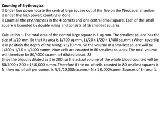
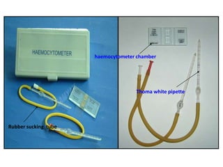
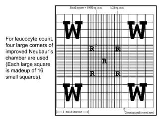
HB estimation and RBC count
HAEMOGLOBINOMETRY
โ€ข It means estimation of hemoglobin.
Methods for Hb estimation:
1. Haemiglobincyanide (cyanmethaemoglobin)
method
2. Acid hematin method (Sahli's method)
3. Oxyhemoglobin method
4. The alkaline-haematin method
5. Direct reading portable haemoglobinometers
Structure of Haemoglobin:
โ€ข Haemoglobin is a chromoprotein consisting of the colourless globin
and four red coloured haem molecules.
โ€ขHaemoglobin is a metal complex, containing an iron atom in the centre
of a porphyrin structure. The globin molecule consists of two peptide
chains (alpha and beta)-each made up of several amino acids. Haem
synthesis occurs in reticuloendothelial cells of the body, except the
mature erythrocytes, but most abundantly in the erythroid precursors.
โ€ข Succinyl co-enzyme A reacts with glycine, which by series of steps form
haem. Globin synthesis occurs in the cytoplasm of the normoblast and
reticulocyte. The polypeptide chains are manufactured on the
ribosomes.
โ€ข In each hemoglobin molecule, one haem group is inserted into a
hydrophobic pocket of one folded - polypeptide chain. Normal adult
HbA consists of four haem groups and four polypeptide chains (2 alpha
& 2 beta). RBCs have 120 days life span. As the cell ages, activity of
certain glycolytic enzymes is diminished and the cells assume a smaller
surface area. These are lysed by the RE system and are removed from
circulation. Hemoglobin also degrades and the components are recycled
.Estimation of haemoglobin
Many methods are available:-
Colorimetric methods:- Cyanmethaemoglobin method: -
Principle:- An aliquot of well mixed whole blood is taken and it is diluted in a solution of
Potassium ferricyanide and potassium cyanide. The potassium ferricyanide oxidizes hemoglobin
to hemiglobin to form hemiglobin cyanide (HiCN) which has a broad absorption maximum at a
wavelength of 540 nm. It is measured in a photometer or a spectro-photometer at 540 nm and
compared with that of a standard HiCN solution.
1. Acid hematin (Sahli's method) and alkali hematin method:
Principle :- Hemoglobin is converted to acid hematin or alkaline hematin and colour is compared
with glass standards in a comparator.
2. In the gasometric method, the oxygen carrying capacity of the blood is measured with the
Von slyke apparatuas and hemoglobin is estimated indirectly.
3. In the specific gravity method, haemoglobin is calculated from the figure for specific gravity
as determined by copper sulphate technique.
This method is employed for blood donors.
4. In chemical method Haemoglobin is determined by measuring iron content of blood. Iron
content of Hb is 0.347%. It is a complex atomic adsorption method.
Very accurate methods are cynamethaemoglobin and chemical method, latter not
used in routine practice. Cyanmethaemoglobin method is recommended by
International committee for standardisation in haemotology & is widely used in
laboratories the world over.
Acid hematin method is simple technique for routine clinical use where automated
counters are not available. (see Pra. No. 17) Acid Hematin Method (Sahli's) Principle :-
Hemoglobin is converted into acid hematin by diluting with weak acids and further
diluted with distilled water until its colour matches with that of the comparator.
Apparatus and reagents:-
Sahli type of hemoglobinometer with comparator with glass standards
Hemoglobin square tube marked both in grams and percentages.
Hemoglobin pipette marked at 20 microlitre
0.1 N HCl
Distilled water
Specimen :- Double oxalated or EDTA venous blood or capillary blood. Anticoagulated
sample must be thoroughly mixed by gentle shaking.
Method :-
1. Fill Hb square tube upto mark 20% with 0.1 N HC1. 24
2. Fill Hb pipette with the blood upto mark 20 (20 microlitre). Excess blood should be
removed with wet gauze.
3. Empty pipette into the acid in the square tube. Rinse pipette atleast three times by
drawing in and discharging the blood acid mixture. Take care to prevent air bubble
formation in acid hematin mixture.
4. Mix the acid hematin mixture in the tube with the glass rod and allow the tube to
stand for 20 minutes.
5. Now dilute the solution by adding distilled water drop by drop and stirring the
mixture all the time with glass rod. The comparator is held against good day light and
distilled water is added till the colour of the solution matches perfectly with that of
standards. Record the reading in GRAMS. Read the result at the bottom of the meniscus.
6. Clean all the apparatus. The hemoglobin pipette is cleaned by sucking and then
expelling water several times.
Errors
:- 1. Technique of collecting blood sample.
2. Manual error: In labelling, in method, in visual comparision.
3. Instrumental: โ€ข Comparator glass standards may fade with time, so dilution is more
in the specimen, hence higher values of Hb are obtained. โ€ข Calibration errors. โ€ข
Improper light can lead to variation in result.
4. All forms of Hb are not measured as only HbA and HbO2 can be converted to acid
hematin. So it does not give the true value of hemoglobin.
Range of Hemoglobin in healthy individuals:-
Adult males :- 14.0 to 17.5 gm/d1.
Adult female:- 12.3 to 15.3gm/d1.
Cord blood or new born :-13.5 to 19.5 gm/dl.
At 1 yr :-11 gm/dl Children upto 10 yrs :- 12 gm/dl
Abnormal values :- Increased - Polycythemia Vera, Dehydration, Poorly compensated
heart disease Decreased - Anemia
ABNORMAL HEMOGLOBINS : The normal adult hemoglobin is hemoglobin A
(alpha2,beta2) and Foetal hemoglobin is hemoglobin F (alpha2 gamma2) At birth HbF is
present in the proportion of 60 to 80% and gradually decreases to 0.5 to 2% in adults.
HbA2 (alpha2 delta2) is in the proportion of 1.5 to 3.5% of the normal adult hemoglobin.
HbA2 levels are increased in the range of 3.5% to 8.0% in Beta Thalassaemia Minor. HbA2
levels are decreased in iron deficiency anemia.
The abnormalities of Hb
In haem molecule disorders, formation of abnormal pigments e.g. carboxyhemoglobin,
methemoglobin,sulphhemglobin and porphyrins, due to disordered porphyrin metabolism
occurs. Except some porphyrias, these abnormalities are acquired detection of abnormal
pigment is done by chemical method & spectroscopic examination.
HEMOGLOBNOPATHIES refer to the abnormal globin molecule disorders.
โ€ข Alpha thalassemia , Beta thalassemia major ,minor
โ€ขSickle cell anemia
RBC
RBC counting
RBCs are circular, homogenous discs of nearly uniform in size, ranging from 6-8 micron in
diameter. The centre of each normal RBC is somewhat paler. They arc flexible, elastic, non-
nucleated, biconcave discs.
The chief chemical constituents are proteins, lipids, minerals and number of enzymes.
The function of the red cell is to transport oxygen and CO2 through the agency of hemoglobin.
After birth, marrow is the only organ concerned in the production of erythrocytes.
The average life span of RBCs is 120 days. The effete red cells are destroyed in the spleen by
R.E. cells.
The tissue oxygen tension controls erythropoiesis and the mass of the red cells in the
circulation.
Other hormonal factors e.g. haematopoietic growth factors like erythropoietin also regulate
the RBC production.
Clinical significance :
(I) To determine whether a person has anaemia or polycythemia and to judge their
degree.
(II) RBC count is required to determine other RBC indices.
(III) RBC count is helpful in differentiating beta thalassemia trait (minor) from iron
deficiency anemia, two important causes of microcytic hypochromic anemia.
Hemocytometer or manual method :
Principle : Blood is diluted exactly 1:200 in a Thoma's pipette, using an isotonic haem's fluid.
Diluted blood is then placed in a hemocytometer chamber. The cells in the measured volume
are then counted. This figure is multiplied by an appropriate factor (10,000) to get the
number of the cells per cu mm of blood.
Specimen :- Capillary blood or EDTA or double oxalated venous blood. Blood with
anticoagulants should be thoroughly mixed by gentle shaking.
Equipments :- 1. Thoma's pipette with red bead (RBC pipette)
2. Neubauer chamber (Improved)
3. Diluting fluid (Haem's solution)
4. Microscope with 10x and 40x objective
5. Hand-tally counter 27
Haem's solution :-
1. Sodium Sulphate-2.5gm Anticoagulant and it lowers the surface tension
2. 2. Sodium chloride - 0.5 gm Provides isotonicity
3. 3. Mercuric chloride - 0.25 gm Acts as a fixative
4. 4. Distilled water upto 100ml
Procedure :- 1. Take blood upto 0.5 mark (or upto the mark 1.0 in case of severe anemia)
in Thomas pipette without any air bubble inside.
2. Remove excess blood from the tip of the pipette. Immediately draw haem's solution to
fill up bulb of the pipette and reach upto 101 mark, taking care that no air bubble gets in.
Dilution is then 1 in 200.
3. Mix fluid in the bulb uniformly by closing the tip of the pipette with forefingers and
then rolling the pipette in horizontal position for 3 min.
4. Expel the first 2 or 3 drops fluid in the capillary portion of the pipette which has not
come in contact with the blood.
5. Then charge chamber properly, place the coverslip on the chamber, so that it covers
the ruled area. Gently put a drop of blood near the edge of a coverslip. It should not
overflow on the other side of chamber and no air bubble should get in. Allow the cells to
settle for three minutes before the counting.
Counting of Erythrocytcs
Under low power locate the central large square out of the five on the Neubauer chamber.
Under the high power, counting is done.
Count all the erythrocytes in the 4 corners and one central small square. Each of the small
square is bounded by double ruling and consists of 16 smallest squares.
Calculation :- The total area of the central large square is 1 sq.mm. The smallest square-has the
size of 1/20 mm. So that its area is 1/400 sq.mm. (1/20 x 1/20 = 1/400 sq.mm.) When coverslip
is in position the depth of the ruling is 1/10 mm. So the volume of a smallest square will be
1/400 x 1/10 = 1/4000 cumm. Now cells are counted in 80 smallest squares. The total volume
will therefore be 80/4000 cu.mm. of diluted blood. 28
Since the blood is diluted as 1 in 200, so the actual volume of the whole blood counted will be
80/4000 x 200 = 1/10,000 cumm. Therefore if the no. of cells counted in 80 smallest squares is
N, then no. of cell per cumm. is N/1/10,000/cu.mm.= N x 1 0,000/cumm Sources of Errors:- 1.
Sampling error: - failure to get free flow of the blood
a) Capillary blood - local circulatory disturbances like cyanosis or oedema.
b) Venous blood - prolonged stasis due to tourniquet. - Inadequate mixture of blood with
anticoagulant.
2. Equipment errors : - Attributed to pipette or hemocytometer. 3. Technical errors : -
Improper technique, improper volume measure, improper charging, counting wrong
calculation, touching of the objective during focussing.
Automated cell counter method (Pra. No. 17)
Erythrocyte count Normal values :- Adult male :- 4.5 to 5.9 millions/cumm
Adult female :- 4.5 to 5.1 millions/cu.mm
Abnormal values :- Increased :- High altitude, Exercise, Pregnancy, Polycythemia vera,
Chronic heart disease, Pulmonary fibrosis Decreased :- Anemias :
(i) Aplastic (ii) Hemorrhagic (iii) Hemolytic ( iv) Nutritional
Manual blood count
haemocytometer chamber
Thoma white pipette
Rubber sucking tube
For leucocyte count,
four large corners of
improved Neubaurโ€™s
chamber are used
(Each large square
is madeup of 16
small squares).

Hb estimation and rbc count

  • 1.
  • 2.
    HAEMOGLOBINOMETRY โ€ข It meansestimation of hemoglobin. Methods for Hb estimation: 1. Haemiglobincyanide (cyanmethaemoglobin) method 2. Acid hematin method (Sahli's method) 3. Oxyhemoglobin method 4. The alkaline-haematin method 5. Direct reading portable haemoglobinometers
  • 3.
    Structure of Haemoglobin: โ€ขHaemoglobin is a chromoprotein consisting of the colourless globin and four red coloured haem molecules. โ€ขHaemoglobin is a metal complex, containing an iron atom in the centre of a porphyrin structure. The globin molecule consists of two peptide chains (alpha and beta)-each made up of several amino acids. Haem synthesis occurs in reticuloendothelial cells of the body, except the mature erythrocytes, but most abundantly in the erythroid precursors. โ€ข Succinyl co-enzyme A reacts with glycine, which by series of steps form haem. Globin synthesis occurs in the cytoplasm of the normoblast and reticulocyte. The polypeptide chains are manufactured on the ribosomes. โ€ข In each hemoglobin molecule, one haem group is inserted into a hydrophobic pocket of one folded - polypeptide chain. Normal adult HbA consists of four haem groups and four polypeptide chains (2 alpha & 2 beta). RBCs have 120 days life span. As the cell ages, activity of certain glycolytic enzymes is diminished and the cells assume a smaller surface area. These are lysed by the RE system and are removed from circulation. Hemoglobin also degrades and the components are recycled
  • 4.
    .Estimation of haemoglobin Manymethods are available:- Colorimetric methods:- Cyanmethaemoglobin method: - Principle:- An aliquot of well mixed whole blood is taken and it is diluted in a solution of Potassium ferricyanide and potassium cyanide. The potassium ferricyanide oxidizes hemoglobin to hemiglobin to form hemiglobin cyanide (HiCN) which has a broad absorption maximum at a wavelength of 540 nm. It is measured in a photometer or a spectro-photometer at 540 nm and compared with that of a standard HiCN solution. 1. Acid hematin (Sahli's method) and alkali hematin method: Principle :- Hemoglobin is converted to acid hematin or alkaline hematin and colour is compared with glass standards in a comparator. 2. In the gasometric method, the oxygen carrying capacity of the blood is measured with the Von slyke apparatuas and hemoglobin is estimated indirectly. 3. In the specific gravity method, haemoglobin is calculated from the figure for specific gravity as determined by copper sulphate technique. This method is employed for blood donors. 4. In chemical method Haemoglobin is determined by measuring iron content of blood. Iron content of Hb is 0.347%. It is a complex atomic adsorption method.
  • 5.
    Very accurate methodsare cynamethaemoglobin and chemical method, latter not used in routine practice. Cyanmethaemoglobin method is recommended by International committee for standardisation in haemotology & is widely used in laboratories the world over. Acid hematin method is simple technique for routine clinical use where automated counters are not available. (see Pra. No. 17) Acid Hematin Method (Sahli's) Principle :- Hemoglobin is converted into acid hematin by diluting with weak acids and further diluted with distilled water until its colour matches with that of the comparator. Apparatus and reagents:- Sahli type of hemoglobinometer with comparator with glass standards Hemoglobin square tube marked both in grams and percentages. Hemoglobin pipette marked at 20 microlitre 0.1 N HCl Distilled water
  • 6.
    Specimen :- Doubleoxalated or EDTA venous blood or capillary blood. Anticoagulated sample must be thoroughly mixed by gentle shaking. Method :- 1. Fill Hb square tube upto mark 20% with 0.1 N HC1. 24 2. Fill Hb pipette with the blood upto mark 20 (20 microlitre). Excess blood should be removed with wet gauze. 3. Empty pipette into the acid in the square tube. Rinse pipette atleast three times by drawing in and discharging the blood acid mixture. Take care to prevent air bubble formation in acid hematin mixture. 4. Mix the acid hematin mixture in the tube with the glass rod and allow the tube to stand for 20 minutes. 5. Now dilute the solution by adding distilled water drop by drop and stirring the mixture all the time with glass rod. The comparator is held against good day light and distilled water is added till the colour of the solution matches perfectly with that of standards. Record the reading in GRAMS. Read the result at the bottom of the meniscus. 6. Clean all the apparatus. The hemoglobin pipette is cleaned by sucking and then expelling water several times.
  • 7.
    Errors :- 1. Techniqueof collecting blood sample. 2. Manual error: In labelling, in method, in visual comparision. 3. Instrumental: โ€ข Comparator glass standards may fade with time, so dilution is more in the specimen, hence higher values of Hb are obtained. โ€ข Calibration errors. โ€ข Improper light can lead to variation in result. 4. All forms of Hb are not measured as only HbA and HbO2 can be converted to acid hematin. So it does not give the true value of hemoglobin. Range of Hemoglobin in healthy individuals:- Adult males :- 14.0 to 17.5 gm/d1. Adult female:- 12.3 to 15.3gm/d1. Cord blood or new born :-13.5 to 19.5 gm/dl. At 1 yr :-11 gm/dl Children upto 10 yrs :- 12 gm/dl Abnormal values :- Increased - Polycythemia Vera, Dehydration, Poorly compensated heart disease Decreased - Anemia
  • 8.
    ABNORMAL HEMOGLOBINS :The normal adult hemoglobin is hemoglobin A (alpha2,beta2) and Foetal hemoglobin is hemoglobin F (alpha2 gamma2) At birth HbF is present in the proportion of 60 to 80% and gradually decreases to 0.5 to 2% in adults. HbA2 (alpha2 delta2) is in the proportion of 1.5 to 3.5% of the normal adult hemoglobin. HbA2 levels are increased in the range of 3.5% to 8.0% in Beta Thalassaemia Minor. HbA2 levels are decreased in iron deficiency anemia. The abnormalities of Hb In haem molecule disorders, formation of abnormal pigments e.g. carboxyhemoglobin, methemoglobin,sulphhemglobin and porphyrins, due to disordered porphyrin metabolism occurs. Except some porphyrias, these abnormalities are acquired detection of abnormal pigment is done by chemical method & spectroscopic examination. HEMOGLOBNOPATHIES refer to the abnormal globin molecule disorders. โ€ข Alpha thalassemia , Beta thalassemia major ,minor โ€ขSickle cell anemia
  • 9.
  • 10.
    RBC counting RBCs arecircular, homogenous discs of nearly uniform in size, ranging from 6-8 micron in diameter. The centre of each normal RBC is somewhat paler. They arc flexible, elastic, non- nucleated, biconcave discs. The chief chemical constituents are proteins, lipids, minerals and number of enzymes. The function of the red cell is to transport oxygen and CO2 through the agency of hemoglobin. After birth, marrow is the only organ concerned in the production of erythrocytes. The average life span of RBCs is 120 days. The effete red cells are destroyed in the spleen by R.E. cells. The tissue oxygen tension controls erythropoiesis and the mass of the red cells in the circulation. Other hormonal factors e.g. haematopoietic growth factors like erythropoietin also regulate the RBC production.
  • 11.
    Clinical significance : (I)To determine whether a person has anaemia or polycythemia and to judge their degree. (II) RBC count is required to determine other RBC indices. (III) RBC count is helpful in differentiating beta thalassemia trait (minor) from iron deficiency anemia, two important causes of microcytic hypochromic anemia.
  • 12.
    Hemocytometer or manualmethod : Principle : Blood is diluted exactly 1:200 in a Thoma's pipette, using an isotonic haem's fluid. Diluted blood is then placed in a hemocytometer chamber. The cells in the measured volume are then counted. This figure is multiplied by an appropriate factor (10,000) to get the number of the cells per cu mm of blood. Specimen :- Capillary blood or EDTA or double oxalated venous blood. Blood with anticoagulants should be thoroughly mixed by gentle shaking. Equipments :- 1. Thoma's pipette with red bead (RBC pipette) 2. Neubauer chamber (Improved) 3. Diluting fluid (Haem's solution) 4. Microscope with 10x and 40x objective 5. Hand-tally counter 27 Haem's solution :- 1. Sodium Sulphate-2.5gm Anticoagulant and it lowers the surface tension 2. 2. Sodium chloride - 0.5 gm Provides isotonicity 3. 3. Mercuric chloride - 0.25 gm Acts as a fixative 4. 4. Distilled water upto 100ml
  • 13.
    Procedure :- 1.Take blood upto 0.5 mark (or upto the mark 1.0 in case of severe anemia) in Thomas pipette without any air bubble inside. 2. Remove excess blood from the tip of the pipette. Immediately draw haem's solution to fill up bulb of the pipette and reach upto 101 mark, taking care that no air bubble gets in. Dilution is then 1 in 200. 3. Mix fluid in the bulb uniformly by closing the tip of the pipette with forefingers and then rolling the pipette in horizontal position for 3 min. 4. Expel the first 2 or 3 drops fluid in the capillary portion of the pipette which has not come in contact with the blood. 5. Then charge chamber properly, place the coverslip on the chamber, so that it covers the ruled area. Gently put a drop of blood near the edge of a coverslip. It should not overflow on the other side of chamber and no air bubble should get in. Allow the cells to settle for three minutes before the counting.
  • 14.
    Counting of Erythrocytcs Underlow power locate the central large square out of the five on the Neubauer chamber. Under the high power, counting is done. Count all the erythrocytes in the 4 corners and one central small square. Each of the small square is bounded by double ruling and consists of 16 smallest squares. Calculation :- The total area of the central large square is 1 sq.mm. The smallest square-has the size of 1/20 mm. So that its area is 1/400 sq.mm. (1/20 x 1/20 = 1/400 sq.mm.) When coverslip is in position the depth of the ruling is 1/10 mm. So the volume of a smallest square will be 1/400 x 1/10 = 1/4000 cumm. Now cells are counted in 80 smallest squares. The total volume will therefore be 80/4000 cu.mm. of diluted blood. 28 Since the blood is diluted as 1 in 200, so the actual volume of the whole blood counted will be 80/4000 x 200 = 1/10,000 cumm. Therefore if the no. of cells counted in 80 smallest squares is N, then no. of cell per cumm. is N/1/10,000/cu.mm.= N x 1 0,000/cumm Sources of Errors:- 1.
  • 16.
    Sampling error: -failure to get free flow of the blood a) Capillary blood - local circulatory disturbances like cyanosis or oedema. b) Venous blood - prolonged stasis due to tourniquet. - Inadequate mixture of blood with anticoagulant. 2. Equipment errors : - Attributed to pipette or hemocytometer. 3. Technical errors : - Improper technique, improper volume measure, improper charging, counting wrong calculation, touching of the objective during focussing. Automated cell counter method (Pra. No. 17) Erythrocyte count Normal values :- Adult male :- 4.5 to 5.9 millions/cumm Adult female :- 4.5 to 5.1 millions/cu.mm Abnormal values :- Increased :- High altitude, Exercise, Pregnancy, Polycythemia vera, Chronic heart disease, Pulmonary fibrosis Decreased :- Anemias : (i) Aplastic (ii) Hemorrhagic (iii) Hemolytic ( iv) Nutritional
  • 17.
  • 18.
    haemocytometer chamber Thoma whitepipette Rubber sucking tube
  • 19.
    For leucocyte count, fourlarge corners of improved Neubaurโ€™s chamber are used (Each large square is madeup of 16 small squares).