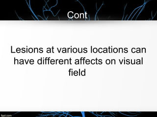
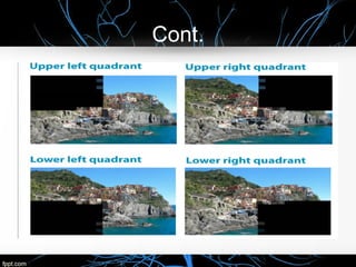
Case 5 : The bumpy lady
By:
Norah alfayez
Nouf almansour
Kholoud alshiha
Introduction
Case 5 : The bumpy lady
• A 66-year-old woman with a history of childhood rheumatic fever abruptly
develops difficulty seeing to the left as she kept bumping on items of
furniture on the left side. On examination her blood pressure is normal, but
her pulse is irregularly irregular at 88/minute, and there is a diastolic murmur
over her left cardiac border and apex. Her only neurological abnormality is a
dense left homonymous hemianopia, which on perimetry is congruent and
spares the macular area. Notably absent are left hemineglect or difficulties
with spatial manipulation (such as copying geometric designs). With her own
glasses her visual acuity is 20/20. An opticokinetic tape produces normal
nystagmus when passed in either direction across her vision. An
electrocardiogram confirms a cardiac arrythmia, and an echocardiogram
shows thickening and stenosis of the mitral valve. Computerized
tomographic (CT) scan of her head is initially normal, but 5 days later it
shows lucency without mass effect in the right occipital lobe .
Objectives
I. Rheumatic Fever.
II. Mitral stenosis.
III. Optic Pathway.
IV. Visual field & visual field defect .
V. Homonymous Hemianopia.
VI. Quadrantanopia Hemianopia.
VII. Heteronymous Hemianopia.
VIII.Treatment.
Rheumatic Fever
Cont.
Cont.
Cont.
Cont.
Cont.
Cont.
Cont.
Cont.
Cont.
Cont.
Cont.
Cont.
Cont.
Cont.
Cont.
Mitral stenosis
Cont.
Cont.
Cont.
Cont.
Cont.
Optic pathway(Anterior)
Cont.
Cont.
Cont.
Cont.(posterior)
Cont.
Cont.
Cont.
Cont.
Left visual field
Temporal right retina
Right visual field
Temporal left retina
Cont.
Cont.
Cont.
Visual Field
• The visual field refers to the total area in
which objects can be seen in the side
(peripheral) vision as you focus your eyes
on a central point.
The normal extent of field of vision
Visual field defect
• a portion of visual field missing.
Cont,
Lesions at various locations can
have different affects on visual
field
Cont.
hemianopia
• is a decreased vision or blindness in half the
visual field, usually on one side of the vertical
midline. The most common causes of this
damage are stroke, brain tumor, and trauma.
1-Homonymous Hemianopia.
• is visual field loss on the left or right side of the vertical midline.
• It can affect one eye but usually affects both eyes.
• homonymous hemianopia is hemianopic visual field loss on the same side
of both eyes.
• occurs because the right half of the brain has visual pathways for the left
hemifield of both eyes, and the left half of the brain has visual pathways for
the right hemifield of both eyes. When one of these pathways is damaged,
the corresponding visual field is lost.
Cont.
Cont.
2-Heteronymous hemianopsia
• is the loss of half of the visual field on
different sides in both eyes.
Binasal hemianopsia
• The loss of the fields surrounding the
nose
Cont.
Binasal hemianopsia
bitemporal heteronymous hemianopsia
 The loss of the fields closest to the temples.
 usually associated with lesions of the
optic chiasm
Bitemporal hemianopsia
3
-
Cont.
• Quadrantanopia is decreased vision or blindness in one quarter of the visual
field.
• depends on 1-the location of the brain damage (temporal or parietal)
2- the side of the lesion.
• For example:
1.left upper (superior) quadrantanopsia: when right temporal lobe damage
specifically to Meyer's loop
2.left lower (inferior) quadrantanopsia: when lesion of the right parietal radiation
is damaged specifically to Baum's loop.
Cont.
Source path information damage
Fibers from the
inferior retina
"Meyer's loop”
pass through the temporal lobe by
looping around the inferior horn of the
lateral ventricle.
Carry information from
the superior part of
the visual field
loss of vision in a
superior quadrant,
quadrantanopia "pie in
the sky" defect.
Fibers from the
superior retina
"Baum's loop”
Travel back through the parietal lobe
to the occipital lobe in the retro
lenticular limb of the internal capsule
to the visual cortex.
Carry information from
the inferior part of the
visual field
Loss of vision in a inferior
quadrant,
quadrantanopia "Pie in
the floor" defect or
inferior.
Cont.
Treatment
• different types of treatments are available, depending on patient
preference, age, type of Hemianopia, and how long they have been
diagnosed.
• None of these treatments 100% will cure Hemianopia, but they will
allow patients to have better perception of the world around them.
1. Audiovisual Stimulation Training.
effective treatment for Homonymous
Hemianopia
multi-sensory stimulation to improve vision
both intact and affected, are stimulated using
sound and light.
4 hours a day for 2 weeks
How it works
• A semicircular structure should be set up
with marks at the directions of
8°, 24°, 40°, and 56° on both right and left of
the central fixation point
Cont.
Cont.
At each point a light and a sound speaker should be fixed.
the patient is asked to move their gaze toward the stimulus
which just has occurred.
The stimulation should be most focused on the patients’
blind hemi-field.
2. Explorative Saccade Training
developed to train patients to make
exploratory saccades (rapid movement of the
eye between fixation points) without head
movement in the area of the visual field which
has been lost
3.Optical Visual Span Expanders
• The main idea behind this treatment is that the patients’ glasses
have a special prism mounted on them to help increase the visual
field.
• The patient must combine the image from the prism with the image
they receive from the rest of the visual field in order to perceive a
larger visual field.
4.Visual Restoration Therapy (VRT)
• Visual Restoration Therapy is another option to treat Hemianopia
patients.
• can be performed on the patient’s home computer.
• The therapy takes half-hour twice a day for two months .
How it works
• The patient focuses on a central fixation
point.
• During each session, patients focus on a
central point displayed on the screen and
respond every time they see light stimuli
appear elsewhere on the screen.
Thank you
Any questions?

hemianopia

  • 1.
    Case 5 :The bumpy lady By: Norah alfayez Nouf almansour Kholoud alshiha
  • 2.
  • 3.
    Case 5 :The bumpy lady • A 66-year-old woman with a history of childhood rheumatic fever abruptly develops difficulty seeing to the left as she kept bumping on items of furniture on the left side. On examination her blood pressure is normal, but her pulse is irregularly irregular at 88/minute, and there is a diastolic murmur over her left cardiac border and apex. Her only neurological abnormality is a dense left homonymous hemianopia, which on perimetry is congruent and spares the macular area. Notably absent are left hemineglect or difficulties with spatial manipulation (such as copying geometric designs). With her own glasses her visual acuity is 20/20. An opticokinetic tape produces normal nystagmus when passed in either direction across her vision. An electrocardiogram confirms a cardiac arrythmia, and an echocardiogram shows thickening and stenosis of the mitral valve. Computerized tomographic (CT) scan of her head is initially normal, but 5 days later it shows lucency without mass effect in the right occipital lobe .
  • 4.
    Objectives I. Rheumatic Fever. II.Mitral stenosis. III. Optic Pathway. IV. Visual field & visual field defect . V. Homonymous Hemianopia. VI. Quadrantanopia Hemianopia. VII. Heteronymous Hemianopia. VIII.Treatment.
  • 5.
  • 6.
  • 7.
  • 8.
  • 9.
  • 10.
  • 11.
  • 12.
  • 13.
  • 14.
  • 15.
  • 16.
  • 17.
  • 18.
  • 19.
  • 20.
  • 21.
  • 22.
  • 23.
  • 24.
  • 25.
  • 26.
  • 27.
  • 28.
  • 29.
  • 30.
  • 31.
  • 32.
  • 33.
  • 34.
  • 35.
    Cont. Left visual field Temporalright retina Right visual field Temporal left retina
  • 36.
  • 37.
  • 38.
  • 39.
    Visual Field • Thevisual field refers to the total area in which objects can be seen in the side (peripheral) vision as you focus your eyes on a central point.
  • 40.
    The normal extentof field of vision
  • 41.
    Visual field defect •a portion of visual field missing.
  • 42.
    Cont, Lesions at variouslocations can have different affects on visual field
  • 43.
  • 44.
    hemianopia • is adecreased vision or blindness in half the visual field, usually on one side of the vertical midline. The most common causes of this damage are stroke, brain tumor, and trauma.
  • 45.
    1-Homonymous Hemianopia. • isvisual field loss on the left or right side of the vertical midline. • It can affect one eye but usually affects both eyes. • homonymous hemianopia is hemianopic visual field loss on the same side of both eyes. • occurs because the right half of the brain has visual pathways for the left hemifield of both eyes, and the left half of the brain has visual pathways for the right hemifield of both eyes. When one of these pathways is damaged, the corresponding visual field is lost.
  • 46.
  • 47.
  • 48.
    2-Heteronymous hemianopsia • isthe loss of half of the visual field on different sides in both eyes.
  • 49.
    Binasal hemianopsia • Theloss of the fields surrounding the nose
  • 50.
  • 51.
  • 52.
    bitemporal heteronymous hemianopsia The loss of the fields closest to the temples.  usually associated with lesions of the optic chiasm
  • 53.
  • 54.
  • 55.
  • 56.
    Cont. • Quadrantanopia isdecreased vision or blindness in one quarter of the visual field. • depends on 1-the location of the brain damage (temporal or parietal) 2- the side of the lesion. • For example: 1.left upper (superior) quadrantanopsia: when right temporal lobe damage specifically to Meyer's loop 2.left lower (inferior) quadrantanopsia: when lesion of the right parietal radiation is damaged specifically to Baum's loop.
  • 57.
    Cont. Source path informationdamage Fibers from the inferior retina "Meyer's loop” pass through the temporal lobe by looping around the inferior horn of the lateral ventricle. Carry information from the superior part of the visual field loss of vision in a superior quadrant, quadrantanopia "pie in the sky" defect. Fibers from the superior retina "Baum's loop” Travel back through the parietal lobe to the occipital lobe in the retro lenticular limb of the internal capsule to the visual cortex. Carry information from the inferior part of the visual field Loss of vision in a inferior quadrant, quadrantanopia "Pie in the floor" defect or inferior.
  • 58.
  • 59.
    Treatment • different typesof treatments are available, depending on patient preference, age, type of Hemianopia, and how long they have been diagnosed. • None of these treatments 100% will cure Hemianopia, but they will allow patients to have better perception of the world around them.
  • 60.
    1. Audiovisual StimulationTraining. effective treatment for Homonymous Hemianopia multi-sensory stimulation to improve vision both intact and affected, are stimulated using sound and light. 4 hours a day for 2 weeks
  • 61.
    How it works •A semicircular structure should be set up with marks at the directions of 8°, 24°, 40°, and 56° on both right and left of the central fixation point
  • 62.
  • 63.
    Cont. At each pointa light and a sound speaker should be fixed. the patient is asked to move their gaze toward the stimulus which just has occurred. The stimulation should be most focused on the patients’ blind hemi-field.
  • 64.
    2. Explorative SaccadeTraining developed to train patients to make exploratory saccades (rapid movement of the eye between fixation points) without head movement in the area of the visual field which has been lost
  • 65.
    3.Optical Visual SpanExpanders • The main idea behind this treatment is that the patients’ glasses have a special prism mounted on them to help increase the visual field. • The patient must combine the image from the prism with the image they receive from the rest of the visual field in order to perceive a larger visual field.
  • 66.
    4.Visual Restoration Therapy(VRT) • Visual Restoration Therapy is another option to treat Hemianopia patients. • can be performed on the patient’s home computer. • The therapy takes half-hour twice a day for two months .
  • 67.
    How it works •The patient focuses on a central fixation point. • During each session, patients focus on a central point displayed on the screen and respond every time they see light stimuli appear elsewhere on the screen.
  • 68.

Editor's Notes

  • #3 https://www.youtube.com/watch?v=juXqG4WiVqE
  • #66 a prism is a transparent optical element with flat, polished surfaces that refract light. At least two of the flat surfaces must have an angle between them. The exact angles between the surfaces depend on the application.