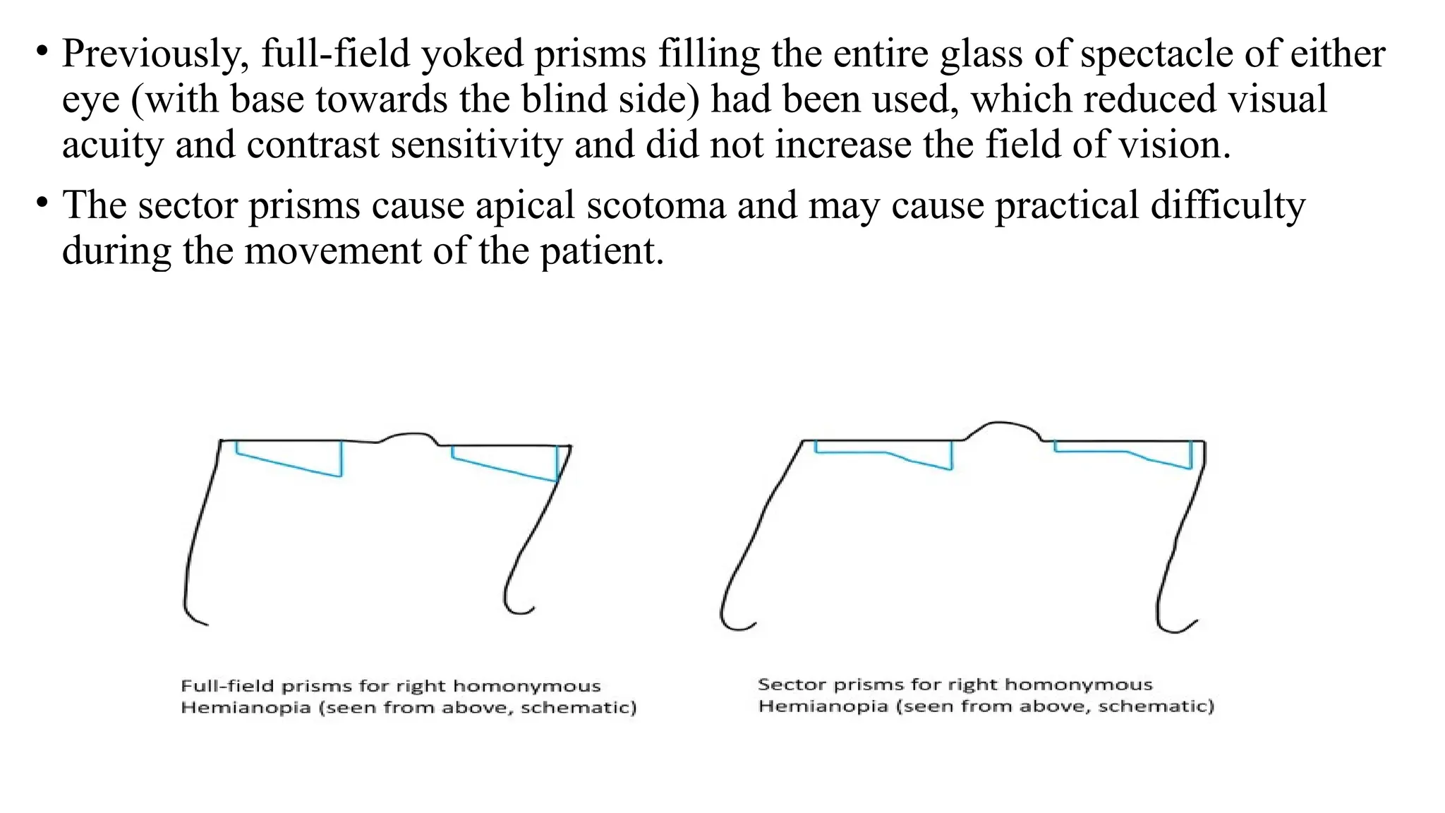
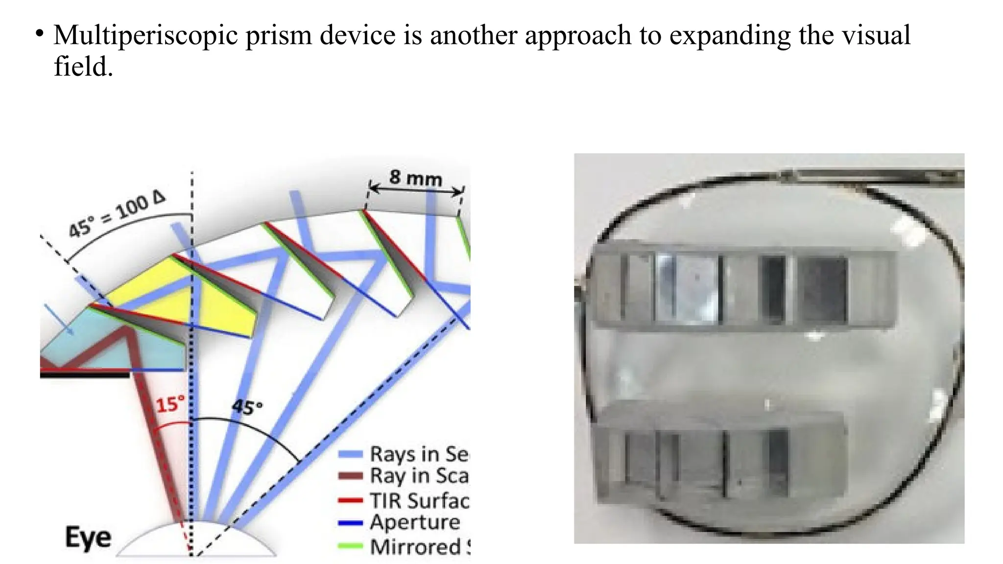
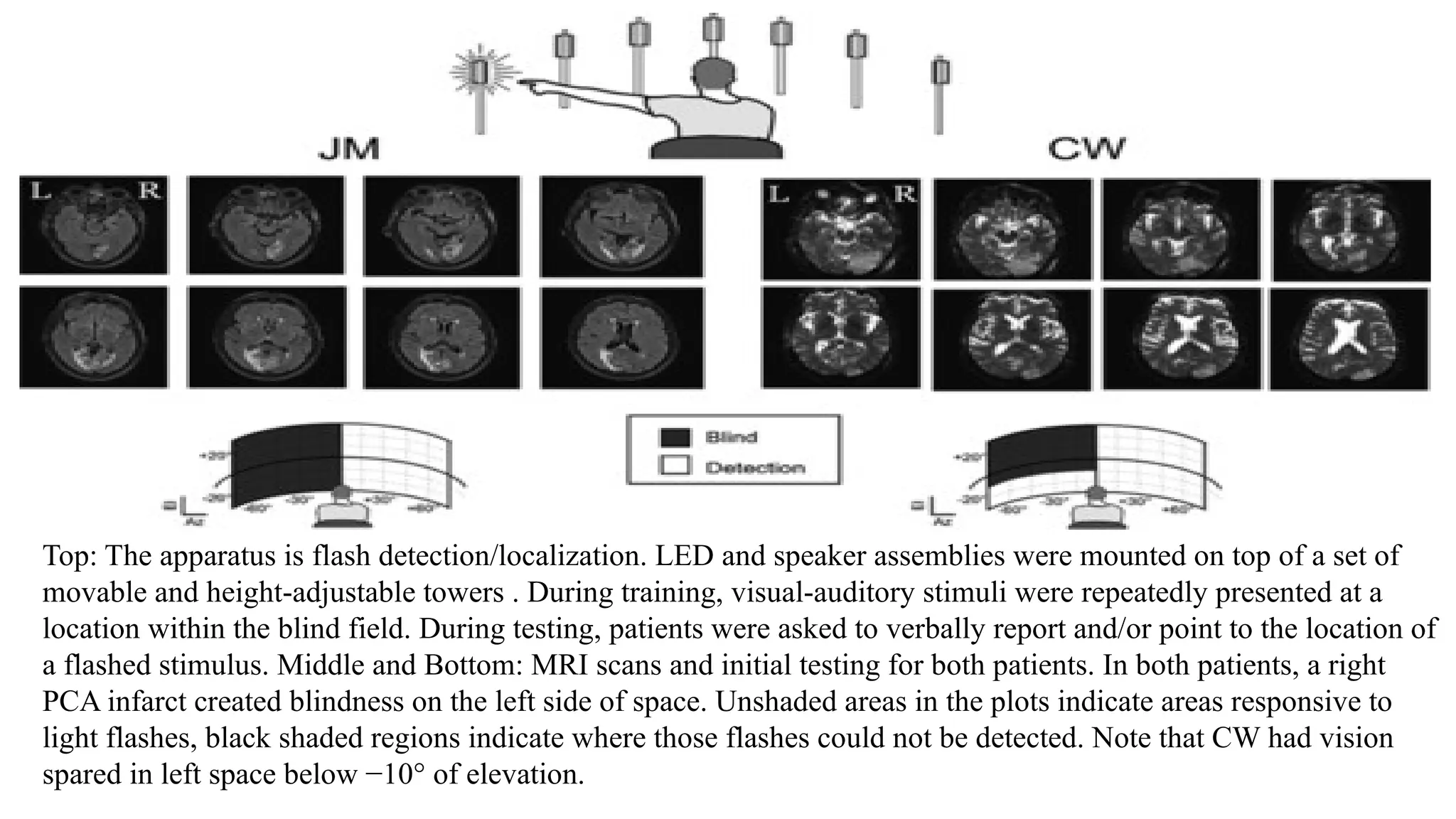
VISUAL FIELD ENHANCING
TECHNIQUES FOR PATIENTS WITH
HEMIANOPIA FIELD
PRESENTER- DR. MANJUNATH
MODERATOR- DR. PALLAVI B.A
OPTIC NERVE PATHWAY
INTRODUCTION
• A condition characterized by loss of vision in half of the visual field in one or
both eyes is known as Hemianopia.
• Homonymous Hemianopia: Same side of the visual field in both eyes.
• Heteronymous Hemianopia: Different sides of the visual field in each eye.
- Causes of Hemianopia;-
• Neurological Disorders: Seizures, posterior cortical atrophy, multiple sclerosis,
sarcoidosis, Alzheimer's disease, CJ disease
• Vascular: Stroke, venous thrombosis, vasculitis, arteriovenous malformation.
• Infectious: Abscess, encephalomyelitis, toxoplasmosis, cysticercosis,
neurosyphilis.
• Inflammatory disease: Demyelinating diseases including multiple sclerosis,
neuromyelitis optica.
• Neoplasm: Metastasis to the brain, lymphoma.
• Trauma: Traumatic brain injury, shaken baby syndrome.
• Metabolic diseases: Mitochondrial encephalomyopathy, lactic acidosis, and
stroke-like episodes (MELAS)
- Causes of transient or temporary HH that resolve spontaneously include:
• Transient ischemic attack
• Migraine
• Occipital, temporal, or parietal lobe seizure
• Nonketotic hyperglycemia
- Locations of lesions causing HH included;-
• Occipital lobe (45%)
• Optic radiation (32.2%)
• Combination of multiple areas (11.4%)
• Optic tract (10.2%)
• Lateral geniculate nucleus (1.3%)
INVESTIGATIONS
• Visual fields-
1) Confrontation visual field testing- most sensitive individual method of
confrontation visual field testing is Static finger wiggle testing.
2) Humphrey automated perimetry, Frequency doubling technology (FDT)
perimetry C20 threshold protocol was found to detect hemianopic field loss less
than half of the time
TREATMENT
• Patients with HH needs a multidisciplinary approach.
• The visual deficit on the hemianopic side can be reduced with 2 prisms that are placed in
the upper and lower part of the spectacle (Peli prisms).These prisms are only placed on
one eye (on the side of HH).
• The base of the prism is toward the hemianopic side (base out). This gives the view of
objects lying on the hemianopic side and expands the intact visual field while avoiding
diplopia, which may occur if prisms are placed in the line of sight. These prisms may
include a single-mounted prism or compound Fresnel press-on prism.
• The prisms work by shifting visual stimuli from the blind field to the seeing field.
• Expansion of the field may be quite satisfying (up to 20° with 40 prism diopter segments
and 30° with 57 prism diopter segments
• Previously, full-field yoked prisms filling the entire glass of spectacle of either
eye (with base towards the blind side) had been used, which reduced visual
acuity and contrast sensitivity and did not increase the field of vision.
• The sector prisms cause apical scotoma and may cause practical difficulty
during the movement of the patient.
• Multiperiscopic prism device is another approach to expanding the visual
field.
An illustration of the new prism correction for left hemianopia. The prisms are worn only over
the left eye. They are restricted to the upper and lower peripheral fields but extend across the
width of the lens so that they are effective at any position of lateral gaze. Illustrated are a simple
mounted prism for the upper segment and a compound Fresnel press-on prism for the lower
segment.
Multisensory therapy
• Effective way to restore visual function in people with homonymous
hemianopia.
• Involves repeatedly exposing patients to visual and auditory stimuli in the part of
their field of vision they can't see.
• The goal is to help patients regain the ability to detect, describe, and locate visual
stimuli in their blind field.
BENEFITS-
• Rapid improvements: Patients may see rapid improvements in their vision.
• Little loss between sessions: Patients may retain the gains they make between
training sessions.
• Increased attention: The therapy may help patients direct more attention to the
edge of their blind field.
Top: The apparatus is flash detection/localization. LED and speaker assemblies were mounted on top of a set of
movable and height-adjustable towers . During training, visual-auditory stimuli were repeatedly presented at a
location within the blind field. During testing, patients were asked to verbally report and/or point to the location of
a flashed stimulus. Middle and Bottom: MRI scans and initial testing for both patients. In both patients, a right
PCA infarct created blindness on the left side of space. Unshaded areas in the plots indicate areas responsive to
light flashes, black shaded regions indicate where those flashes could not be detected. Note that CW had vision
spared in left space below −10° of elevation.
BIOFEEDBACK TRAINING
• Biofeedback training (BT) is a compensatory rehabilitation technique that
emerged three decades ago .
• BT is one of the newest and more modern low vision rehabilitation techniques.
• By increasing the oculomotor control and relocating the visual fields through a
change in the patient’s locus of fixation, BT improves the visual acuity for
distance, near vision, contrast sensitivity, retinal sensitivity, reading speed, and
quality of life in many eye conditions.
• Mounting evidence has highlighted the benefits from BT in age-related macular
degeneration, myopic degeneration, Stargardt’s disease, glaucoma, and
nystagmus.
VISION RESTORATION THERAPY
• VRT is the only commercially available FDA-cleared therapy designed to restore
vision loss due to neurological brain damage.
• VRT does not require surgery or medication of any kind.
• Therapy is done at home on the patient’s own computer and on their own
schedule.
• VRT is designed to strengthen the visual information processing of residual
neuronal structures that have survived following acute lesions of the nervous
system resulting from trauma, stroke, inflammation, or elective surgery for
removal of brain tumors.
• By repeated activation through the course of the therapy, patients use the
program to train and improve their impaired visual functions, and thus regain
useful vision in the area of the visual field deficit
• VRT is performed on a computer at home twice daily, six days a week for six
months.
• During each session, patients focus on a central point displayed on the screen
and respond every time they see light stimuli appear elsewhere on the screen.
• The light stimuli are typically presented along the border of the intact and
damaged visual field

VISUAL FIELD ENHANCING TECHNIQUES FOR PATIENTS WITH HEMIANOPIA FIELD.pptx

  • 1.
    VISUAL FIELD ENHANCING TECHNIQUESFOR PATIENTS WITH HEMIANOPIA FIELD PRESENTER- DR. MANJUNATH MODERATOR- DR. PALLAVI B.A
  • 2.
  • 3.
    INTRODUCTION • A conditioncharacterized by loss of vision in half of the visual field in one or both eyes is known as Hemianopia. • Homonymous Hemianopia: Same side of the visual field in both eyes. • Heteronymous Hemianopia: Different sides of the visual field in each eye. - Causes of Hemianopia;- • Neurological Disorders: Seizures, posterior cortical atrophy, multiple sclerosis, sarcoidosis, Alzheimer's disease, CJ disease • Vascular: Stroke, venous thrombosis, vasculitis, arteriovenous malformation. • Infectious: Abscess, encephalomyelitis, toxoplasmosis, cysticercosis, neurosyphilis.
  • 4.
    • Inflammatory disease:Demyelinating diseases including multiple sclerosis, neuromyelitis optica. • Neoplasm: Metastasis to the brain, lymphoma. • Trauma: Traumatic brain injury, shaken baby syndrome. • Metabolic diseases: Mitochondrial encephalomyopathy, lactic acidosis, and stroke-like episodes (MELAS) - Causes of transient or temporary HH that resolve spontaneously include: • Transient ischemic attack • Migraine • Occipital, temporal, or parietal lobe seizure • Nonketotic hyperglycemia
  • 5.
    - Locations oflesions causing HH included;- • Occipital lobe (45%) • Optic radiation (32.2%) • Combination of multiple areas (11.4%) • Optic tract (10.2%) • Lateral geniculate nucleus (1.3%)
  • 6.
    INVESTIGATIONS • Visual fields- 1)Confrontation visual field testing- most sensitive individual method of confrontation visual field testing is Static finger wiggle testing. 2) Humphrey automated perimetry, Frequency doubling technology (FDT) perimetry C20 threshold protocol was found to detect hemianopic field loss less than half of the time
  • 7.
    TREATMENT • Patients withHH needs a multidisciplinary approach. • The visual deficit on the hemianopic side can be reduced with 2 prisms that are placed in the upper and lower part of the spectacle (Peli prisms).These prisms are only placed on one eye (on the side of HH). • The base of the prism is toward the hemianopic side (base out). This gives the view of objects lying on the hemianopic side and expands the intact visual field while avoiding diplopia, which may occur if prisms are placed in the line of sight. These prisms may include a single-mounted prism or compound Fresnel press-on prism. • The prisms work by shifting visual stimuli from the blind field to the seeing field. • Expansion of the field may be quite satisfying (up to 20° with 40 prism diopter segments and 30° with 57 prism diopter segments
  • 9.
    • Previously, full-fieldyoked prisms filling the entire glass of spectacle of either eye (with base towards the blind side) had been used, which reduced visual acuity and contrast sensitivity and did not increase the field of vision. • The sector prisms cause apical scotoma and may cause practical difficulty during the movement of the patient.
  • 10.
    • Multiperiscopic prismdevice is another approach to expanding the visual field.
  • 11.
    An illustration ofthe new prism correction for left hemianopia. The prisms are worn only over the left eye. They are restricted to the upper and lower peripheral fields but extend across the width of the lens so that they are effective at any position of lateral gaze. Illustrated are a simple mounted prism for the upper segment and a compound Fresnel press-on prism for the lower segment.
  • 13.
    Multisensory therapy • Effectiveway to restore visual function in people with homonymous hemianopia. • Involves repeatedly exposing patients to visual and auditory stimuli in the part of their field of vision they can't see. • The goal is to help patients regain the ability to detect, describe, and locate visual stimuli in their blind field. BENEFITS- • Rapid improvements: Patients may see rapid improvements in their vision. • Little loss between sessions: Patients may retain the gains they make between training sessions. • Increased attention: The therapy may help patients direct more attention to the edge of their blind field.
  • 14.
    Top: The apparatusis flash detection/localization. LED and speaker assemblies were mounted on top of a set of movable and height-adjustable towers . During training, visual-auditory stimuli were repeatedly presented at a location within the blind field. During testing, patients were asked to verbally report and/or point to the location of a flashed stimulus. Middle and Bottom: MRI scans and initial testing for both patients. In both patients, a right PCA infarct created blindness on the left side of space. Unshaded areas in the plots indicate areas responsive to light flashes, black shaded regions indicate where those flashes could not be detected. Note that CW had vision spared in left space below −10° of elevation.
  • 15.
    BIOFEEDBACK TRAINING • Biofeedbacktraining (BT) is a compensatory rehabilitation technique that emerged three decades ago . • BT is one of the newest and more modern low vision rehabilitation techniques. • By increasing the oculomotor control and relocating the visual fields through a change in the patient’s locus of fixation, BT improves the visual acuity for distance, near vision, contrast sensitivity, retinal sensitivity, reading speed, and quality of life in many eye conditions. • Mounting evidence has highlighted the benefits from BT in age-related macular degeneration, myopic degeneration, Stargardt’s disease, glaucoma, and nystagmus.
  • 17.
    VISION RESTORATION THERAPY •VRT is the only commercially available FDA-cleared therapy designed to restore vision loss due to neurological brain damage. • VRT does not require surgery or medication of any kind. • Therapy is done at home on the patient’s own computer and on their own schedule. • VRT is designed to strengthen the visual information processing of residual neuronal structures that have survived following acute lesions of the nervous system resulting from trauma, stroke, inflammation, or elective surgery for removal of brain tumors. • By repeated activation through the course of the therapy, patients use the program to train and improve their impaired visual functions, and thus regain useful vision in the area of the visual field deficit
  • 18.
    • VRT isperformed on a computer at home twice daily, six days a week for six months. • During each session, patients focus on a central point displayed on the screen and respond every time they see light stimuli appear elsewhere on the screen. • The light stimuli are typically presented along the border of the intact and damaged visual field