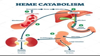
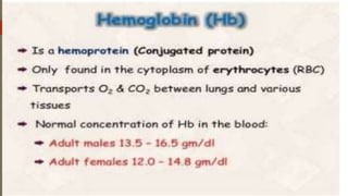
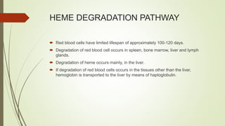
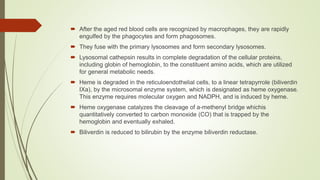
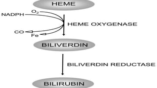
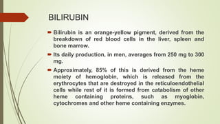
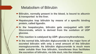
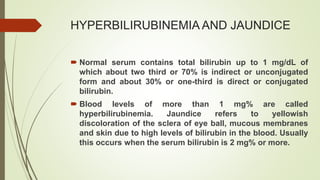
3. HEME DEGRADATION PATHWAY
Red blood cells have limited lifespan of approximately 100-120 days.
Degradation of red blood cell occurs in spleen, bone marrow, liver and lymph
glands.
Degradation of heme occurs mainly, in the liver.
If degradation of red blood cells occurs in the tissues other than the liver,
hemoglobin is transported to the liver by means of haptoglobulin.
4. After the aged red blood cells are recognized by macrophages, they are rapidly
engulfed by the phagocytes and form phagosomes.
They fuse with the primary lysosomes and form secondary lysosomes.
Lysosomal cathepsin results in complete degradation of the cellular proteins,
including globin of hemoglobin, to the constituent amino acids, which are utilized
for general metabolic needs.
Heme is degraded in the reticuloendothelial cells, to a linear tetrapyrrole (biliverdin
IXa), by the microsomal enzyme system, which is designated as heme oxygenase.
This enzyme requires molecular oxygen and NADPH, and is induced by heme.
Heme oxygenase catalyzes the cleavage of a-methenyl bridge whichis
quantitatively converted to carbon monoxide (CO) that is trapped by the
hemoglobin and eventually exhaled.
Biliverdin is reduced to bilirubin by the enzyme biliverdin reductase.
5.
6. BILIRUBIN
Bilirubin is an orange-yellow pigment, derived from the
breakdown of red blood cells in the liver, spleen and
bone marrow.
Its daily production, in men, averages from 250 mg to 300
mg.
Approximately, 85% of this is derived from the heme
moiety of hemoglobin, which is released from the
erythrocytes that are destroyed in the reticuloendothelial
cells while rest of it is formed from catabolism of other
heme containing proteins, such as myoglobin,
cytochromes and other heme containing enzymes.
7. Metabolism of Bilirubin
Bilirubin, normally present in the blood, is bound to albumin
& transported to the liver.
Hepatocytes trap bilirubin by means of a specific binding
protein, called ligandin.
In the heapatocytes, bilirubin gets conjugated with UDP
Glucuronate, which is derived from the oxidation of UDP
glucose.
This reaction is catalysed by UDP- glucuronyltransferase.
In the normal bile, bilirubin diglucuronide is the major form of
excreted bilirubin with only a smal amount of the bilirubin
monoglucuronide. As bilirubin diglucuronide is much more
water soluble than free bilirubin, transferase thus facilitates
the excretion of bilirubin, via bile duct, into the intestine
9. HYPERBILIRUBINEMIA AND JAUNDICE
Normal serum contains total bilirubin up to 1 mg/dL of
which about two third or 70% is indirect or unconjugated
form and about 30% or one-third is direct or conjugated
bilirubin.
Blood levels of more than 1 mg% are called
hyperbilirubinemia. Jaundice refers to yellowish
discoloration of the sclera of eye ball, mucous membranes
and skin due to high levels of bilirubin in the blood. Usually
this occurs when the serum bilirubin is 2 mg% or more.
10. Latent jaundice
• It refers to the stateof hyperbilirubinemia when serum
bilirubin level is between 1-2 mg%.
Usually there is no yellowish discoloration of sclera or mucous
membrane in this state. As the level increases, jaundice
appears.
11. Types of Jaundice
Three types of jaundice are seen depending on the cause of increased bilirubin in
the blood.
1. Hemolytic jaundice
2. Hepatic jaundice
3. Obstructive jaundice
12. Hemolytic Jaundice
Haemolytic jaundice is also called retention or acholuric
jaundice.
In typical hemolytic jaundice, the basic abnormality is over
production of bilirubin due to excessive heme catabolism.
13. Causes
Abnormal shape of RBCs such as in sickle cell anaemia
Deficiencies of some enzymes like pyruvate kinase,
glucose-6-phosphate dehydrogenase, etc.
Mismatched blood transfusion is the other common
cause leading to hemolysis of RBCs.
Abnormal RBCs are prone to undergo hemolysis.
Excessive bilirubin goes to the hepatocytes which
conjugate bilirubin to its maximum capacity. But the
hemolysis is usually much more and so ultimately the
unconjugated bilirubin dominates in blood. This is water
insoluble and hence it is not excreted in urine.
14. Conjugated bilirubin is also more than the normal.
It leads to excessive urobilinogen production and its
excretion in urine and stools.
This is because proportionately more urobilinogen is
absorbed in enterohepatic circulation.
15. Hepatic Jaundice
Hepatic jaundice is due to diminished hepatic function due to liver
diseases.
Commonly in typical hepatic jaundice there is deficiency or complete
absence of glucuronosyl transferase enzyme.
So the conjugation of bilirubin is absent or decreased.
Causes
Acute and chronic hepatitis caused by hepatitis viruses A, B, C, D and
E; infection with B and C viruses is dangerous and should be treated
early.
Long term use of hepatotoxic drugs like antitubercular drugs etc.
Congenital defects e.g. Gilbert syndrome, Crigler Najjar syndrome, etc.
16. Metabolic Abnormality
Since bilirubin is not adequately conjugated, increase in
unconjugated bilirubin is seen.
Some conjugated bilirubin will also be present due to residual
conjugation activity in the liver.
Since in hepatitis there is usually narrowing of hepatic canaliculi
due to swelling of hepatocytes, there is intrahepatic obstruction
to bilirubin secretion into the bile.
In such cases, the conjugated bilirubin is reabsorbed into blood
circulation.
It is then excreted in urine because this bilirubin is water soluble.
So bilirubin may be seen in urine in hepatic jaundice due to
hepatitis also in active phase of disease. Normally, no bilirubin is
present in urine.
17. Obstructive Jaundice
Obstructive jaundice is also called regurgitation or
choluric jaundice.
Conjugation occurs normally in the liver, but there is a
defect in secretion.
This type of jaundice occurs due to extrahepatic
obstruction to the flow of bile. So the conjugated bilirubin
is not secreted into the intestine.