1. Cardiac failure, also known as heart failure, occurs when the heart is unable to pump sufficiently to maintain blood flow to meet the body's needs. It can result from several cardiac causes related to problems with the heart muscle, valves, or pericardium, as well as non-cardiac causes like anemia or hypertension that overload the heart.
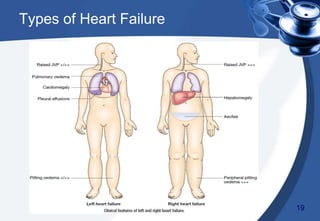
2. Symptoms depend on whether the left side or right side of the heart is predominantly affected but commonly include fatigue, breathlessness, and fluid retention. Signs involve findings related to congestion in the lungs or periphery.
3. Treatment involves identifying and treating the underlying cause, restricting salt and fluid intake, medications like diuretics and A