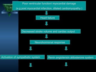
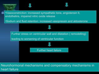
1. Heart failure occurs when the heart cannot pump enough blood to meet the body's needs or can only do so with increased filling pressure.
2. Common causes of heart failure include coronary artery disease, hypertension, cardiomyopathy, valvular heart disease, and arrhythmias.
3. Symptoms of heart failure depend on whether it affects the left side, right side, or both ventricles, and include fatigue, breathlessness, leg swelling, and liver congestion.
This document summarizes the pathophysiology of heart failure (HF). It discusses how HF results from abnormalities in cardiac structure/function that limit oxygen delivery to tissues, despite normal filling pressures. The progression of HF is driven by neurohumoral activation of the sympathetic nervous system and renin-angiotensin-aldosterone system, which initially help compensate but eventually exacerbate cardiac remodeling and dysfunction. The document outlines the effects of various neurohormones involved in HF, including their normal and maladaptive roles in the progression of disease. Management of HF focuses on interrupting the harmful effects of long-term neurohumoral activation.
1. Heart failure is the inability of the heart to pump sufficient blood to meet the tissues' needs for oxygen and nutrients.
2. It can be caused by mechanical abnormalities of the heart, myocardial abnormalities, or altered cardiac rhythm/conduction disturbances.
3. Symptoms of heart failure depend on whether the left or right side of the heart is affected and include shortness of breath, edema, fatigue, cough, and liver/spleen enlargement.
Heart failure occurs when the heart cannot pump enough blood to meet the body's needs. It can be caused by problems with either the left or right side of the heart. Common causes include heart disease and hypertension. Symptoms depend on whether the left or right side is affected. The left side controls blood flow to the lungs, so left heart failure causes shortness of breath and coughing up blood. The right side controls blood returning from the body, so right heart failure causes fatigue, leg swelling and liver/kidney congestion. Over time the heart tries to compensate through enlargement but eventually decompensates leading to further symptoms.
Heart failure occurs when the heart is unable to pump sufficiently to meet the body's needs. It can be systolic, caused by the ventricles' inability to contract properly, or diastolic, caused by the ventricles' inability to relax and fill normally. Symptoms depend on whether the left or right ventricle is affected. Management involves risk factor control, lifestyle changes, diuretics, ACE inhibitors, beta blockers, and sometimes surgical procedures like defibrillators or transplantation for severe cases.
This document provides information on congestive cardiac failure (CCF), including its definition, pathophysiology, clinical features, investigations, and management. CCF occurs when the heart muscle is weakened and cannot maintain adequate cardiac output. The pathophysiology involves changes in preload, afterload, and contractility that decrease cardiac output. Compensatory mechanisms initially help but later worsen symptoms. Clinically, CCF presents with dyspnea, edema, elevated JVP, hepatomegaly, and other signs. Investigations include BNP, ECG, echocardiogram. Management focuses on treating the underlying cause, reducing preload/afterload, and improving contractility. Diuretics, ACE inhibitors, beta
Congestive heart failure (CHF) results from an inadequate cardiac output which causes increased blood volume and congestion. Fluid accumulates in tissues causing edema. CHF can be caused by ventricular failure where the heart's contractions become weak, or mechanical failure where the ventricles fail to fill with blood properly. Common causes include coronary artery disease, high blood pressure, and valve problems.
This document provides an overview of cardiac failure (congestive heart failure), including its pathophysiology, manifestations, causes, investigations, treatment and management. Some key points are:
- Cardiac failure occurs when the heart cannot maintain sufficient output to meet bodily demands. It increases with age and many patients are repeatedly admitted.
- It can manifest as left or right heart failure, depending on which side of the heart is affected, with different symptoms and signs for each.
- Common causes include hypertension, ischemic heart disease and valvular issues.
- Investigations include chest x-ray, ECG, echocardiogram and blood tests to determine the underlying cause.
- Treatment involves
This document discusses heart failure, including its definition, causes, types, and compensatory mechanisms. Heart failure occurs when the heart is unable to pump enough blood to meet the body's needs. It can be caused by intrinsic pump failure, an increased workload on the heart, or impaired filling of the cardiac chambers. The types of heart failure include acute or chronic, right-sided or left-sided, and forward or backward failure. When the heart begins to fail, compensatory mechanisms such as cardiac hypertrophy, dilation, and increased heart rate attempt to maintain adequate blood circulation.
This document discusses valvular heart disorders, focusing on mitral valve stenosis and regurgitation. It defines stenosis as a narrowing of the valve orifice, while regurgitation is the incomplete closure of the mitral valve, allowing blood to flow back into the left atrium. The most common cause of both is rheumatic heart disease. Symptoms of mitral stenosis include dyspnea and fatigue, while regurgitation causes fatigue, dyspnea, and palpitations. Diagnosis is via echocardiogram. Treatment depends on severity but may include medications, balloon valvuloplasty, or valve replacement surgery.
This document discusses heart failure, including its definitions, types, causes, clinical manifestations, and management from a dental perspective. It defines heart failure as the heart's inability to supply sufficient oxygenated blood to the body's needs. The types include left ventricle, right ventricle, and combined failure. Causes include congenital heart defects and conditions like cardiomyopathy. Clinical manifestations vary depending on which ventricle is affected but may include edema, dyspnea, fatigue, and cyanosis. Dental management involves recognizing signs of failure, terminating procedures if needed, positioning the patient comfortably, and providing post-procedure care and follow-up.
Valvular heart disease can be caused by conditions like rheumatic fever, infections, and aging. The main types are aortic and mitral stenosis, and aortic and mitral regurgitation. Symptoms depend on the valve affected and include shortness of breath, chest pain, and fatigue. Exams may reveal murmurs. Tests like echocardiograms can evaluate the severity of stenosis or regurgitation. Treatment involves medications, valve repair or replacement surgery, depending on symptoms and severity. Early surgery is often recommended for severe aortic stenosis to prevent heart failure.
- Right heart failure is characterized by low cardiac output, hypotension, hepatic enlargement and raised jugular venous pressure. It has a high mortality rate comparable to left heart failure.
- Failure occurs when the right ventricle can no longer compensate for increased volume. Determining preload is difficult but high right atrial pressures indicate elevated right ventricular pressures and volume.
- Treatment aims to reduce afterload and optimize preload. Afterload reduction can be achieved through selective pulmonary vasodilation using inhaled nitric oxide, prostacyclins or phosphodiesterase inhibitors.
This document provides an overview of heart failure, including its causes, pathophysiology, clinical presentation, diagnosis and management. Heart failure occurs when the heart cannot pump enough blood to meet the body's needs. It may be due to conditions that weaken the heart muscle such as coronary artery disease. Symptoms depend on whether the left, right or both ventricles are affected and include shortness of breath, fatigue, swelling and fluid retention. Treatment focuses on managing symptoms, improving cardiac function and preventing future damage through medications, lifestyle changes and device-based therapies.
This document provides an overview of cardiac failure (heart failure) including:
- Causes such as heart attacks, high blood pressure, and heart muscle diseases
- Types including left vs right heart failure and systolic vs diastolic dysfunction
- Signs and symptoms like shortness of breath, edema, and fatigue
- Diagnostic tests including chest X-rays, electrocardiograms, and blood tests
- Treatment options such as medications, lifestyle changes, and surgeries
- Prognosis which remains poor despite advances, with 50-60% mortality for severe cases within one year.
Cardiomegaly is a condition where the heart is enlarged. It can be caused by conditions that make the heart work harder like high blood pressure, heart disease, or heart valve problems. Symptoms include shortness of breath, fatigue, chest pain, and swelling. Diagnosis involves tests like chest x-rays, echocardiograms, and blood tests. Treatment may include medications to reduce blood pressure and swelling, surgery, lifestyle changes, and sometimes a heart transplant for severe cases.
This document discusses the pathophysiology of heart failure. It describes how heart failure can result from abnormalities in systolic or diastolic cardiac function. In heart failure with reduced ejection fraction (HFrEF), the left ventricle contracts poorly and cannot adequately pump blood. In heart failure with preserved ejection fraction (HFpEF), left ventricular filling is impaired. Both types result in inadequate blood flow and organ congestion. The document discusses neurohormonal changes, organ dysfunction, and other compensatory mechanisms involved in heart failure.
Cardiac failure refers to the heart's inability to pump enough blood to meet metabolic demands. It can be classified in several ways, including based on severity, the side of heart involved, cardiac output level, duration, and type of function affected. Compensated heart failure involves mechanisms that return cardiac output to normal levels, while decompensated failure results in fluid retention and pulmonary or peripheral edema as compensatory mechanisms fail. Ejection fraction is normally over 50% and remains relatively normal in diastolic heart failure.
Nursing care of clients with disorders of cardiac function part ICarmela Domocmat
This document discusses disorders of cardiac function and heart failure. It defines heart failure as when the heart is unable to pump enough blood to meet the body's needs at rest or during exercise. It then covers the etiology, pathophysiology, signs and symptoms, diagnosis, and classification of heart failure. Some key points include that heart failure results from conditions that impair the structure or function of the heart, common causes include coronary artery disease and cardiomyopathy, and heart failure can present as either left or right sided failure with different clinical manifestations.
Heart failure occurs when the heart is unable to pump enough blood to meet the body's needs. It results from conditions that damage or weaken the heart muscle such as hypertension, heart attack, or cardiomyopathy. Symptoms include dyspnea, fatigue, swelling, and weakness. Diagnosis involves chest x-rays, ECGs, and echocardiograms. Treatment focuses on managing symptoms with diuretics, ACE inhibitors, beta blockers, and addressing the underlying heart condition if possible through surgery or lifestyle changes.
Heart failure (HF), often used to mean chronic heart failure (CHF), occurs when the heart is unable to pump sufficiently to maintain blood flow to meet the needs of the body.
Valvular Heart Diseases - Final Year LectureMr Adeel Abbas
Rheumatic fever is caused by a streptococcal infection and commonly presents in children and young adults in developing countries. It can cause valvular heart disease by generating an immune response that damages the heart valves. The mitral and aortic valves are most often affected, leading to stenosis or regurgitation and symptoms like dyspnea and palpitations. Treatment involves antibiotics to prevent initial infection, lifestyle changes, and potentially valve repair or replacement surgery for severe cases.
The document discusses various heart valve conditions including mitral valve stenosis, mitral valve regurgitation, tricuspid stenosis, tricuspid regurgitation, aortic valve stenosis, aortic valve regurgitation, pulmonary valve stenosis, and pulmonary valve regurgitation. It defines each condition, discusses their causes, symptoms, diagnostic evaluations, and potential treatments which may include medications, balloon valvuloplasty procedures, or valve repair/replacement surgeries.
This document discusses diseases of the heart valves, including causes, pathophysiology, clinical features, investigations, and management. It focuses on rheumatic heart disease as a common cause of valve disease. Rheumatic fever results from an immune response to streptococcal infection and can cause inflammation of the heart valves and tissues. Over time, this leads to valve stenosis or regurgitation. The major valves affected are the mitral and aortic valves. Clinical exam findings and imaging tests like echocardiogram are used to diagnose valve disease and determine severity. Treatment involves medications for symptoms as well as surgical procedures like valve repair or replacement in severe cases.
This document discusses different types of valvular heart disease including stenosis, regurgitation, and functional regurgitation. It provides details on specific valve diseases like mitral stenosis, mitral regurgitation, aortic stenosis, and aortic regurgitation. For each condition, it outlines the causes, pathophysiology or effects, which depend on the degree and duration of the disease. Long term effects can include hypertrophy and failure of the left or right ventricle leading to pulmonary congestion, edema, and heart failure.
This document discusses drugs used to treat congestive cardiac failure (CCF). It describes how CCF causes the heart to fail in pumping adequate blood to organs. Common symptoms include shortness of breath, edema, and fatigue. Causes include conditions like coronary artery disease, hypertension, and diabetes. The main drug classes used to treat CCF are vasodilators like ACE inhibitors, diuretics, beta blockers, and digoxin. ACE inhibitors improve outcomes by reducing afterload and preload on the heart. Loop diuretics are effective but can cause hypokalemia, addressed by potassium sparing diuretics. Beta blockers inhibit sympathetic activity and improve function. Digoxin increases contractility
1. The document discusses the management of acute coronary syndrome (ACS), including risk stratification, reperfusion therapy options like fibrinolysis and percutaneous coronary intervention (PCI), and antithrombotic and antiplatelet therapies.
2. It highlights the importance of rapid reperfusion through fibrinolysis or PCI to restore blood flow and reduce mortality. PCI is generally preferred over fibrinolysis when it can be performed quickly by an experienced center.
3. Antiplatelet therapies with aspirin and clopidogrel are recommended, along with anticoagulants like unfractionated heparin or low molecular weight heparin to prevent clotting in ACS patients.
1. Stable angina is caused by fixed stenosis, while unstable angina involves dynamic obstruction from plaque rupture and thrombosis. Acute myocardial infarction results from acute coronary artery occlusion and necrosis.
2. Management involves controlling risk factors, treating symptoms, and improving prognosis. Symptomatic treatment includes nitrates, beta blockers, or calcium channel blockers. Prognostic treatment consists of aspirin, other antiplatelets, and statins. Invasive options are percutaneous coronary intervention or coronary artery bypass grafting.
3. For acute myocardial infarction, treatment focuses on reperfusion through thrombolysis or angioplasty, pain management, and prevention of complications. Long-term management emphasizes secondary prevention with
This document discusses diseases of the heart valves, focusing on rheumatic heart disease and mitral valve disease. It describes the causes, pathogenesis, clinical features, investigations, and management of acute rheumatic fever and chronic rheumatic heart disease, with a specific focus on mitral stenosis due to valve fibrosis and calcification from repeated rheumatic fever attacks. Key points include the immunological mechanism leading to valve damage, Jones criteria for diagnosing acute rheumatic fever, and the resulting mitral stenosis pathology of restricted blood flow from the left atrium to ventricle.
This document discusses palpitations, which refer to abnormal awareness of one's heartbeat. Palpitations can be caused by rapid, slow, or irregular heart rhythms and may result from primary cardiac diseases or systemic conditions affecting the heart. Common causes include anxiety, hyperthyroidism, caffeine, smoking, sinus tachycardia, supraventricular tachycardia, ventricular tachycardia, atrial fibrillation, extrasystoles, and Wolff-Parkinson-White syndrome. A thorough history and electrocardiogram can help diagnose the underlying rhythm abnormality.
Cor pulmonale is right heart failure caused by chronic pulmonary hypertension. It is often caused by lung diseases like COPD and asthma, or pulmonary vascular diseases like pulmonary embolism. Symptoms include dyspnea, fatigue and syncope. Signs include elevated jugular venous pressure, heart murmurs, hepatomegaly and edema. Investigations show signs of pulmonary hypertension and right heart strain on tests like chest x-ray and ECG. Management focuses on treating the underlying cause, respiratory failure with oxygen, and cardiac failure with diuretics. The prognosis is generally poor with 50% of patients dying within 5 years.
This document provides an overview of electrocardiography (ECG or EKG):
- The ECG is essential for diagnosing cardiac rhythm abnormalities and chest pain, and guides treatment like thrombolysis for heart attacks.
- The history of ECG development is traced from early experiments in the 1800s to William Einthoven's invention of the first clinical ECG machine in the early 1900s.
- A normal ECG shows a regular rhythm between 60-100 beats per minute, visible P waves before each QRS complex, and normal durations for the P-R interval, QRS complex, and T wave.
This document provides a summary of basics of electrocardiography (ECG/EKG). It discusses the history and development of ECG technology. It describes the components of a normal ECG waveform including the P, QRS, and T waves. It explains how to determine heart rate from an ECG and identify different arrhythmias based on the waveform. Key anatomical structures involved in heart's electrical conduction system are also outlined.
This document discusses heart failure, including its pathophysiology, types, and causes. Heart failure occurs when the heart cannot maintain adequate output or can only do so at the expense of elevated ventricular pressures. It may result from systolic or diastolic dysfunction. Types include left, right, and bi-ventricular failure. Acute pulmonary edema is treated with oxygen, nitrates, and diuretics. Chronic heart failure is managed with drugs like diuretics, ACE inhibitors, ARBs, and beta-blockers to improve outcomes.
This document provides an overview of heart failure, including its causes, pathophysiology, clinical presentation, diagnosis and management. Heart failure occurs when the heart cannot pump enough blood to meet the body's needs. It may be due to conditions that weaken the heart muscle such as coronary artery disease. Symptoms depend on whether the left, right or both ventricles are affected and include shortness of breath, fatigue, swelling and fluid retention. Treatment focuses on managing symptoms, improving cardiac function and preventing future damage through medications, lifestyle changes and device therapies.
This document discusses heart failure, including its classification, pathophysiology, clinical manifestations, investigations, and clinical syndromes. It describes how heart failure occurs when the heart is overloaded or the heart muscle is disordered. It discusses the neuroendocrine and cellular changes that occur in heart failure and how this impacts fluid retention, circulatory pressures, and organ function. Specifically, it outlines the features of left heart failure including common causes, symptoms of pulmonary congestion, physical exam findings, investigations such as echocardiography and natriuretic peptide levels, and how to differentiate it from other conditions like pulmonary disease.
This document provides an overview of heart failure, including its pathophysiology, types, clinical presentation, investigations, and management. Heart failure occurs when the heart cannot pump enough blood to meet the body's needs and can develop due to conditions that weaken the heart such as heart attacks or high blood pressure. Symptoms depend on whether the left side, right side, or both sides of the heart are affected. Management involves treating the underlying cause, reducing symptoms through medications, lifestyle changes, and addressing complications.
This document summarizes heart failure, including its classification, pathophysiology, clinical manifestations, investigations, and management. Heart failure means the heart cannot pump sufficient blood for the body's needs. It can affect the left side, right side, or both sides of the heart. Management involves correcting underlying causes, reducing demands on the heart through diet and exercise, and pharmacological therapy including diuretics, ACE inhibitors, and other drugs to modify neuroendocrine and renal responses. The goals of treatment are to alleviate symptoms and improve prognosis.
This document defines cardiac failure and heart failure, describes the types and causes, and discusses the pathophysiology, clinical features, investigations, and treatment. Heart failure is a clinical syndrome where the heart cannot pump enough blood to meet the body's needs, or can only do so with elevated filling pressures. It can be systolic or diastolic in nature. Common causes include ischemic heart disease, cardiomyopathy, valvular disease, and hypertension. Symptoms include breathlessness, fatigue, and fluid retention. Echocardiography, biomarkers like BNP, and cardiac imaging are used in diagnosis and assessment. Treatment aims to relieve symptoms, improve quality of life, and reduce mortality through medications, device therapies, and lifestyle changes.
This document provides information about heart failure including its definition, pathophysiology, types, causes, symptoms, diagnosis, and management. Heart failure is defined as impaired cardiac function that cannot maintain adequate circulation. The heart fails to pump sufficient blood due to reduced cardiac output and venous congestion. There are various compensatory mechanisms that work during early stages of heart failure to maintain blood flow such as the Frank-Starling mechanism and increased sympathetic activity. Common causes of heart failure include myocardial infarction, hypertension, valvular heart disease, and cardiomyopathy. Symptoms include shortness of breath, exercise intolerance, and swelling of the legs or abdomen. Diagnosis involves imaging like echocardiography and electrocardiography to evaluate cardiac
This document defines and describes heart failure, its causes, forms, and pathophysiology. Heart failure is defined as the inability of the heart to pump enough blood to meet the body's needs. It is most often caused by impaired contractility from conditions like ischemic heart disease or cardiomyopathy. Heart failure can present as systolic or diastolic dysfunction and can affect the left or right ventricle. The body undergoes adaptive and maladaptive changes like neurohormonal activation to try to maintain cardiac output as heart function declines.
Heart failure, also known as cardiac decompensation or cardiac insufficiency, occurs when the heart is unable to pump enough blood to meet the body's needs. It can be caused by conditions that impair the heart muscle's ability to contract effectively or limit ventricular filling. Symptoms vary depending on whether the left or right ventricle is primarily affected and include dyspnea, fatigue, edema and others. Diagnostic tests may include echocardiography, ECG, chest x-ray and BNP level. Treatment focuses on managing symptoms, slowing disease progression, and preventing hospitalizations through lifestyle changes and medication.
Heart failure is defined as the heart's inability to pump enough blood to meet the body's needs. It can be caused by conditions that impair the heart muscle or overload it. Heart failure is classified based on location (right, left, or both ventricles), timing (acute or chronic), and pumping ability (systolic or diastolic). Signs and symptoms include dyspnea, fatigue, fluid retention, and reduced exercise tolerance. Treatment involves lifestyle changes, medications to relieve symptoms and improve pumping ability, and treating the underlying cause.
The document defines heart failure as a clinical syndrome characterized by typical symptoms such as breathlessness and swelling caused by structural or functional abnormalities of the heart. This results in reduced cardiac output and elevated pressures in the heart at rest or during stress. Heart failure is classified based on ejection fraction and other factors, and can involve either the left or right side of the heart. Long term, heart failure leads to neurohormonal activation and pathological remodeling of the heart muscle over time.
Heart failure occurs when the heart is unable to pump enough blood to meet the body's needs. It affects approximately 2% of the developed world and is most commonly caused by coronary artery disease or hypertension damaging the heart muscle. Symptoms vary depending on whether the left or right side of the heart is affected but may include fatigue, shortness of breath, swelling, and confusion. Treatment involves medications to reduce fluid retention, lower blood pressure and heart rate, and devices or surgery for more severe cases.
This document outlines the different types of heart failure:
- Systolic and diastolic heart failure refer to the ventricle's ability to contract or relax. Right and left ventricular failure affect blood flow to the lungs or body.
- Heart failure can be acute or chronic, high or low output, and cause blood to back up (backward) or tissues to receive less blood flow (forward).
- The types are distinguished by their effects on cardiac output and the parts of the circulatory system they impact.
Diastolic heart failure occurs when the ventricles become stiff and cannot relax fully during diastole. This prevents full ventricular filling and blood backs up in the organs. Around half of heart failure patients have diastolic heart failure. Diagnosis relies on echocardiogram showing diastolic dysfunction. Treatment focuses on controlling hypertension, volume overload, and other causes through medications like ACE inhibitors, diuretics and beta blockers.
The Advanced Cardiovascular Life Support (ACLS) algorithm is a systematic, evidence-based approach designed to guide healthcare providers in the urgent treatment of: Cardiac arrest. Arrhythmias. Stroke. Other life-threatening cardiovascular emergencies.
This document discusses heart failure, including its causes, types, signs and symptoms, diagnosis, and treatment. Heart failure occurs when the heart cannot pump effectively to meet the body's needs. It can be caused by problems with the heart muscle itself or with the heart's workload. Treatment focuses on improving heart function through medications like diuretics, ACE inhibitors, and beta blockers to manage symptoms and prevent worsening of the condition.
This document provides guidance on cardiovascular (CVS) examination for dental practice. It discusses CVS diseases that can be induced or exacerbated by dental procedures. The examination involves taking a history of present illness, past medical history, drug history, family history, and social history. The physical examination includes inspection of general appearance and vital signs, along with detailed examination of pulses, heart sounds, lungs, abdomen, edema, and jugular veins. Differential diagnoses are provided for common CVS symptoms.
This document summarizes a study comparing 3 months versus 6 months of anticoagulation treatment in patients with deep vein thrombosis (DVT) or pulmonary embolism (PE) or both. The study found that 3 months of treatment was as effective as 6 months, with a lower risk of major bleeding events during treatment. Based on this and other studies, 3 months of warfarin is recommended for first episode VTE, while 6 weeks may be adequate for distal VTE. Treatment duration may need to be longer if reversible risk factors are still present.
AMI is caused by formation of an occlusive thrombus at the site of a ruptured or eroded atherosclerotic plaque. It presents with chest pain and symptoms of ischemia. Investigations show changes on ECG, elevated cardiac enzymes, and echocardiogram may show regional wall motion abnormalities. Treatment involves oxygen, aspirin, anticoagulants, reperfusion with thrombolysis or PCI, and adjunctive therapies like beta blockers. Goals are to limit damage and prevent complications through risk factor modification and medical management.
Shock is a physiological state characterized by a significant systemic reduction in perfusion resulting in decreased tissue oxygen delivery. The causes of shock include hypovolumic, cardiogenic, neurogenic, anaphylactic, septic, and obstructive shock. The management of shock involves initial assessment of vital signs, resuscitation focusing on airway, breathing, and circulation, urgent investigation to identify the underlying cause, and appropriate treatment of the cause such as fluids for hypovolumic shock, inotropes for septic shock, and adrenaline with antihistamines for anaphylactic shock. The mortality from septic and cardiogenic shock remains high.
Central chest pain can be caused by cardiac issues like angina or myocardial infarction, or non-cardiac issues like pulmonary embolism or esophageal disorders. A thorough history of the characteristics of the pain, including location, duration, aggravating/relieving factors, and associated symptoms can help determine the underlying cause. Key factors that help differentiate cardiac from non-cardiac chest pain include relationship to exertion and response to nitroglycerin.
This document provides guidance on evaluating and diagnosing causes of acute breathlessness. It lists potential conditions based on presenting symptoms such as wheezing, stridor, crepitations, or a clear chest. Priority is given to pulse oximetry, ECG, chest x-ray, and blood tests. Common diagnoses are acute asthma, pulmonary edema, pneumonia, or COPD exacerbation. Features, urgent investigations, and expected blood gas results for each condition are outlined to aid clinical assessment.
This document discusses early pregnancy bleeding and disorders. It covers causes of early pregnancy bleeding including spontaneous miscarriage, ectopic pregnancy, and gestational trophoblastic disease. It then discusses management of different types of abortions including threatened abortion, inevitable abortion, incomplete abortion, missed abortion, septic abortion, and habitual abortion. Specific treatments covered include antibiotics, evacuation and curettage, peritoneal drainage, and laparotomy depending on the situation.
The document is about an iRead certification ceremony held at the Baldwin Library, American Center in Yangon, Myanmar on October 12, 2012. It provides the location and date of the event where certifications were given out for completing iRead trainings.
2. Heart Failure
Heart Failure is the state that develops when the
heart cannot maintain an adequate cardiac output
or can do so only at the expense of an elevated
filling pressure. In the mildest forms of heart
failure, cardiac output is adequate at rest becomes
inadequate only when the metabolic demand
increases during exercise or some other form of
stress. Heart failure may be diagnosed whenever a
patient with significant heart disease develops the
signs or symptoms of a low cardiac output,
pulmonary congestion or systemic venous
congestion. 2
3. Cause of heart failure
Coronary artery disease
Myocardial infarction
Ischaemia
Hyoertension
Cardiomyopathy
Dilated (congestive)
Hypertrophic/ obstructive
Restrictive – for example, amyloidosis,
sarcoidosis, haemochromatosis
Obliterative
3
4. Valvar and congenital heart disease
Mitral valve disease
Aortic valve disease
Atrial septal defect, ventricular septal defect
Arrhythmias
Tachycardiac
Bradycardia (complete heart block, the sick
sinus syndrome)
Loss of atrial transport – for example, atrial
fibrillation
4
6. Pericardial disease
Constrictive pericarditis
Pericardial effusion
Primary right heart failure
Pulmonary hypertension – for example,
pulmonary embolism, cor pilmonale
Tricuspid incompetence
6
7. Poor ventricular function/ myocardial damage
(e.g post myocardial infarction, dilated cardiomyopathy )
Heart failure
Decreased stroke volume and cardiac output
Neurohormonal response
Activation of sympathetic system Renin angiotensin aldosterone system
7
8. •Vasoconstriction: increased sympathetic tone, angiotensin II,
endothelins, impaired nitric oxide release
•Sodium and fluid retention: increased vasopressin and aldosterone
Further stress on ventricular wall and dilatation ( remodelling)
leading to worsening of ventricular function
Further heart failure
Neurohormonal mechanisms and compensatory mechanisms in
heart failure
8
9. Liver Vessels Brain
Renin substrate ( angiotensinogen)
Renin
(kidney)
Angiotensin I
Angiotensin converting enzyme
(lungs and vasculature )
9
10. Angiotenin II
Vasoconstriction Aldosterone release Enhanced
sympathetic activity
Salt and water retention
Renin-angiotensin-aldosterone axis in heart failure
10
11. Types of heart failure
Heart failure can be classified in several ways.
Acute and chronic heart failure
Heart failure may develop suddenly, as in myocardial
infarction, or gradually, as in progressive valvular heart
disease. When there is gradual impairment of cardiac
function, a variety of compensatory changes may take
place.
11
12. The phrase 'compensated heart failure' is
sometimes used to described a patient with
impaired cardiac function in whom adaptive
changes have prevented the development of
overt heart failure. A minor event such as an
intercurrent infection or development of atrial
fibrillation, may precipitate overt or acute
heart failure. Patients with chronic heart
failure commonly experience a relapsing and
remitting course, with periods of stability and
episodes of decompensation.
12
13. FACTORS THAT MAY PRECIPITATE OR
AGGRAVATE HEART FAILURE IN
PATIENTS WITH PRE-EXISTING HEART
DISEASE
Myocardial ischaemia or infarction
Intercurrent illness (e.g. infection)
Arrhythmia ( e.g. atrial fibrillation)
Inappropriate reduction of therapy
Administration of a drug with negative inotropic properties
(e.g. β - blocker ) or fluid-retaining properties ( e.g. non-
steroidal anti-inflammatory drugs, corticosteroids)
Pulmonary embolism
Conditions associated with increased metabolic demand
(e.g. pregnancy, thyrotoxicosis, anaemia)
Intravenous fluid overload (e.g. post-operative i.v.
infusion) 13
14. Left right and biventricular heart failure
The left side of the heart is a term for the functional
unit of the left atrium and left ventricle, together
with the mitral and aortic valves; the right heart
comprises the right atrium, right ventricle, tricuspid
and pulmonary valves.
Left-sided heart failure. In this condition there is a
reduction in the left ventricular output and /or an
increase in the left atrial or pulmonary venous
pressure. An acute increase in left atrial pressure
may cause pulmonary congestion or pulmonary
oedema; a more gradual increase in left atrial
pressure, however, may lead to reflex pulmonary
vasconstriction, which protects the patient from
pulmonary oedema at the cost of increasing
pulmonary hypertension.
14
15. Right – sided heart failure . In this there is a reduction in the right
ventricular output for any given right atrial pressure. Causes of
isolated right heart failure include chronic lung disease
( corpulmonale ), multiple pulmonary emboli and pulmonary
valvular stenosis.
Bventricular heart failure.Failure of the left and right heart may
develop because the disease preocess ( e.g. dilated
cardiomyopathy or ischaemic heart disease) affects both
ventricles, or because disease of the left heart leads to chronic
elevation of the left atrial pressure, pulmonary hypertension and
subsequent right heart failure.
Forward and backward heart failure.
In some patient with heart failure the predominant problem is an
inadequate cardiac output ( forward failure), whilst other
patients may have a normal or near-normal cardiac output with
marked salt and water retention causing pulmonary and
systemic venous congestion ( backward failure).15
16. Diastolic and systolic dysfunction
Heart failure may develop as a result of impaired
myocardial contraction ( systolic dysfunction) but can
also be due to poor ventricular filling and high filling
pressures caused by abnormal ventricular relaxation
(disastolic dysfunction ). The latter is commonly
found in patient with left ventricular hypertrophy and
occurs in many forms of heart disease, notably
hypertension and ischaemic heart disease. Systolic
and diastolic dysfunction often coexist, particularly in
patents with coronary artery disease.
16
17. High-output failure
Conditions that are associated with a very high
cardiac output (e.g. a large AV shunt, beri-
beri, severe anaemia or thyrotoxicosis) can
occasionally cause heart failure.
Clinical features
The clinical picture depends on the nature of the
underlying heart disease, the type of heart
failure that it has evoked, and the neural and
endocrine changes that have developed.
17
18. A low cardiac output causes fatigue, listlessness
and a poor effort tolerance, the peripheries are
cold and the blood pressure is low. To
maintain perfusion of vital organs blood flow
may be diverted away from skeletal muscle
and this may contribute to symptoms of
fatigue. Poor renal perfusion may lead to
oliguria and ureaemia.
Pulmonary oedema due to left heart failure may
present with breathlessness, orthorpnoea,
paroxysmal nocturnal dyspnoea and
inspiratory crepitations over the lung bases.
The chest radiograph show characteristic
abnormalities and is usually a more sensitive
indicator of pulmonary venous congestion than
the physical signs. 18
19. In contrast, right heart failure produces a high
jugular venous pressure, with hepatic
congestion and dependent peripheral oedema.
In ambulant patients the oedema affects the
ankles, whereas in bed-bound patients it
collects around the thighs and sacurum.
Massive accumulation of fluid may cause
ascites or pleural effusion.
Chronic heart failure is sometimes associated
with marked weight loss (cardiac cachexia)
caused by a combination of anorexia and
impaired absorption due to gastrointestinal
congestion; poor tissue perfusion due to a low
cardiac output; and skeletal muscle atrophy
due to immobility. Increase circulation levels
of cytokine tumour necrosis factor have been
found in patients with cardiac cachexia.
19
20. Complications
In advanced heart failure a number of non-specific
complications may occur.
Ureamia. This reflects poor renal prefusion due to
the effects of diuretic therapy and allow cardiac
output. Treatment with vasodilators or dopamine
may improve renal perfusion.
Hypokalaemia. This may be the result of treatment
with potassium-losing diuretics or
hyperaldosteronism caused by activation of the
renin-angiotensin system and impaired
aldosterone metabolism due to hepatic congestion.
20
21. Most of the body's potassium is intracellular, and
there may be substantial depletion of potassium
stores even when the plasma potassium
concentration is in the normal range.
Hyperkalaemia-This may be due to the effects of
drug treatment, particularly the combination of
ACE inhibitors and spironolactone (which both
promote potassium retention), and renal
dysfunction.
Hyponatraemia This is feature of severe heart
failure and may be caused by diuretic therapy,
inappropriate water retention, or failure of the cell
membrane ion pump.
21
22. Impaired liver function. Hepatic venous
congestion and poor arterial perfusion frequently
cause mild jaundice and abnormal liver function
tests; reduced synthesis of clotting factors may
make anticoagulant control difficult.
Tbromboembolism.Deep vein thrombosis and
pulmonary embolism may occur due to the effect
of a low cardiac output and enforced immobility,
whereas systemic emboli may be related to
arrhythmias, particularly atrial fibrillation, or
intracardiac thrombus complication conditions
such as mitral stenosis or LV aneurysm.
22
23. Arrhythmias. Atrial and ventricular arrhythmias
are very common and may be related to electrolyte
changes ( e.g hypokalaemia, hypomagnesaemia ),
the underlying structural heart disease, and the
pro-arrhythmic effects of increased circulation
catecholamines and some drugs (e.g. digoxin).
Sudden death occur in up ot 50% of patients with
heart failure and is often due to a ventricular
arrhythmia. Frequent ventricular ectopic beats and
runs of non-sustained ventricular tachycardia are
common findings in patients with heart failure and
are associated with an adverse prognosis.
23
24. Investigation
Clinical assessment is mandatory before
detailed investigations are conducted in
patients with suspected heart failure,
although specific clinical features are often
absent and the condition can be diagnosed
accurately only in conjunction with more
objective investigation, particularly
echocardiography.
24
25. Investigations if heart failure is suspected
Initial investigations
Chest radiography
Electrocardiography
Echocardiography, including Doppler studies
Haematology tests
Serum biochemistry, including renal function and
glucose concentrations, liver function tests, and thyroid
function tests
Cardiac enzymes ( if recent infarction is suspected )
Other Investigation
Redionuclide imaging
Cardiopulmonary exercise testing
Cardic catheterisation
Myocardial biopsy-for example, in suspected
myocarditis.
25
26. Chest X rays examination
The chest x ray examination has an important role in
the routine investigation of patients with suspected
heart failure, and it may also be useful in
monitoring the response to treatment. Cardiac
enlargement (cardiothoracis ratio > 50%) may be
present but there is a poor correlation between the
cardiothoracic ratio and left ventricular function.
Cardiomegaly is frequently absent, for example, in
acute left ventricular failure secondary to acute
myocardial infarction, acute vavular regurgition,
or an acquired ventricular septal defect. An
increased cardiothoracic ratio may be related to
left or right ventricular dilatation, left ventricular
hypertrophy, and occasionally a pericardial
26
27. In left sides failure, pulmonary venous congestion occur,
initially in the upper zones (referred to as upper lobe
diversion or congestion). When the pulmonary venous
pressure increases further, usually above 20 mmHg, fluid
may be present in the horizontal fissure and Kerley may
be present in the costophrenic angles. In the presence of
pulmonary venous pressure above 25 mmHg, frank
pulmonary oedema occurs, with a " bats wing"
appearance in the lungs, although this is also dependent
on the rate at which the pulmonary oedema has
developed. In addition ,
pleural effusion occur, normally bilaterally ,but if they
are unilateral the right side is more commonly affected.
27
28. Rarely ,chest radiography may also show
valvar calcification, a left ventricular
aneurysm, and the typical pericardial
calcification of constrictive pericarditis.
Chest radiography may also provide
valuable information about non-cardiac
cause of dyspnoea.
28
29. 12 lead electrocardiography
The 12 lead electrocardiographic tracing is
abnormal in most patients with heart failure,
although it can be normal in up to 10% of cases.
Common abnormalities include Q waves,
abnormalities in the T wave and ST segment, left
ventricular hypertrophy, bundle branch block, and
atrial fibrillation.
29
30. The combination of a normal chest x ray finding
and a normal electrocardiographic tracing makes a
cardiac cause of dyspnoea very unlikely.
In patents with symptoms (palpitations or
dizziness), 24 hors electrocardiographic (Holter)
monitoring or a Cardiomemo divice will detect
paroxysmal arrhythmias or other abnormalities,
such as ventricular extrasystoles, sustained or non-
sustained ventricular tachycardia, and abnormal
atrial rhythmas (extrasystoles, supraventricular
tachycardia, and paroxysmal atrial fibrillation).
Many patients with heart failure, however, show
complex ventricular extrasystoles on 24 hour
monitoring
30
31. Echocardiography
Echocardiography is the single most useful non-
invasive test in the assessment of left ventricular
function; ideally it should be conducted in all
patients with suspected heart failure. Left
ventricular dilatation and impairment of
contraction is observed in patients with systolic
dysfunction related to ischaemic heart disease
(where a regional wall motion abnormality may
be detected) or in dilated cardiomyopathy (with
global impairment of systolic contraction)> The
left ventricular ejection fraction has been
correlated with outcome and surcival in patients
with heart failure.
31
32. Echocardiography may also show other abnormalities,
including valvar disease, left ventricular aneurysm,
intracardiac thrombus, and pericardial disease.
Doppler echocardiography allows the quantitative
assessment of flow across valves and the identification of
valve stenosis, in addition to the assessment of right
ventricular systolic pressure and allowing the indirect
diagnosis of pulmonary hypertension. Doppler studies have
been used in the assessment of diastolic function. Colour
flow Doppler techniques are particularly sensitive in
detecting the direction of blood flow and the presence of
valve incompletence.
Transoesophageal echocardiography allows the detailed
assessment of the atrial, valves, pulmonary veins, and any
cardiac massess, including thrombi.
32
33. Haematology and biochemistry
Routine haematology and biochemistry
investigations are recommended to exclude
anaemia as a cause of breathlessness and high
output heart failure. In mild and moderate heart
failure, renal function and electrolytes are usually
normal. In severs ( New York Heart Association,
class IV) heart failure, however, as a result of
reduced renal perfusion, high dose diuretics,
sodium restriction, and activition of the
neurohormonal mechanisms (including
vasopressin), there is an inability to present.
Hyponatraemia is, therefore, a marker of the
severity of chronic heart failure.
33
34. Hypokalaemia occurs when high dose diuretics are
used without potassium supplementation or
potassium sparing agents. Hyperkalaemia can also
occur in severe congestive heart failure with a low
glomerular filtration rate, particularly with the
concurrent use of angiotensin converting enzyme
inhivitors and potassium sparing diuretics. Both
hypokalaemia and hyperkalemia increase the risk
of cardiac arrhythmias; hypomagnesaemia, with
long term diuretic treatment, increases the risk of
ventricular arrhythmias. Thyroid function tests are
also recommended in all patients, in view of the
association between thyroid disease and the heart.
34
35. Radionuclide methods
Radionuclide inaging- or multigated
ventriculography- allows the assessment of
the global left and right ventricular
function. This allows the assessment of
ejection fraction, systolic filling rate,
diastolic emptying rate, and wall motion
abnormalities.
35
36. Angiography, cardiac catheterisation, and myocardial
biopsy
Angiography should be considered in patients with
recurrent ischaemic chest pain associated with heart
failure and in those with evidence of severe reversible
ischaemia or hibernating myocardium. Cardiac
catheterisation with myocardial biopsy can be valuable in
more difficult cases where there is diagnostic doubt-for
example, in restrictive and infiltrating cardiomyopathies
(amyloid heart disease, sarcoidosis ), myocarditis, and
pericardial disease
Pulmonary function tests
Objective measurement of lung function is useful in
excluding respiratory causes of breathlessness, although
respiratory and cardiac disease commonly36 coexist.