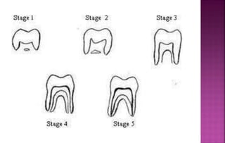
The document discusses various methods used to assess skeletal maturity indicators including hand-wrist radiographs, cervical vertebrae morphology, and tooth development stages. It describes in detail the Greulich and Pyle atlas method of comparing radiographs to standardized images, the nine stages of the Bjork, Brown, Grave method, Singer's six stage system, and Fishman's 11 skeletal maturity indicators. Assessment of cervical vertebrae morphology uses the six stage Cervical Vertebral Maturation Index. Skeletal maturity assessment is useful for orthodontic treatment planning, predicting growth, and research studies.
The document discusses various skeletal maturity indicators used to assess skeletal maturity, including hand-wrist radiographs, cervical vertebrae, and dental indicators. It provides details on the anatomy of the hand and wrist bones and stages of ossification visible in hand-wrist radiographs according to different methods. It also describes the six stages of cervical vertebral maturation as seen on lateral cephalograms according to Lamparski. Comparing the stages of ossification seen in the middle phalanx of the third finger (MP3) to the cervical vertebral maturation stages shows similarities between the MP3-F stage and initiation stage, MP3-FG stage and acceleration stage, and MP3-G stage and transition stage.
The document discusses various methods for assessing skeletal maturity and growth, including hand-wrist radiographs. It describes the bones seen in hand-wrist radiographs and several methods for analyzing skeletal maturity based on stages of ossification, including the Greulich and Pyle atlas method, Bjork method, Fishman method, and Hagg and Taranger method. The document also discusses other indicators of skeletal maturity such as cervical vertebrae and their relationship to skeletal age assessment.
The Indian Dental Academy is the Leader in continuing dental education , training dentists in all aspects of dentistry and
offering a wide range of dental certified courses in different formats.for more details please visit
www.indiandentalacademy.com
This document summarizes Dr. Pratik Yadav's journal club presentation on Downs WB Analysis of the dento-facial profile. It discusses the 10 parameters in Downs analysis, which includes 5 skeletal and 5 dental measurements. The parameters are measured based on landmarks and reference planes on lateral cephalograms. Downs analysis is one of the most commonly used cephalometric analyses originally developed based on Caucasian patients with excellent occlusion.
The Indian Dental Academy is the Leader in continuing dental education , training dentists in all aspects of dentistry and
offering a wide range of dental certified courses in different formats.for more details please visit
www.indiandentalacademy.com
The Indian Dental Academy is the Leader in continuing dental education , training dentists in all aspects of dentistry and
offering a wide range of dental certified courses in different formats.for more details please visit
www.indiandentalacademy.com
This document discusses several orthodontic appliances including the Nance appliance, transpalatal arch, quad helix, lip bumper, and tongue crib. It provides details on the design, indications, mechanisms of action, advantages and disadvantages of each appliance. The document is intended as an educational guide for orthodontic residents, as it is presented by several orthodontists and covers the key aspects of these common fixed functional appliances.
1) The document discusses smile esthetics in orthodontics, including the anatomy of the smile, ideal smile characteristics, smile classifications, and considerations for macroesthetics, miniesthetics, and microesthetics in treatment.
2) It describes the key components of the smile, including the lips, teeth, gingiva, and their proportions. Ideal smile characteristics include the smile arc, tooth width-height ratios, spacing, gingival levels, and lip fullness.
3) Smiles are classified based on the involved muscles and tooth display, including posed/social, unposed/enjoyment, and specific patterns involving the commissures or cuspids. Treatment must consider the patient's
The Indian Dental Academy is the Leader in continuing dental education , training dentists in all aspects of dentistry and
offering a wide range of dental certified courses in different formats.for more details please visit
www.indiandentalacademy.com
The document provides a history and overview of rapid maxillary expansion (RME). It discusses:
- The early history of RME dating back to the 1800s and its reintroduction by Haas in the 1960s.
- Classification of RME based on factors like rate of expansion, direction, and type of appliance.
- Indications for RME including dental issues like posterior crossbites and medical issues like poor nasal breathing.
- Contraindications such as single tooth crossbites or severe anteroposterior skeletal discrepancies.
- Examples of RME appliances including the Hyrax expander and bonded expanders, discussing their advantages.
1. There are several methods to assess skeletal maturity including hand-wrist radiographs, cervical vertebrae shape assessment, and tooth development stages.
2. Hand-wrist radiographs can be assessed using the Greulich-Pyle atlas method or the Bjork, Grave, and Brown method which divides skeletal development into 9 stages.
3. Cervical vertebrae shape changes through 6 stages of maturation and can indicate how much growth remains.
4. Tooth development through 8 stages of calcification as shown in the Demirjian Index also corresponds to skeletal maturity.
The Indian Dental Academy is the Leader in continuing dental education , training dentists in all aspects of dentistry and
offering a wide range of dental certified courses in different formats.for more details please visit
www.indiandentalacademy.com
The Indian Dental Academy is the Leader in continuing dental education , training dentists in all aspects of dentistry and
offering a wide range of dental certified courses in different formats.for more details please visit
www.indiandentalacademy.com
This document discusses methods for assessing human growth, including direct measurements like anthropometry and vital staining, as well as indirect measurements like dental casts, photographs, and radiographs. It also covers assessing a patient's age based on chronological, somatotypic, morphologic, dental, sexual, facial, and skeletal age. Key methods discussed include hand-wrist radiographs, cervical vertebrae maturation, midpalatal suture closure, and frontal sinus development. The timing of growth spurts and their clinical importance in orthodontic treatment planning is also summarized.
The Indian Dental Academy is the Leader in continuing dental education , training dentists in all aspects of dentistry and
offering a wide range of dental certified courses in different formats.for more details please visit
www.indiandentalacademy.com
The document discusses various concepts related to mandibular growth rotations proposed by different orthodontic researchers over time. It begins by introducing Arne Bjork who first described growth rotations in 1955. It then covers Enlow's concept of remodeling and displacement rotations. Bjork's 1969 classification of forward and backward mandibular rotations is described, including his identification of three types of forward and two types of backward rotations based on their center of rotation. The document also discusses concepts by Bjork and Skieller on total, matrix, and intramatrix rotations. Fred Schudy's concept relating rotation to the disharmony between vertical, anteroposterior and horizontal growth is summarized. Finally, Dibbets' re
The Indian Dental Academy is the Leader in continuing dental education , training dentists in all aspects of dentistry and
offering a wide range of dental certified courses in different formats.for more details please visit
www.indiandentalacademy.com
Canine Impaction and Its Importance in OrthodonticsAnalhaq Shaikh
Canine Impaction, Its Importance in Orthodontics, Etiology, Diagnosis and Management.
by Dr Analhaq Shaikh, 2nd year Postgraduate student, Sharavathi Dental College and Hospital, Shimoga, Karnataka
Canine Impaction can also be termed as Shy Canine.
This document provides an overview of rapid maxillary expansion (RME) in orthodontics. It discusses the history of RME dating back to 1860. It also covers anatomy related to RME, including the midpalatal suture. Key topics covered include indications and contraindications for RME, types of expansion screws used, jackscrew turn schedules, and different types of RME appliances such as the Haas expander and Hyrax expander. The document is an educational resource on the clinical use and mechanics of RME.
This document discusses growth spurts and their significance in orthodontics. It begins by defining growth and development, and describing the major developmental growth periods. It then discusses methods for studying growth, including longitudinal, cross-sectional, and semi-longitudinal studies. Key factors that influence growth and maturation are genetic, hormonal, nutritional, environmental and socioeconomic factors. The document explores concepts of growth including normality, growth rhythms, differential growth, and growth spurts. It concludes by noting the significance of growth spurts is important for orthodontic treatment planning.
The Indian Dental Academy is the Leader in continuing dental education , training dentists in all aspects of dentistry and
offering a wide range of dental certified courses in different formats.for more details please visit
www.indiandentalacademy.com
The Indian Dental Academy is the Leader in continuing dental education , training dentists in all aspects of dentistry and
offering a wide range of dental certified courses in different formats.for more details please visit
www.indiandentalacademy.com
The Indian Dental Academy is the Leader in continuing dental education , training dentists in all aspects of dentistry and
offering a wide range of dental certified courses in different formats.for more details please visit
www.indiandentalacademy.com
This document summarizes Bjork's analysis, a method developed by orthodontist Arne Bjork to analyze craniofacial growth and development using lateral cephalograms. It describes Bjork's landmarks, angular and linear measurements used to construct a facial diagram. Bjork conducted studies on Scandinavian children to establish norms for comparison. His analysis helps determine the amount and distribution of facial prognathism based on configurations in the facial diagram.
Digital technologies have greatly advanced orthodontic diagnostic aids. Digital imaging uses electronic sensors instead of film and allows for image enhancement techniques like contrast optimization. Digitalized surface imaging uses laser scanning or stereophotogrammetry to create 3D surface models of the teeth and jaws. Digital casts can be obtained via direct interior scanning or indirect external scanning and offer advantages over physical casts like elimination of breakage and easy sharing. 3D occlusograms combine lateral ceph images and occlusal views to model the 3D occlusal relationship. These recent diagnostic technologies provide more detailed information to aid orthodontic treatment planning.
Influence of Drugs on Orthodontic Tooth MovementMahmoud Shaheen
This document summarizes the effects of various medications on orthodontic tooth movement. It discusses how analgesics like NSAIDs inhibit prostaglandin synthesis and can slow tooth movement. Corticosteroids increase bone resorption and can accelerate movement. Bisphosphonates, fluorides, estrogens, and androgens inhibit osteoclast activity and bone resorption, potentially delaying movement. Thyroid hormones and vitamin D may increase tooth movement by stimulating osteoclasts. Anti-convulsants can induce gingival issues complicating treatment. The conclusion emphasizes the importance for orthodontists to be aware of how medications can influence treatment outcomes and discuss potential complications with patients.
Skeletal Age Assessment and Maturity IndicatorsAIIMS New Delhi
Skeletal age assessment is important for determining an individual's maturity level and appropriate treatment plans. There are several methods to assess skeletal age using radiographs of the hand-wrist or cervical vertebrae. The Greulich and Pyle atlas method compares a patient's hand-wrist radiograph to standardized photographs. The Bjork, Grave and Brown method stages skeletal maturity based on ossification of various hand bones. Other methods include Fishman's skeletal maturity indicators, Hagg and Taranger staging based on the ulnar sesamoid and phalanges, and Hassel and Farman's method using cervical vertebrae morphology. Accurate skeletal age assessment provides insight into a patient's growth and development.
skeletal maturity indicators in orthodontics /certified fixed orthodontic cou...Indian dental academy
The document discusses biological age assessment methods for orthodontic treatment planning, focusing on hand-wrist radiograph analysis. It describes 9 stages of skeletal maturity assessment using ossification of bones in the hand and wrist. Specific indicators are defined, such as the sesamoid bone, stages in the third finger, and union of epiphyses. Assessing a patient's skeletal age using hand-wrist radiographs can help determine their growth stage and remaining growth potential to optimize orthodontic treatment.
The Indian Dental Academy is the Leader in continuing dental education , training dentists in all aspects of dentistry and
offering a wide range of dental certified courses in different formats.for more details please visit
www.indiandentalacademy.com
The document provides a history and overview of rapid maxillary expansion (RME). It discusses:
- The early history of RME dating back to the 1800s and its reintroduction by Haas in the 1960s.
- Classification of RME based on factors like rate of expansion, direction, and type of appliance.
- Indications for RME including dental issues like posterior crossbites and medical issues like poor nasal breathing.
- Contraindications such as single tooth crossbites or severe anteroposterior skeletal discrepancies.
- Examples of RME appliances including the Hyrax expander and bonded expanders, discussing their advantages.
1. There are several methods to assess skeletal maturity including hand-wrist radiographs, cervical vertebrae shape assessment, and tooth development stages.
2. Hand-wrist radiographs can be assessed using the Greulich-Pyle atlas method or the Bjork, Grave, and Brown method which divides skeletal development into 9 stages.
3. Cervical vertebrae shape changes through 6 stages of maturation and can indicate how much growth remains.
4. Tooth development through 8 stages of calcification as shown in the Demirjian Index also corresponds to skeletal maturity.
The Indian Dental Academy is the Leader in continuing dental education , training dentists in all aspects of dentistry and
offering a wide range of dental certified courses in different formats.for more details please visit
www.indiandentalacademy.com
The Indian Dental Academy is the Leader in continuing dental education , training dentists in all aspects of dentistry and
offering a wide range of dental certified courses in different formats.for more details please visit
www.indiandentalacademy.com
This document discusses methods for assessing human growth, including direct measurements like anthropometry and vital staining, as well as indirect measurements like dental casts, photographs, and radiographs. It also covers assessing a patient's age based on chronological, somatotypic, morphologic, dental, sexual, facial, and skeletal age. Key methods discussed include hand-wrist radiographs, cervical vertebrae maturation, midpalatal suture closure, and frontal sinus development. The timing of growth spurts and their clinical importance in orthodontic treatment planning is also summarized.
The Indian Dental Academy is the Leader in continuing dental education , training dentists in all aspects of dentistry and
offering a wide range of dental certified courses in different formats.for more details please visit
www.indiandentalacademy.com
The document discusses various concepts related to mandibular growth rotations proposed by different orthodontic researchers over time. It begins by introducing Arne Bjork who first described growth rotations in 1955. It then covers Enlow's concept of remodeling and displacement rotations. Bjork's 1969 classification of forward and backward mandibular rotations is described, including his identification of three types of forward and two types of backward rotations based on their center of rotation. The document also discusses concepts by Bjork and Skieller on total, matrix, and intramatrix rotations. Fred Schudy's concept relating rotation to the disharmony between vertical, anteroposterior and horizontal growth is summarized. Finally, Dibbets' re
The Indian Dental Academy is the Leader in continuing dental education , training dentists in all aspects of dentistry and
offering a wide range of dental certified courses in different formats.for more details please visit
www.indiandentalacademy.com
Canine Impaction and Its Importance in OrthodonticsAnalhaq Shaikh
Canine Impaction, Its Importance in Orthodontics, Etiology, Diagnosis and Management.
by Dr Analhaq Shaikh, 2nd year Postgraduate student, Sharavathi Dental College and Hospital, Shimoga, Karnataka
Canine Impaction can also be termed as Shy Canine.
This document provides an overview of rapid maxillary expansion (RME) in orthodontics. It discusses the history of RME dating back to 1860. It also covers anatomy related to RME, including the midpalatal suture. Key topics covered include indications and contraindications for RME, types of expansion screws used, jackscrew turn schedules, and different types of RME appliances such as the Haas expander and Hyrax expander. The document is an educational resource on the clinical use and mechanics of RME.
This document discusses growth spurts and their significance in orthodontics. It begins by defining growth and development, and describing the major developmental growth periods. It then discusses methods for studying growth, including longitudinal, cross-sectional, and semi-longitudinal studies. Key factors that influence growth and maturation are genetic, hormonal, nutritional, environmental and socioeconomic factors. The document explores concepts of growth including normality, growth rhythms, differential growth, and growth spurts. It concludes by noting the significance of growth spurts is important for orthodontic treatment planning.
The Indian Dental Academy is the Leader in continuing dental education , training dentists in all aspects of dentistry and
offering a wide range of dental certified courses in different formats.for more details please visit
www.indiandentalacademy.com
The Indian Dental Academy is the Leader in continuing dental education , training dentists in all aspects of dentistry and
offering a wide range of dental certified courses in different formats.for more details please visit
www.indiandentalacademy.com
The Indian Dental Academy is the Leader in continuing dental education , training dentists in all aspects of dentistry and
offering a wide range of dental certified courses in different formats.for more details please visit
www.indiandentalacademy.com
This document summarizes Bjork's analysis, a method developed by orthodontist Arne Bjork to analyze craniofacial growth and development using lateral cephalograms. It describes Bjork's landmarks, angular and linear measurements used to construct a facial diagram. Bjork conducted studies on Scandinavian children to establish norms for comparison. His analysis helps determine the amount and distribution of facial prognathism based on configurations in the facial diagram.
Digital technologies have greatly advanced orthodontic diagnostic aids. Digital imaging uses electronic sensors instead of film and allows for image enhancement techniques like contrast optimization. Digitalized surface imaging uses laser scanning or stereophotogrammetry to create 3D surface models of the teeth and jaws. Digital casts can be obtained via direct interior scanning or indirect external scanning and offer advantages over physical casts like elimination of breakage and easy sharing. 3D occlusograms combine lateral ceph images and occlusal views to model the 3D occlusal relationship. These recent diagnostic technologies provide more detailed information to aid orthodontic treatment planning.
Influence of Drugs on Orthodontic Tooth MovementMahmoud Shaheen
This document summarizes the effects of various medications on orthodontic tooth movement. It discusses how analgesics like NSAIDs inhibit prostaglandin synthesis and can slow tooth movement. Corticosteroids increase bone resorption and can accelerate movement. Bisphosphonates, fluorides, estrogens, and androgens inhibit osteoclast activity and bone resorption, potentially delaying movement. Thyroid hormones and vitamin D may increase tooth movement by stimulating osteoclasts. Anti-convulsants can induce gingival issues complicating treatment. The conclusion emphasizes the importance for orthodontists to be aware of how medications can influence treatment outcomes and discuss potential complications with patients.
Skeletal Age Assessment and Maturity IndicatorsAIIMS New Delhi
Skeletal age assessment is important for determining an individual's maturity level and appropriate treatment plans. There are several methods to assess skeletal age using radiographs of the hand-wrist or cervical vertebrae. The Greulich and Pyle atlas method compares a patient's hand-wrist radiograph to standardized photographs. The Bjork, Grave and Brown method stages skeletal maturity based on ossification of various hand bones. Other methods include Fishman's skeletal maturity indicators, Hagg and Taranger staging based on the ulnar sesamoid and phalanges, and Hassel and Farman's method using cervical vertebrae morphology. Accurate skeletal age assessment provides insight into a patient's growth and development.
skeletal maturity indicators in orthodontics /certified fixed orthodontic cou...Indian dental academy
The document discusses biological age assessment methods for orthodontic treatment planning, focusing on hand-wrist radiograph analysis. It describes 9 stages of skeletal maturity assessment using ossification of bones in the hand and wrist. Specific indicators are defined, such as the sesamoid bone, stages in the third finger, and union of epiphyses. Assessing a patient's skeletal age using hand-wrist radiographs can help determine their growth stage and remaining growth potential to optimize orthodontic treatment.
Skeletal maturity can be estimated using hand-wrist radiographs which show the ossification and fusion of bones over time. There are several methods for assessing skeletal maturity based on indicators seen on hand-wrist radiographs, including the Greulich and Pyle atlas method, Bjork-Grave-Brown method using 9 stages of maturity, Singer's 6-stage method, and Fishman's skeletal maturity indicators method using 11 indicators across 4 anatomical sites. These methods allow clinicians to estimate a patient's skeletal age and determine their growth potential for orthodontic treatment planning.
The Indian Dental Academy is the Leader in continuing dental education , training dentists in all aspects of dentistry and
offering a wide range of dental certified courses in different formats.for more details please visit
www.indiandentalacademy.com
skeletal maturity indicators /certified fixed orthodontic courses by Indian d...Indian dental academy
The Indian Dental Academy is the Leader in continuing dental education , training dentists in all aspects of dentistry and offering a wide range of dental certified courses in different formats.
Indian dental academy provides dental crown & Bridge,rotary endodontics,fixed orthodontics,
Dental implants courses.for details pls visit www.indiandentalacademy.com ,or call
00919248678078
Skeletal maturity indicators /certified fixed orthodontic courses by Indian d...Indian dental academy
This document discusses skeletal maturity indicators and describes Fishman's skeletal maturity indicator (SMI) system. The SMI system uses 11 indicators seen on hand-wrist radiographs to assess skeletal maturity. The indicators include widening, capping, and fusion of epiphyses. A longitudinal study found average ages for each SMI. Other systems for assessing skeletal maturity using hand-wrist radiographs and cervical vertebrae on lateral cephalograms are also described. The cervical vertebrae system identifies 6 categories of skeletal maturity based on the shape of C3. Assessing skeletal maturity is important for evaluating growth potential and orthodontic treatment planning.
Growth assessment in orthodontics using radiograph by dr aghimienAghimien Osaronse
This document discusses the use of hand and wrist radiographs, cephalometric radiographs, and panoramic radiographs for assessing growth in orthodontic patients. It outlines the key indicators and stages seen on each radiograph that correlate with skeletal maturity and predict remaining growth. Hand-wrist radiographs assess epiphyseal development to determine skeletal age. Cephalometric radiographs examine cervical vertebrae maturation to predict timing and direction of growth. Panoramic radiographs evaluate dental development to assess dental age. Understanding a patient's skeletal and dental maturity from these radiographs helps orthodontists determine appropriate treatment timing.
THE USE OF HAND AND WRIST RADIOGRAPH, OPG AND CEPHALOMETRIC RADIOGRAPH FOR TH...Aghimien Osaronse
This document discusses the use of hand and wrist radiographs, cephalometric radiographs, and panoramic radiographs for assessing growth in orthodontic patients. It covers the indications, methods, and clinical relevance of each radiograph type. Hand-wrist radiographs can be used to determine skeletal maturity stages and predict timing of growth spurts. Cephalometric radiographs allow assessment of cervical vertebrae maturation stages, which correlate with remaining growth. Panoramic radiographs provide dental age by evaluating tooth calcification stages. Together these radiographs provide useful information for orthodontic treatment planning and timing.
This document discusses various methods of assessing skeletal maturity from radiographs, which is important for orthodontic treatment planning. It describes five main methods: 1) Greulich & Pyle atlas method comparing hand-wrist radiographs to standard images 2) Bjork, Grave & Brown method assessing nine stages of ossification in the hand and wrist 3) Fishman's Skeletal Maturity Indicators using four stages and six anatomical sites 4) Hassel and Farman method evaluating six stages of cervical vertebrae development 5) Assessment of tooth mineralization stages on panoramic radiographs. Evaluating a patient's skeletal maturity is crucial for determining prognosis, treatment goals and timing of growth modification therapies.
Skeletal maturity can be assessed using hand-wrist radiographs. Several methods exist including Greulich and Pyle (1959), Bjork (1972), and Fishman (1982). Greulich and Pyle use an atlas to compare maturity. Bjork identifies 9 stages of ossification. Fishman's Skeletal Maturation Assessment identifies 11 stages based on ossification events in the fingers, wrist, and radius. Assessment of skeletal maturity is important for orthodontic treatment planning to determine facial growth status and the timing of growth modification interventions.
This document discusses various skeletal maturity indicators used to assess skeletal age and predict growth spurts. It describes methods using hand-wrist radiographs, tooth mineralization of the mandibular canine, and cervical vertebrae morphology. For hand-wrist radiographs, it outlines the anatomy and several methods to evaluate skeletal maturity stages based on ossification of carpals, metacarpals, and phalanges. Tooth mineralization focuses on mandibular canine root development. Cervical vertebrae maturation is assessed using morphological changes that occur in predictable sequences.
Growth prediction and age estimation /certified fixed orthodontic courses by...Indian dental academy
The Indian Dental Academy is the Leader in continuing dental education , training dentists in all aspects of dentistry and offering a wide range of dental certified courses in different formats.
Indian dental academy provides dental crown & Bridge,rotary endodontics,fixed orthodontics,
Dental implants courses.for details pls visit www.indiandentalacademy.com ,or call
00919248678078
Growth predictions /certified fixed orthodontic courses by Indian dental acad...Indian dental academy
The Indian Dental Academy is the Leader in continuing dental education , training dentists in all aspects of dentistry and offering a wide range of dental certified courses in different formats.
Indian dental academy provides dental crown & Bridge,rotary endodontics,fixed orthodontics,
Dental implants courses.for details pls visit www.indiandentalacademy.com ,or call
0091-9248678078
This document discusses methods for assessing skeletal age through radiographic examination of bones and structures. It describes using hand-wrist radiographs according to Greulich and Pyle's atlas to determine skeletal age based on ossification patterns. Cervical vertebrae morphology is also assessed in six stages of maturation. Additional methods examined are frontal sinus size relative to growth velocity and midpalatal suture approximation correlated to hand-wrist development. Skeletal age assessment provides a more reliable indicator of maturation than chronological age alone for orthodontic treatment planning.
Skeletal age assesment /certified fixed orthodontic courses by Indian dental ...Indian dental academy
The Indian Dental Academy is the Leader in continuing dental education , training dentists in all aspects of dentistry and offering a wide range of dental certified courses in different formats.
Indian dental academy provides dental crown & Bridge,rotary endodontics,fixed orthodontics,
Dental implants courses.for details pls visit www.indiandentalacademy.com ,or call
0091-9248678078
Brief description of Skeletal Maturity indicators and advancements in this aspect .
Biochemical markers as new advancements in AGE assessment
Realtionship between SMI and CVMI , MP3 AND CVMI
This document discusses skeletal maturity indicators that can be used to assess a patient's biological age and remaining growth potential. It describes how chronological age alone is not enough, and that skeletal age determined from hand wrist radiographs provides a more accurate assessment. The document outlines several methods for assessing skeletal age, including the Fishman skeletal maturity indicators and the Modified MP3 Cervical Vertebrae Maturation Index. It explains what each method evaluates and the stages involved. Assessing skeletal age is important for orthodontic treatment planning to determine treatment timing and prognosis.
Bone age assessment is used to evaluate growth and diagnose endocrine disorders in pediatric patients. It is determined by examining X-rays of the bones and assessing the appearance and fusion of ossification centers, which occurs at characteristic ages. Various factors like genetics and nutrition can cause some variability between populations, with Indian bone maturation generally occurring earlier than Western standards. The document provides detailed tables outlining the appearance and fusion of ossification centers from infancy through adulthood in the bones of the hands, feet, pelvis and other regions. It also describes the stages of epiphyseal fusion used to determine bone age during adolescence.
Skeletal maturity is assessed through examination of ossification centers in bones like the hand and wrist. The Greulich and Pyle atlas and Bjork method involve comparing radiographs to standardized images to determine skeletal age. Singer's method stages skeletal maturity based on characteristics like the width of epiphyses compared to diaphyses and appearance of sesamoid bones. Assessing skeletal maturity is important for orthodontic treatment planning by indicating remaining growth potential.
Similar to Skeletal maturity indicators.pptx modified (20)
Histololgy of Female Reproductive System.pptxAyeshaZaid1
Dive into an in-depth exploration of the histological structure of female reproductive system with this comprehensive lecture. Presented by Dr. Ayesha Irfan, Assistant Professor of Anatomy, this presentation covers the Gross anatomy and functional histology of the female reproductive organs. Ideal for students, educators, and anyone interested in medical science, this lecture provides clear explanations, detailed diagrams, and valuable insights into female reproductive system. Enhance your knowledge and understanding of this essential aspect of human biology.
Rasamanikya is a excellent preparation in the field of Rasashastra, it is used in various Kushtha Roga, Shwasa, Vicharchika, Bhagandara, Vatarakta, and Phiranga Roga. In this article Preparation& Comparative analytical profile for both Formulationon i.e Rasamanikya prepared by Kushmanda swarasa & Churnodhaka Shodita Haratala. The study aims to provide insights into the comparative efficacy and analytical aspects of these formulations for enhanced therapeutic outcomes.
share - Lions, tigers, AI and health misinformation, oh my!.pptxTina Purnat
• Pitfalls and pivots needed to use AI effectively in public health
• Evidence-based strategies to address health misinformation effectively
• Building trust with communities online and offline
• Equipping health professionals to address questions, concerns and health misinformation
• Assessing risk and mitigating harm from adverse health narratives in communities, health workforce and health system
Cell Therapy Expansion and Challenges in Autoimmune DiseaseHealth Advances
There is increasing confidence that cell therapies will soon play a role in the treatment of autoimmune disorders, but the extent of this impact remains to be seen. Early readouts on autologous CAR-Ts in lupus are encouraging, but manufacturing and cost limitations are likely to restrict access to highly refractory patients. Allogeneic CAR-Ts have the potential to broaden access to earlier lines of treatment due to their inherent cost benefits, however they will need to demonstrate comparable or improved efficacy to established modalities.
In addition to infrastructure and capacity constraints, CAR-Ts face a very different risk-benefit dynamic in autoimmune compared to oncology, highlighting the need for tolerable therapies with low adverse event risk. CAR-NK and Treg-based therapies are also being developed in certain autoimmune disorders and may demonstrate favorable safety profiles. Several novel non-cell therapies such as bispecific antibodies, nanobodies, and RNAi drugs, may also offer future alternative competitive solutions with variable value propositions.
Widespread adoption of cell therapies will not only require strong efficacy and safety data, but also adapted pricing and access strategies. At oncology-based price points, CAR-Ts are unlikely to achieve broad market access in autoimmune disorders, with eligible patient populations that are potentially orders of magnitude greater than the number of currently addressable cancer patients. Developers have made strides towards reducing cell therapy COGS while improving manufacturing efficiency, but payors will inevitably restrict access until more sustainable pricing is achieved.
Despite these headwinds, industry leaders and investors remain confident that cell therapies are poised to address significant unmet need in patients suffering from autoimmune disorders. However, the extent of this impact on the treatment landscape remains to be seen, as the industry rapidly approaches an inflection point.
Local Advanced Lung Cancer: Artificial Intelligence, Synergetics, Complex Sys...Oleg Kshivets
Overall life span (LS) was 1671.7±1721.6 days and cumulative 5YS reached 62.4%, 10 years – 50.4%, 20 years – 44.6%. 94 LCP lived more than 5 years without cancer (LS=2958.6±1723.6 days), 22 – more than 10 years (LS=5571±1841.8 days). 67 LCP died because of LC (LS=471.9±344 days). AT significantly improved 5YS (68% vs. 53.7%) (P=0.028 by log-rank test). Cox modeling displayed that 5YS of LCP significantly depended on: N0-N12, T3-4, blood cell circuit, cell ratio factors (ratio between cancer cells-CC and blood cells subpopulations), LC cell dynamics, recalcification time, heparin tolerance, prothrombin index, protein, AT, procedure type (P=0.000-0.031). Neural networks, genetic algorithm selection and bootstrap simulation revealed relationships between 5YS and N0-12 (rank=1), thrombocytes/CC (rank=2), segmented neutrophils/CC (3), eosinophils/CC (4), erythrocytes/CC (5), healthy cells/CC (6), lymphocytes/CC (7), stick neutrophils/CC (8), leucocytes/CC (9), monocytes/CC (10). Correct prediction of 5YS was 100% by neural networks computing (error=0.000; area under ROC curve=1.0).
TEST BANK For Basic and Clinical Pharmacology, 14th Edition by Bertram G. Kat...rightmanforbloodline
TEST BANK For Basic and Clinical Pharmacology, 14th Edition by Bertram G. Katzung, Verified Chapters 1 - 66, Complete Newest Version.
8 Surprising Reasons To Meditate 40 Minutes A Day That Can Change Your Life.pptxHolistified Wellness
We’re talking about Vedic Meditation, a form of meditation that has been around for at least 5,000 years. Back then, the people who lived in the Indus Valley, now known as India and Pakistan, practised meditation as a fundamental part of daily life. This knowledge that has given us yoga and Ayurveda, was known as Veda, hence the name Vedic. And though there are some written records, the practice has been passed down verbally from generation to generation.
2. 1.Introduction
2.History
3.Hand-wrist Radiograph
4.Methods of Hand-wrist Radiograph as SMI
a. Greulich and Pyle Method
b. Bjork, Grave and Brown Method
c. Singer’s Method
d. Fishman’s SMI
e. Hagg and Taranger Method
f. Modification of Hagg and Taranger Method
5. Cervical vertebrae as SMI and its Modification
6. Tooth mineralization as SMI
7. Applications
8. Conclusion
9.References
3. Since orthodontist works primarily with teeth and
bone, the skeletal age or bone age can provide reliable
information while helping in accurate growth prediction.
The level of maturity attained and the amount of growth
potential remaining is an important consideration while
treating malocclusion.
4. A number of methods are available to assess the
skeletal maturity indicators. They include :-
a. Use of hand-wrist radiographs
b. Evaluation of skeletal maturation using cervical
vertebrae
c. Assessment of maturity by clinical and radiographic
examination of different stages of tooth development.
5. 1. 1896- Ranke is considered to have been the first to
study skeletal development progress by means of wrist
roentgenogram.
2. 1910-
i. Rotch recognized that weight, height, and tooth eruption
were at best only rough estimates of physical maturity in
terms of chronological age.
ii. Bayley found a correlation between maturation of the
knee as seen on the roentgenogram up to 13 years.
iii.Garn and Rohmann concluded that hand-wrist ossification
is useful in detecting growth abnormalities but it is not a
precise method of measuring developmental progress in
normal individuals.
6. 1931- Todd TW started a longitudinal study by taking
a series of periodic hand and wrist radiographs of
growing children in Cleveland, Ohio, USA.
Unfortunately , Professor Todd died in 1938 after
publishing the initial data of his study in 1937.
1959- After his death , the study was continued and
William Greulich and Idell Pyle compiled the
Radiographic atlas of skeletal development of hand
and wrist , which was published in 1950 and revised.
8. 1965- Lampirski developed 6 stages of CVM.
1976-Bjork, Brown, Grave discovered 9
developmental stages of skeletal maturation.
1980-Julian Singer proposed 6 stages of Hand-wrist
development.
1982- Leonardo S. Fishman’s 11 SMI
1995-Hassel and Farman proposed CVMI 6 stages of
skeletal maturation.
2005-Bacietti et al modified Hagg and Taranger
method of Skeletal maturation
9. Indications-
In patients who exhibit major discrepancy between dental
and chronological age.
It is indicated in patients with skeletal malocclusion
needing orthognathic surgery, if undertaken between 16-
20 years so as to assess the growth status.
To predict the pubertal growth spurt.
It is valuable aid in research aimed at studying the role of
heredity ,environment, nutrition etc, on the skeletal
maturation pattern.
10. Hand-wrist region is made up of 4 groups of bone ;-
1. Distal ends of long bones of forearm( Distal
epiphysis of Radius and Ulna)
2. Carpals(8 carpal bones)
3. Meta carpals(5 in no)
4. Phalanges(14 in no)
13. Most commonly used methods:
1. Atlas Method by Greulich and Pyle
2. Bjork, Grave and Brown Method
3. Singer’s method
4. Fishman’s Skeletal Maturity Indicators
5. Hagg and Taranger Method
14. Greulich and Pyle published an atlas containing ideal
skeletal age pictures of the hand-wrist for different
chronological ages and for each sex.
Each photograph in the atlas is representative of a
particular skeletal age. The patient’s radiograph is
matched on an overall basis with one of the photographs
in the atlas.
15. STAGE-I (PP2) (Males 10.6 yrs ,
Females 8.1 yrs)
The epiphysis and diaphysis of proximal
phalanx of Index finger are equal.
It occurs approximately 3 years before the
peak of pubertal growth spurt.
They divided skeletal development in to 9 stages. Each
of these represents a skeletal maturity. Appropriate
chronological age for each of the stages was given by
Schopf in 1976.
16. STAGE II-(MP3)-
(males 12.0 yr, females 8.1yr):-
The epiphysis and diaphysis of the middle phalanx of the
middle finger are equal.
This stage is noticed prior to the beginning of the
pubertal growth spurt.
17. STAGE III:-(Pisi-stage)(males 12.6 yr, females 9.6
yr)-
This stage is characterized by presence of 3 areas of
ossification:
The hamular process of the hamate exhibits ossification.
Ossification of psiform
The epiphysis and diaphysis of radius are equal.
18.
19. Stage-IV:-(S, H2 Stage)(males 13.0 yr, females
10.6 yr)-
This stage marks the beginning of the pubertal growth
spurt. It is characterized by:
Initial mineralization of the ulnar sesamoid of the
thumb.
Increased ossification of the hamular process of the
hamate bone.
20.
21. Stage V;-(MP3 CAP)(males 14.0 yr, females
10yr)-
This stage heralds the peak of the pubertal growth
spurt. Capping of diaphysis by the epiphysis is seen in:
Middle phalanx of third finger
Proximal phalanx of the thumb
Radius
22.
23. Stage VI;- (DP3u)(males 15.0 yr, females 13.0
yr):
This stage signifies the end of pubertal growth spurt.
It is characterized by union between epiphysis and
diaphysis of the distal phalanx of the middle finger.
24.
25. Stage VII;-(PP3u)(males 15.9 yr, females 13.3
yr)-
Union of epiphysis and diaphysis of the proximal
phalanx of the third finger occurs.
It is seen a year after the growth spurt.
26.
27. It shows fusion between the epiphysis and diaphysis of
the middle phalanx of the middle finger.
28. Stage IX;-(Ru)(males 18.5 yr, females 16.0 yr)-
This is the last stage and it signifies the end of skeletal
growth.
It is characterized by fusion of epiphysis and diaphysis
of the radius.
29.
30. Julian Singer in 1980 proposed system of hand wrist
radiographic assessment. It helps the clinician to
rapidly determine maturational status of the
adoloscent patient. This system has 6 stages. The
stages and their characteristics are:
Stage 1(early)-
It is characterized by absence of hook of the hamate
and epiphysis of proximal phalanx of second finger
being narrower than its diaphysis.
31.
32. Stage 2(pre-pubertal)
• It characterized by initial ossification of hook of the
hamate , initial ossification of pisiform and proximal
phalanx of second finger being equal to its epiphysis.
• It represents that period prior to the adolescent growth
spurt during which significant amounts of mandibular
growth are possible.
• Maxillary orthodontic therapy in conjuction with
mandibular growth might aid correction of a Class II
relationship with considerable speed and ease.
33.
34. Stage III(pubertal onset)-
This stage is characterised by beginning of
calcification of ulnar sesamoid, increased width of
epiphysis of proximal phalanx of the 2nd finger and
increased calcification of hook of hamate and pisiform
.
Signifies the onset of pubertal growth spurt.
35.
36. Stage IV(Pubertal)
It is characterized by calcified ulnar sesamoid and
capping of the diaphysis of the middle phalanx of the
third finger by its epiphysis .
This stage signifies the accelerating phase of pubertal
growth spurt.
37.
38. Stage V(Pubertal deceleration)-
1. This stage is characterized by fully calcified ulnar
sesamoid , fusion of epiphysis of distal phalanx of
third finger with its shaft, and epiphyses of radius and
ulna not fully fused with respective shafts.
2. Stage 5 represents that period of growth when
orthodontic treatment might be completed and the
patient is in retention therapy.
42. Leonard S. Fishman in 1982 proposed a system for
evaluation of skeletal maturation.
It uses anatomical sites located on the thumb, third
finger ,fifth finger and radius.
Fishman’s system for interpretation uses 4 stages for
bone maturation.
1. Epiphysis equal in width to diaphysis.
2. Apperance of Adductor Sesamoid of Thumb
3. Capping of epiphysis
4. Fusion of epiphysis
43. Eleven discrete adolescent SMI covering the entire period of
adolescent development have been described .
Epiphysis as width as diaphysis;-
SMI I;- Third finger – Proximal phalanx(PP3)
SMI II;- Third finger – Middle Phalanx(MP3)
SMI III;- Fifth finger - Middle Phalanx(MP5)
44. Ossification;-
SMI IV;- Appearance of Adductor Sesamoid of
Thumb(4S)
Capping of Epiphysis ;-
SMI V ;- Third finger – distal phalanx(DP3 CAP)
SMI VI;- Third Finger– middle phalanx(MP3 CAP)
SMI VII;- Fifth Finger-middle phalanx (MP5 CAP)
45.
46. Fusion of epiphysis and diaphysis ;-
SMI VIII- Third finger- distal phalanx(DP3U)
SMI IX- Third finger- proximal phalanx(PP3)
SMI X- Third finger- middle phalanx(MP3u)
SMI XI- Radius(Ru)
47. Skeletal development in the hand and wrist is analysed
from annual radiogrphs, taken between ages of 6
and 18 years.
1. By assessment of ossification of ulnar sesamoid of the
metacarpophalangeal joint of the first finger (S)
2. By certain specified stages of 3 epiphyseal bones:
a. the middle and distal phalanges of the third
finger(MP3 and DP3)
b. the distal epiphysis of the radius (R).
48. Sesamoid is attained during the acceleration period of
the pubertal growth spurt.
49.
50. MP3-F;-The epiphysis is as wide as the metaphysis.
MP3-FG;-The epiphysis is as wide as metaphysis and
there is distinct medial or lateral border of the
epiphysis forming a line of demarcation at right angles
to the distal border.
MP3-G;- The sides of the epiphysis have thickened
and also cap its metaphysis, forming a sharp edge
distally at one or both sides.
MP3-H;-Fusion of epiphysis and metaphysis has
begun .
MP3-I:-Fusion of epiphysis and metaphysis is
completed.
51. Fusion of the epiphysis and metaphysis is
completed.
This stage signifies the fusion of the epiphysis and
metaphysis and is attained during the deceleration
period of the pubertal growth spurt by all subjects.
52.
53. R-I;- Fusion of epiphysis and metaphysis on radius
has begun. Stage R-I is attained one year before or at
end of pubertal growth spurt.
R-IJ;- Fusion almost completed.
R-J;- Complete fusion of epiphysis and metaphysis.
R-IJ, R-J are not attained before the end of pubertal
growth spurts.
54.
55. MP3-HI stage:-
Features of this “new” stage observed in this study :
1. Superior suface of epiphysis shows smooth concavity.
2. Metaphysis shows smooth, convex surface, almost
fitting in to reciprocal concavity of epiphysis.
3. Radiolucent gap between epiphysis and metaphysis
is insignificant.
56.
57. CVMI was given by Lamparski in 1965.Later it was
modified by Hassel and Farman in 1995 and Baccetti et al
in 2005.
The following six stages were put forward in vertebral
development.
58. 1.This stage is called initiation
corresponds to beginning of
adolescent growth with 80-
100 percent growth expected.
2. Inferior border of C2, C3
and C4 were flat at this stage.
3.The vertebrae were wedge
shaped, the superior
vertebrae borders were
tapered from posterior to
anterior.
59. 1. The second stage is called
acceleration .
2. Growth acceleration begins at
this stage with 65-75 percent
adolescent growth expected.
3. Concavities were developing in
the inferior border of C2 and C3.
4. The inferior border of C4 was
flat.
5. The bodies of C3 and C4 were
nearly rectangular in shape.
60. 1.This stage is called transition .
2.It corresponds to acceleration of
growth towards peak height
velocity with 25-65 percent of
adolescent growth expected.
3.Distinct concavities were
developing in the inferior border of
C2 and C3 .
4.A concavity was beginning to
develop in the inferior border of
C4.
5.The bodies of C3 and C4 are
rectangular in shape.
61. 1.The stage is called Deceleration,
corresponds to deceleration of
adolescent growth spurt with 10-25
percent of growth expected.
2. Discint concavities were seen
in the inferior border of C2, C3 and
C4.
3. The vertebral bodies of C3 and
C4 were more squarer in shape.
62. 1. The fifth stage is called
maturation.
2.Final maturation of vertebrae
took place during this stage
with 5-10 percent adolescent
growth expected.
3. More accentuated concavities
were seen in the inferior
borders of C2, C3 and C4.
4. The bodies of C3 and C4
were nearly square in shape
63. 1. The stage is called completion
of growth.
2. Little or no adolescent growth
expected.
3. Deep concavities were seen in
the inferior borders of C2 , C3
and C4.
4. The bodies of C3 and C4 were
square or were greater in
vertical dimension than the
horizontal dimension.
64. 1. Class II treatment is most effective when it includes
the peak in Mandibular growth.(Stage III)
2. Class III treatment with maxillary expansion and
protraction is effective
When performed before the peak ( Stage I and II ).
3. Class III treatment is effective in mandible during both
pubertal and pre-pubertal stage.
65.
66.
67. The shapes of the cervical vertebral bodies of C3 and C4
changes from wedge shape to rectangle followed by
square shape.
In addition, they become taller as skeletal maturity
progressed .The inferior vertebral borders were flat when
immature and became concave with maturity.
68. CS1;- The lower borders of all the three vertebrae
(c2-c4) are flat . The bodies of both C3 and C4 are
trapezoid in shape. The peak mandibular growth will
occur on average 2 years after this stage.
CS2;- A concavity is present at the lower border of
C2.The bodies of both C3 and C4 are still trapezoid in
shape. The peak in mandibular growth will occur on
average 1 year after this stage.
69. CS3;- Concavities at the lower borders of both C2
and C3 are present. The bodies of C3 and C4 may be
either trapezoid or rectangular horizontal in shape .
The peak in mandibular growth will occur during the
year after this stage.
CS4;- Concavities at the lower borders of C2, C3 and
C4 now are present. The bodies of both C3 and C4
are rectangular horizontal in shape. The peak in
mandibular growth has occurred within 1 or 2 years
before this stage.
70. CS5;-The concavities at the lower borders of C2, C3, and
C4 still are present .At least one of the bodies of C3 and
C4 is squred in shape . If not squared , the body of the
other cervical vertebrae still is rectangular horizontal .The
peak in mandibular growth has ended at least 1 year
before this stage.
CS6;-The concavities at the lower borders of C2, C3 and
C4 still are evident .At least one of the bodies of C3 and
C4 is rectangular vertical in shape .If not rectangular
vertical , the body of the other cervical vertebra is
squared. The peak in mandibular growth has ended at
least 2 years before this stage.
71. The calcification patterns and stage of mineralization
of the teeth is believed to have a close relationship
with the skeletal maturation of the individual.
Dental development can be assessed by either the
tooth eruption or the stage of tooth calcification ,with
the latter being more reliable.
To assess the developmental stage of dentition
through examination of panoromic radiograph offers
a several advantages.
72.
73. STAGE A;- Calcification of single occlusal points with
out fusion of different calcifications.
STAGE B;-. Fusion of mineralization points ; the
contour of occlusal surface is recognizable.
STAGE C;- Enamel formation has been completed at
the occlusal surface , the dentine formation has
commenced, the pulp chamber is curved and no pulp
horns are visible.
74. STAGE D;-Crown formation has completed to the level
of CEJ. Root formation has commenced. The pulp
horns are beginning to differentiate, but the walls of
the pulp chamber remain curved.
STAGE E;- The root length remains shorter than the
crown height. The walls of pulp chamber are straight
and the pulp horns become more differentiated than
the previous stage.
STAGE F:- The walls of the pulp chamber now form an
isoscleles triangle , and the root length is equal to or
greater than the crown height. In molars the
bifurcation has developed sufficiently to give the roots
a distinct form.
75. STAGE G;- The walls of root canals are now parallel
but apical end is partially open in molars only the
distal root is rated.
STAGE H:-The root apex is completely closed (
distal roots in molars). The periodontal membrane
surrounding the root and apex is uniform in width
through out.
78. Engstrom et al in 1983 did a study correlating the
developmental stages of mandibular 3rd molar with skeletal
age assessed by hand wrist radiographs.
The development stages of 3rd molar were categorized in
to one of the following classes.
A. Tooth germ visible as a rounded radiolucency.
B. Cusp mineralization complete.
C. Crown formation is complete.
D. Root half formed.
E. Root formation complete, but apex not closed.
79.
80. After comparing the stages of formation of lower 3rd
molar with hand-wrist radiographs , the following
points were concluded.
PP2- Lower 3rd molar showed complete crown
mineralization.
MP3 CAP- The lower 3rd molar crown formation was
complete in majority of subjects.
DP3 U- The lower 3rd molar crown was completed
with full root length in others and some cases it was
not completed.
RU- The crown was completed in one-third of subjects
and root had reached full length in rest.
81. Correlation between Chronological Age, Cervical
Vertebrae Maturation and Fishman’s Skeletal
Maturity Indicators in Southern Chinese. Alkhal et
al. Angle Orthodontist. 2008; 78(4):591-6.
OBJECTIVE:-
To investigate the correlation between chronological
age, cervical vertebral maturation(CVM), and Fishman’s
hand-wrist skeletal maturity indicators in southern
chinese.
82. MATERIALS AND METHODS;-
1.400 Contemporary hand-wrist and lateral cephalometric
radiographs of Southern Chienese subjects were
randomly selected and analysed.
2.The female subjects were between 10-15 years of age,
and the male subjects were between 12-17 years of
age; all subjects were in circumpubertal period .
3.The CVM was assessed using the method developed by
Baccetti and coworkers, but the hand-wrist maturation
was assessed using the method developed by Fishman.
83. RESULTS;-
1. The CVM was significantly correlated with the hand-
wrist skeletal age.
2. All patients in the cervical maturation stage (CS3) of
CVM were discovered to be skeletal maturation
indicator(SMI 2 and SMI 3 ) stages of hand-wrist
maturation, which was around peak of growth spurt.
3.Low correlations were found between the CVM and
chronological age and between the HWM and
chronological age.
84. CONCLUSION:-
1. CVM is a valid indicator of skeletal growth during the
circumpubertal and has a high correlation with the
HWM for the Southern Chienese population .
2. However, the low correlations found between the
chronological age and both CVM and HWM showed that
the chronological age was not suitable to measure
skeletal maturity.
85. Association between frontal sinus morphology and
cervical vertebral maturation for the assessment
of skeletal maturity. Mahmood et al. AJODO .
2016;150:637-42.
INTRODUCTION;-
The aim of the study to evaluate the association
between frontal sinus morphology and cervical maturation
for the assessment of skeletal maturity and to determine
its validity in assessing the different stages of the
adoloscent growth spurts.
86. METHODS;-
1. A cross sectional study was performed on the
pretreatment lateral cephalograms of 252 subjects
aged 8-21 years.
2. The sample was divided in to 6 groups based on the
cervical vertebral maturation stages.
3. The frontal sinus index was calculated by dividing the
frontal sinus height and width and the cervical stages
were evaluated on the same radiograph.
87. Fig. Assessment of frontal sinus morphology on a lateral cephalometric
radiograph. SH-Highest point of the frontal sinus ; SL-Lowest point of
the frontal sinus; SPP-Posterior point on the frontal sinus; SAP-Anterior
point on the frontal sinus; B-Line joining SPP and SAP denoting the
maximum frontal sinus width perpendicular to line A.
88. RESULTS:-
The height and width of the frontal sinus were
significantly larger in the male subjects than in the
females.
A significant association between frontal sinus
height and width and cervical stages in both sexes.
89. CONCLUSION:-
Frontal sinus height and width are significantly
associated with the cervical vertebral maturation stages
in both sexes.
However, the frontal sinus index failed to
differentiate between the pre-pubertal, pubertal and
postpubertal growth periods.
Hence, the cervical vertebral maturation method
remains the standard to evaluate the skeletal maturity
of patients undergoing orthodontic treatment.
96. 1.Orthodontics principles and practice 2nd edition by
Phulari .
2.Color atlas of Dental Medicine by Thomas Rakosi.
3.Text book of Craniofacial Growth. Sridhar Premkumar.
4.Correlation between Chronological age , Cervical
vertebrae Maturation and Fishman’s Skeletal Maturity
Indicators in Southern Chinese. Alkhal et al. Angle
Orthodontist. 2008; 78(4):591-6.
97. 5.Association between frontal sinus morphology and
cervical vertebral maturation for the assessment of
skeletal maturity. Mahmood et al. AJODO.
2016:150:637-42.
6. Skeletal maturation evaluation using mandibular third
molar development in adoloscents. Cho SM, Hwang CJ.
KJO. 2009; 39(2):120-9.
7. Assessing skeletal Maturity by using blood spot
insulin-like growth factor I (IGF-I) testing. Am J Orthod
Dentofacial Orthop . 2008; 134:209-16.
98. 8.Insulin like growth factor 1:A biologic maturation
Indicator. Ishaq et al. AJODO.2012; 142:654-61.
9. Relationship between Dental and Skeletal Maturity in
Turkish Subjects. UT et al. Angle Orthod . 2004;
74:657-664.
10.Hassel and Farman : Skeletal maturation evaluation
.AJODO. 1995; vol:58-66.
11. Julian Singer : Physiologic timing of Orthodontic
treatment.Angle Orthod.1980; vol: 322-333.
12. Grave and Brown: Skeletal ossification and
adoloscent growth spurt.AJODO. 1976; vol:69-80.