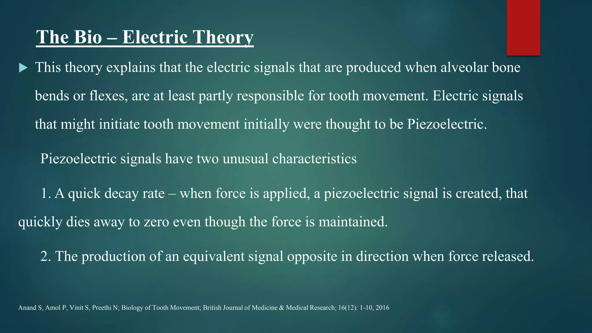
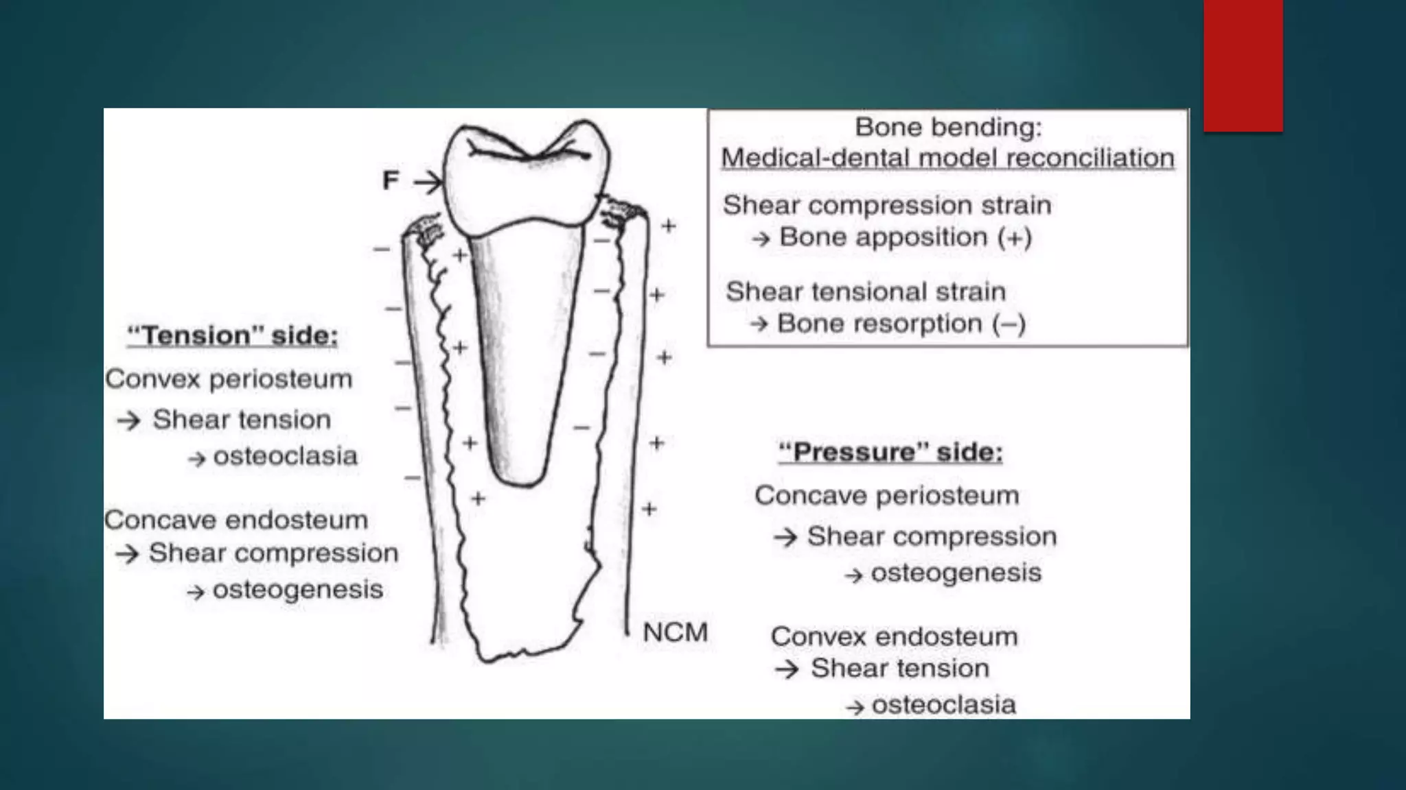
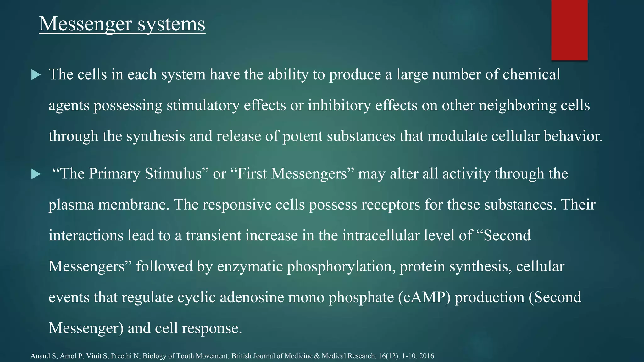
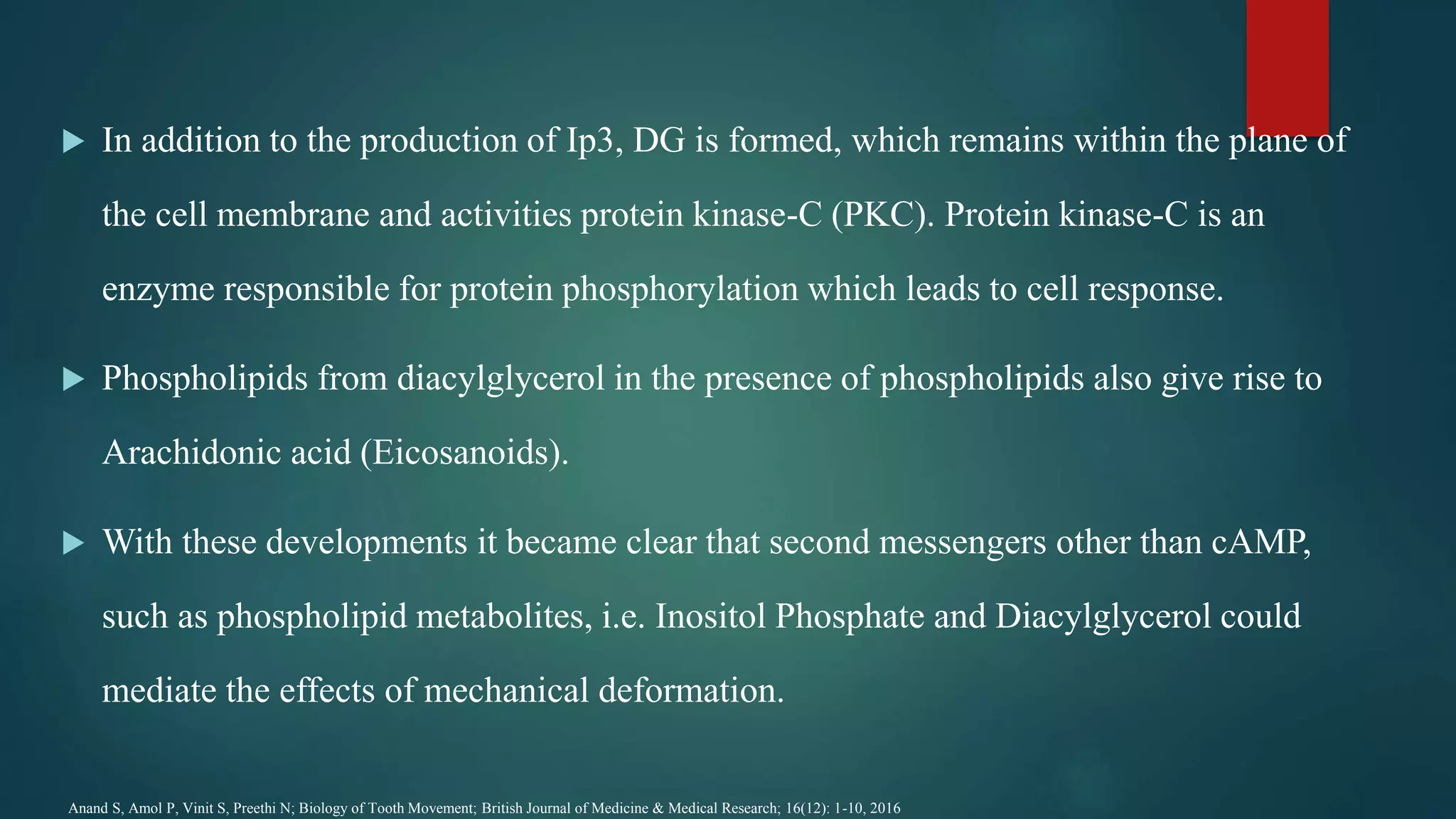
PROSTSAGLANDINS
IN
ORTHODONTIC TOOTH
MOVEMENT
-UNDER THE GUIDANCE OF DR. MRIDULA TREHAN
PRESENTED BY DR DEEKSHA BHANOTIA
PG 1ST YEAR
DEPARTMENT OF ORTHODONTICS AND DENTOFACIAL ORTHOPAEDICS
CONTENTS
 INTRODUCTION
 DRUGS ALTERING TOOTH MOVEMENT
 BIOLOGY OF TOOTH MOVEMENT
 PROSTAGLANDINS
 MECHANISM OF ACTION
 EFFECT ON TOOTH MOVEMENT
 CONCLUSION
Introduction
 Orthodontic tooth movement is a complex procedure in which changes occur from the
tissue level to the molecular level.
 Orthodontic force when applied gives rise to a cascade of inflammatory steps in which
chemical mediators are involved.
 Various metabolites in human body like prostaglandins, cytokines, interleukins are
involved in the molecular level of tooth movement.
Drugs in Orthodontics: A Review; Indian Journal of Forensic Medicine and Toxicology, July 2018, Vol. 12, No. 3.
 The constituents of nutrients of food as well as drugs alike, consumed both regularly and
occasionally by patients reach the periodontal tissues involved with orthodontic
movement and target local cells.
 The cumulative effects could be inhibitory, additive, or synergistic.
 During orthodontic treatment, drugs are prescribed to manage pain from force
application to biological tissues, manage temporomandibular joint (TMJ) problems and
tackle some infection throughout the course of treatment.
Drugs in Orthodontics: A Review; Indian Journal of Forensic Medicine and Toxicology, July 2018, Vol. 12, No. 3.
 Drugs increasing rate of tooth movement
 Prostaglandins
 Vitamin D
 PTH
 Thyroid hormone
 Corticosteroids
 Relaxin
Drugs in Orthodontics: A Review; Indian Journal of Forensic Medicine and Toxicology, July 2018, Vol. 12, No. 3.
 Drugs decreasing rate of tooth movement
 NSAID’s
 Fluorides
 Bis-phosphonates
 Estrogen
 Calcitonin
 No effect on tooth movement-Acetaminophen
Drugs in Orthodontics: A Review; Indian Journal of Forensic Medicine and Toxicology, July 2018, Vol. 12, No. 3.
The flowchart represents a brief outline of the molecular level of
orthodontic tooth movement and effect of drugs on it.
THEORIES OF TOOTH MOVEMENT
 Alveolar bone resorption and deposition during orthodontic tooth movement is a cell –
mediated process regulated by various factors. However the mechanisms involved in
conversion of OF(Orthodontic Force) into biologic activity are not completely
understood.
 There are two possible control elements that form two major theories of orthodontic tooth
movement. They are:
1) Biological Electricity (Farrar 1876).
2) Pressure – Tension in the periodontal ligament (PDL) Oppenheim(1911),
Sandstedt(1905), and Schwarz(1932).
3) Fluid dynamic theory (Bien 1966).
Anand S, Amol P, Vinit S, Preethi N; Biology of Tooth Movement; British Journal of Medicine & Medical Research; 16(12): 1-10, 2016
 Fluid Dynamic Theory: (Bien 1966)
This theory is also called the Blood Flow Theory as proposed by Bien. According to
this theory tooth movement occurs as a result of alterations in fluid dynamics in the
PDL.
 The periodontal space contains a fluid system made up of interstitial fluid, cellular
elements, blood vessels and viscous ground substance in addition to periodontal
fibres.
 It is a confined space and passage of fluid in and out of this space is limited. The
contents of the PDL thus create a unique hydrodynamic condition resembling a
hydraulic mechanism and a shock absorber.
 When the force is removed, the fluid is replenished by diffusion from capillary walls
and recirculation of interstitial fluid.
 But when a force of greater magnitude and duration is applied such as during
orthodontic tooth movement, the interstitial fluid in the periodontal space gets
squeezed out and moves towards the apex and cervical margins and results in
decreased tooth movement.
 This is called the “squeeze film effect” as proposed by Bien.
The Bio – Electric Theory
 This theory explains that the electric signals that are produced when alveolar bone
bends or flexes, are at least partly responsible for tooth movement. Electric signals
that might initiate tooth movement initially were thought to be Piezoelectric.
Piezoelectric signals have two unusual characteristics
1. A quick decay rate – when force is applied, a piezoelectric signal is created, that
quickly dies away to zero even though the force is maintained.
2. The production of an equivalent signal opposite in direction when force released.
Anand S, Amol P, Vinit S, Preethi N; Biology of Tooth Movement; British Journal of Medicine & Medical Research; 16(12): 1-10, 2016
 Zengo and associates investigated the nature of electro-chemical relationship
associated with the dentoalveolar complex using simulated OF.
 Both in-vivo studies indicated that areas that are electro-negative were characterized
by elevated osteoblastic, activity and areas of electro positivity were characterized by
elevated osteoclastic activity.
 Davidovitch and associates were able to show that accelerated Orthodontic tooth
movement resulted when exogenous electric current was administrated in conjunction
with orthodontic forces, which further increased cellular response to electrical
stimulation.
 This suggests that the piezoelectric response propagated by bone bending incident to
of application may be functioning as “Cellular first messenger.”
Anand S, Amol P, Vinit S, Preethi N; Biology of Tooth Movement; British Journal of Medicine & Medical Research; 16(12): 1-10, 2016
The Pressure Tension Theory
 This theory explains the cellular changes produced by chemical messengers during tooth
movement. This is mainly because of alteration in blood flow through the PDL. The
alteration in blood flow quickly creates changes in the environment. For example, oxygen
levels would fall in the compressed area but might increase on the tension side and the
relative proportions of other metabolites would also change in a matter of minutes.
 Orhan Tuncay and Daphane observed that low oxygen tension causes increased cellular
proliferation and decreased Adenosine triphosphate (ATP) activity and partial pressure of
oxygen (Po2) while hypoxic conditions result in suppressed cellular proliferation and
increased ATP activity.
Anand S, Amol P, Vinit S, Preethi N; Biology of Tooth Movement; British Journal of Medicine & Medical Research; 16(12): 1-10, 2016
 These chemical changes acting directly or by stimulating the release of other biologically
active agents then would stimulate cellular differentiation and activity.
 In essence, this view of tooth movement shows three stages.
a) Alteration in blood flow in the PDL.
b) The formation and / or release of Chemical Messengers.
c) Cell response.
Anand S, Amol P, Vinit S, Preethi N; Biology of Tooth Movement; British Journal of Medicine & Medical Research; 16(12): 1-10, 2016
Messenger systems
 The cells in each system have the ability to produce a large number of chemical
agents possessing stimulatory effects or inhibitory effects on other neighboring cells
through the synthesis and release of potent substances that modulate cellular behavior.
 “The Primary Stimulus” or “First Messengers” may alter all activity through the
plasma membrane. The responsive cells possess receptors for these substances. Their
interactions lead to a transient increase in the intracellular level of “Second
Messengers” followed by enzymatic phosphorylation, protein synthesis, cellular
events that regulate cyclic adenosine mono phosphate (cAMP) production (Second
Messenger) and cell response.
Anand S, Amol P, Vinit S, Preethi N; Biology of Tooth Movement; British Journal of Medicine & Medical Research; 16(12): 1-10, 2016
 The agonists, primary stimulus or first
messenger such as hormones or
mechanical forces (Orthodontic Force)
may alter activity through the plasma
membrane.
 The production of second messenger
(cAMP) pathway is regulated by
stimulatory (Rs) or inhibitory (Ri)
receptors.
Anand S, Amol P, Vinit S, Preethi N; Biology of Tooth Movement; British Journal of Medicine &
Medical Research; 16(12): 1-10, 2016
 “Second Messenger” system was investigated in 1950’s by Hokin and Hokin who
showed an increase in Phosphate incorporation into cell membrane phospholipids in
response to many stimuli.
 In this pathway, agonist binds cell surface receptors followed by receptor-G-protein
interaction, resulting in the formation of Inositol phosphate. Inositol phosphate in the
presence of phospholipids gets converted to phosphotidylinositol biphosphate.
 The Phosphodiesterase then cleaves IP2 into diacylglycerol (DG) and Inositol
Triphosphate with the subsequent release of calcium from intracellular stores.
Anand S, Amol P, Vinit S, Preethi N; Biology of Tooth Movement; British Journal of Medicine & Medical Research; 16(12): 1-10, 2016
 IP3 in turn is either dephosphorylated to free
Inositol, which is then recycled into the
poshosphatidyl inositol (P.I.) Pathway or
phosphorylated to form Tetra Inositol
Phosphate.
 This IP4 may have a role in gating calcium
through calcium channels of the membrane.
IP4 is a proven mediator of mitogenesis in a
variety of cell types.
Anand S, Amol P, Vinit S, Preethi N; Biology of Tooth Movement; British Journal of Medicine &
Medical Research; 16(12): 1-10, 2016
 In addition to the production of Ip3, DG is formed, which remains within the plane of
the cell membrane and activities protein kinase-C (PKC). Protein kinase-C is an
enzyme responsible for protein phosphorylation which leads to cell response.
 Phospholipids from diacylglycerol in the presence of phospholipids also give rise to
Arachidonic acid (Eicosanoids).
 With these developments it became clear that second messengers other than cAMP,
such as phospholipid metabolites, i.e. Inositol Phosphate and Diacylglycerol could
mediate the effects of mechanical deformation.
Anand S, Amol P, Vinit S, Preethi N; Biology of Tooth Movement; British Journal of Medicine & Medical Research; 16(12): 1-10, 2016
 Prostaglandins (PGs) are basically active derivatives of 20 carbon atom
polyunsaturated essential fatty acids that are released from cell membrane
phospholipids.
 They are the major lipid autacoids.
 In the body PGs are all derived from eicosa (referring to 20 carbon atoms),
therefore, they are also called as (eicosanoids).
Tripathi KD; Essentials of Medical Pharmacology; 5th Edition, 2003, Chapter 12
 Eicosanoids are the most universally distributed autocoids in the body.
 Practically every cell and tissue is capable of synthesizing one or more types
of PGs.
 There are no preformed stores of PGs. They are synthesized locally at rates
governed by the release pf arachidonic acid from membrane lipids in response
to appropriate stimuli.
Tripathi KD; Essentials of Medical Pharmacology; 5th Edition, 2003, Chapter 12
Biosynthesis of prostaglandins
 The cyclooxygenase (COX) pathway generates eicosanoids with a ring structure.
 All tissues have COX and can form endoperoxides PGG2 and PGH2 which are
unstable compounds.
 PGE2 and PGE2α are the primary prostaglandins.
 Lung and spleen can synthesize the whole range of COX products.
Tripathi KD; Essentials of Medical Pharmacology; 5th Edition, 2003, Chapter 12
 Cyclooxygenase is now known to exist in two isoforms COX-1 and COX-2 . While
both isoforms catalyse the same reactions.
 COX1 is a constitutive enzyme in most cells-its activity is not changed once the cell
is fully grown.
 COX2 is normally present in insignificant amounts but is inducible by cytokines,
growth factors and other stimuli during the inflammatory response.
Tripathi KD; Essentials of Medical Pharmacology; 5th Edition, 2003, Chapter 12
 It is believed that eicosanoids produced by COX1 participate in physiologic
functions such as secretion of mucus for protection of gastric mucosa, haemostasis
and maintenance of renal functions.
 COX2 leads to inflammatory and other pathological changes.
Tripathi KD; Essentials of Medical Pharmacology; 5th Edition, 2003, Chapter 12
Inhibition of synthesis
 Synthesis of cyclooxygenase products can be initiated by nonsteroidal anti-
inflammatory (NSAIDs).
 Aspirin acetylates COX at a serine residue and causes irreversible inhibition while
other NSAIDs are competitive and reversible inhibitors.
 Most NSAIDs are nonselective for COX-1 and COX-2 inhibitors, but some newer
ones like celecoxib, refecoxib are selective for CoX-2.
Tripathi KD; Essentials of Medical Pharmacology; 5th Edition, 2003, Chapter 12
Degradation
 Degradation of PGs occur rapidly in most tissues, but fastest in lungs. Most PGs have plasma
half life of few seconds to few minutes.
 A specific carrier mediated uptake into cells occur, the side changes are oxidised and double
bonds are reduced in stepwise manner to yield inactive metabolites.
 Metabolites are excreted in urine. PGI2 is catabolized mainly in kidney.
Tripathi KD; Essentials of Medical Pharmacology; 5th Edition, 2003, Chapter 12
 Prostaglandins were first discovered by Von Euler [16] in 1934. The compound
was isolated from human semen and it was believed at that time that the prostate
gland was major source. It is now known that prostaglandins are produced by
nearly all tissues, but the name has been retained.
 The PGs, which are synthesized and secreted by local cells in response to orthodontic
mechanical stress, have been shown to stimulate the osteoclastic process of bone
resorption.
Effect of misoprostol, a prostaglandin E1 analog, on orthodontic tooth movement in rats;Ali Reza S, Kazem M, Hamid RP and Fatemeh SA; American Journal of Orthodontics and Dentofacial
Orthopedics; Vol. 122, No. 5
Tripathi KD; Essentials of Medical Pharmacology; 5th Edition, 2003, Chapter 12
 PGE1 and PGE2 have been shown to stimulate bone and root resorption. However, the
prevalence of root resorption varies among investigations.
 A major disadvantage of the local administration of PG is pain at the site of injection,
which was shown to be alleviated by dissolving PG in dental lidocaine.
 However, dental lidocaïne solutions induce peripheral vasoconstriction at the site of
administration and suppress local inflammatory reaction, which is necessary for bone
resorption.
Effect of misoprostol, a prostaglandin E1 analog, on orthodontic tooth movement in rats;Ali Reza S, Kazem M, Hamid RP and Fatemeh SA; American Journal of
Orthodontics and Dentofacial Orthopedics; Volume 122, Number 5
 The local injection of PGs has drawbacks, such as pain and leakage of the drug at
the site of injection.
 Systemic intravenous administration of PGs also has limitations, such as phlebitis
and local irritation.
 Therefore, oral administration of a PG analog, such as misoprostol, might be a
simple way to increase orthodontic tooth movement without encountering these
limitations.
Effect of misoprostol, a prostaglandin E1 analog, on orthodontic tooth movement in rats;Ali Reza S, Kazem M, Hamid RP and Fatemeh SA; American Journal of Orthodontics and
Dentofacial Orthopedics; Volume 122, Number 5
Arachidonic acid pathway
 An earlier report in a rabbit model found only a significant decrease in
osteoclast number and not in the degree of tooth movement.
 Sandy and Harris suggested that prostaglandins alone do not account for
bone remodeling associated with tooth movement.
 Leukotrienes and Hydroxy Eicosa Tetra-enoic acid (HETE’s) produced
from the same substrate (Arachidonic acid) could account for this
discrepancy. It has been demonstrated that these inflammatory modulators
potentially resorb bone.
Anand S, Amol P, Vinit S, Preethi N; Biology of Tooth Movement; British Journal of Medicine & Medical Research; 16(12): 1-10, 2016
 Leukotrienes, which are also metabolites of Arachidonic acid, were originally demonstrated
in leukocytes and were called leukotrienes. It is possible then, since prostaglandins are not
fully responsible for bone remodeling associated with tooth movement, lipoxygenase
products may also be involved.
 However, factors controlling these events may both be mediated by intracellular alterations in
the cyclic nucleotides.
 The following sequence of events is considered.
First, activation of the enzyme phospholipase with subsequent release of Arachidonic acid
results in increased cAMP production.
Anand S, Amol P, Vinit S, Preethi N; Biology of Tooth Movement; British Journal of Medicine & Medical Research; 16(12): 1-10, 2016
 Second, there is also an increase in intracellular
calcium and stimulation of DNA Synthesis.
Arachidonic acid is metabolized by cyclo-
oxygenase enzymes producing PG and
Thromboxanes. Metabolism through lipo-
oxygenase pathway results in production of
leukotrienes and HETE’s (Hydroxy Eicosa-
Tetraenoic acid).
Anand S, Amol P, Vinit S, Preethi N; Biology of Tooth Movement; British Journal of Medicine & Medical
Research; 16(12): 1-10, 2016
 To sum up the event, it is possible that there are
two biologic pathways generated by
orthodontic forces.
 Pathway – I: Represents a more physiologic
response that may be associated with normal
growth and remodeling.
 Pathway – II: Represents the production of a
tissue inflammatory response generated by the
Orthodontic Force.
Anand S, Amol P, Vinit S, Preethi N; Biology of Tooth Movement; British Journal of Medicine &
Medical Research; 16(12): 1-10, 2016
Pathway – I:
 From the Pathway – I It can be explained that Orthodontic Force creates pressure
and tension ultimately leading to bone bending.
 Since collagen fibers possess piezoelectric properties, the primary response to
orthodontic force is the generation of tissue bioelectric polarization in response to
bone bending.
 Alternatively, it has recently been demonstrated by Somjen et al. that bone cells
maintained in culture release prostaglandins in response to pressure.
 It is not clear whether piezoelectric effects themselves stimulate the observed
prostaglandin synthesis or whether alternative independent events are involved.
Anand S, Amol P, Vinit S, Preethi N; Biology of Tooth Movement; British Journal of Medicine & Medical Research; 16(12): 1-10, 2016
Pathway – II:
 In pathway – II the tissue injury generated by OF elicits a classic inflammatory response.
Inflammatory processes are triggered along with the classic vascular and cellular
infiltration.
 Lymphocytes, monocytes and macrophages invade the inflamed tissue and in all
likelihood contribute to prostaglandin release and hydrolytic enzyme secretion. It has
been well documented that local inflammatory responses stimulate osteoclastic activity.
 This increase in osteoclastic activity is believed to be generated by local elevation of
prostaglandin and subsequent increase in cellular cAMP.
Anand S, Amol P, Vinit S, Preethi N; Biology of Tooth Movement; British Journal of Medicine & Medical Research; 16(12): 1-10, 2016
BIOCHEMICAL REACTIONS TO
ORTHODONTIC TOOTHMOVEMENT
ORTHODONTIC FORCE
BIO-PHYSICAL REACTIONS
Bone deformation
Compression of PDL
Tissue Injury
PRODUCTION OF FIRST MESSENGERS
Hormones (e.g.. PTH)
Prostaglandins
Neurotransmitters
PRODUCTION OF SECOND MESSENGERS
C amp, C gmp, Ca++
Increase in cells of Resorption (osteoclasts)
Increase in cells of Deposition (osteoblasts)
Bone Remodelling
ORTHODONTIC TOOTH MOVEMENT
Inflammation
due to tissue
injury
Activation of
Collagenase
 Yamasaki and associates (JDR 1980) were among the earliest researchers to investigate the
role of prostaglandins in bone resorption associated with orthodontic tooth movement. They
conducted experiments on rats to investigate whether the synthesis of prostaglandins is
induced by orthodontic force, and whether exogenous prostaglandins can produce bone
resorption similar to orthodontic force. They reported that the application of orthodontic
force did indeed cause increased synthesis of prostaglandins, which in turn
stimulated osteoclastic bone resorption.
Yamasaki K, Shibata Y, Fukuhara T. The Effect of Prostaglandins on Experimental Tooth Movement in Monkeys (Macaca fuscata). Journal of Dental Research, Issue 12, volume 61,December 1, 1982.
Prostaglandins and orthodontics
 Research into the properties of prostaglandins by Klein and Raisz, demonstrated that
prostaglandins had an important role in promoting the resorption of bone in the human
body. Though the exact role of prostaglandins in bone resorption is not clear, it is thought to
do so by stimulating cells to produce cyclic AMP, which is an important chemical
messenger for bone resorption.
 A study on cats by Davidovitch et al.(1988) also showed increased levels of PGE2 in the
alveolar bone, as a result of application of orthodontic force. The histological data were
supported by the finding of Chumbley et al.(AJO 1986),that Indomethacin, an inhibitor of
prostaglandin synthesis, also inhibited orthodontic tooth movement.
Prostaglandins and Bone Bone Regulators and Osteoporosis Therapy, 2020, Vol. 262
Davidovitch Z, Olivier F.N, Peter WN.Joseph LN; Neurotransmitters, Cytokines, and the Control of Alveolar Bone Remodelling in Orthodontics; Volume 32, Issue 3, July 1988.
 These indications led Yamasaki et al.(JDR 1982) to attempt to clarify the effect of
prostaglandins on the rate of orthodontic tooth movement in monkeys and also to examine
possible side effects on gingival tissues. Another aim was to explore the possibility of local
administration of prostaglandins in conjunction with orthodontic tooth movement to
increase the rate of tooth movement as well as decrease the treatment time. The results of
experiments on two Macaca fuscata monkeys showed that the local administration of PGE1
or PGE2 in gingiva near the distal area of canines to be retracted, caused double the rate of
tooth movement compared to the opposite, control side. Also, no side effects were seen in the
gingiva.
Yamasaki K, Shibata Y, Fukuhara T. The Effect of Prostaglandins on Experimental Tooth Movement in Monkeys (Macaca fuscata). Journal of Dental Research, Issue 12, volume 61,December 1, 1982
 As a result of these favorable findings, Yamasaki et al.(AJO 1984) were encouraged
to study the effects of PGE1 administration on orthodontic tooth movement in humans.
 This study was performed in two phases. In the first phase, buccal movement of the
first premolars scheduled for extraction was examined with and without PGE1
administration.
 In the second stage, PGE1 was administered in canine retraction for up to three weeks
in first premolar extraction cases. In both phases, the rate of tooth movement was
doubled compared to control sides.
Yamasaki K, Shibata Y, Imai S, Tani Y, Shibasaki Y, Fukuhara T. Clinical application of prostaglandin E1 (PGE1) upon orthodontic tooth movement. Am J Orthod. 1984 Jun;85(6):508-18.
 The authors reported no side effects macroscopically or radiographically, except for a
slight pain reaction consistent with tooth movement. This lack of pain was presumably
due to the fact that the PGE1 was injected in a lidocaine vehicle, which exerted an
anesthetic effect.
 Leiker et al.(AJO 1995), studied the long-term effects of varying concentrations and
frequencies of injectable, exogenous PGE2 on the rate of tooth movement in rats, and
reported that injections of exogenous PGE2 over an extended period of time in rats did enhance
the amount of tooth movement.
 However, there was an increase in the amount of root resorption with increasing numbers and
concentrations of the PGE2 injections.
Leiker BJ, Nanda RS, Currier GF, Howes RI, Sinha PK. The effects of exogenous prostaglandins on orthodontic tooth movement in rats. Am J Orthod Dentofacial Orthop. 1995 Oct;108(4):380-8.
 In India, Bhalajhi and Shetty(JIO 1996) studied the effect of
exogenous administration of PGE2 in young rabbits and reported an increase in the
rate of tooth movement clinically and an increase in the number of osteoclasts and
resorption lacunae, microscopically. Their results supported the theory that PGE2
increases tooth movement. The two major drawbacks associated with the use of
prostaglandins as reported by them are: pain reaction and the need for
frequent administration because of rapid metabolism of PGE2 in the lungs.
Synthetic alternatives
 The local injection of PGs has drawbacks, such as pain and leakage of the drug at the site
of injection. Systemic intravenous administration of PGs also has limitations, such as
phlebitis and local irritation.
 The major disadvantages associated with the use of prostaglandins led researchers to test
the applicability of Misoprostol, a synthetic PGE1 analogue, in enhancing orthodontic
tooth movement.
 Misoprostol is a synthetic PGE1 analog. Nonsteroidal anti-inflammatory drugs inhibit
PG synthesis within the gastric mucosa, which may contribute to mucosal damage.
Effect of misoprostol, a prostaglandin E1 analog, on orthodontic tooth movement in rats;Ali Reza S, Kazem M, Hamid RP and Fatemeh SA; American Journal of Orthodontics and Dentofacial
Orthopedics; Vol. 122, No. 5
 Kehoe et al.(AO 1996) administered Misoprostol twice daily in guinea pigs
and noted a significant increase in the degree and rate of orthodontic tooth
movement . In a later study by it was reported that Misoprostol was effective in
enhancing tooth movement.
 In a later study by Sekhavat et al.(AJO 2002), it was reported that Misoprostol
did not significantly increase the amount of root resorption. They suggested
that oral Misoprostol could be used to enhance orthodontic tooth movement with
minimal root resorption.
Effect of misoprostol, a prostaglandin E1 analog, on orthodontic tooth movement in rats;Ali Reza S, Kazem M, Hamid RP and Fatemeh SA; American Journal of Orthodontics and Dentofacial
Orthopedics; Volume 122, Number 5
 The results of the only study in which the effect of misoprostol (100.0 g/kg/12 h) on
orthodontic tooth movement has been evaluated showed that the drug did significantly
enhance the degree and rate of orthodontic tooth movement.
 The optimum dose of misoprostol to enhance orthodontic tooth movement with minimal
root resorption in rats appears to be 25.0 g/kg/d.
Effect of misoprostol, a prostaglandin E1 analog, on orthodontic tooth movement in rats;Ali Reza S, Kazem M, Hamid RP and Fatemeh SA; American Journal of Orthodontics
and Dentofacial Orthopedics; Volume 122, Number 5
CONCLUSION
 Orthodontists have long observed that teeth move at different rates , and that
individuals have differing responses to treatment.
 Certain pharmacological agents PGs, Corticosteroids that affect bone tissue
metabolism can influence the velocity of tooth movement.
 Estrogen, androgen, calcitonin, bisphosphonates, vitamin D, fluoride, and
salicylates may decrease the velocity of tooth movement.
 There has been a rapid increase in the interest levels of product companies to enhance
the effects of biology in orthodontics. These new approaches have the potential to be
the next frontier for orthodontics and its resources.
 An acceleration of orthodontic tooth movement in conjunction with exogenous PG
administration has been reported previously.
 Administration of oral misoprostol, a PGE1 analog, enhanced orthodontic tooth
movement with minimal root resorption in rats.
REFERENCES
 Tripathi KD; Essentials of Medical Pharmacology; 5th Edition, 2003, Chapter 12
 Drugs in Orthodontics: A Review; Indian Journal of Forensic Medicine and Toxicology, July 2018, Vol.
12, No. 3.
 Anand S, Amol P, Vinit S, Preethi N; Biology of Tooth Movement; British Journal of Medicine &
Medical Research; 16(12): 1-10, 2016
 Effect of misoprostol, a prostaglandin E1 analog, on orthodontic tooth movement in rats;Ali Reza S,
Kazem M, Hamid RP and Fatemeh SA; American Journal of Orthodontics and Dentofacial Orthopedics;
Vol. 122, No. 5
 Yamasaki K, Shibata Y, Imai S, Tani Y, Shibasaki Y, Fukuhara T. Clinical application of prostaglandin
E1 (PGE1) upon orthodontic tooth movement. Am J Orthod. 1984 Jun;85(6):508-18.
 Leiker BJ, Nanda RS, Currier GF, Howes RI, Sinha PK. The effects of exogenous prostaglandins on
orthodontic tooth movement in rats. Am J Orthod Dentofacial Orthop. 1995 Oct;108(4):380-8.
 Yamasaki K, Shibata Y, Fukuhara T. The Effect of Prostaglandins on Experimental Tooth Movement in
Monkeys (Macaca fuscata). Journal of Dental Research, Issue 12, volume 61,December 1, 1982
 Davidovitch Z, Olivier F.N, Peter WN.Joseph LN; Neurotransmitters, Cytokines, and the Control of
Alveolar Bone Remodelling in Orthodontics; Volume 32, Issue 3, July 1988.
 Prostaglandins and Bone Bone Regulators and Osteoporosis Therapy, 2020, Vol. 262
Thankyou

PROSTSAGLANDINS

  • 1.
    PROSTSAGLANDINS IN ORTHODONTIC TOOTH MOVEMENT -UNDER THEGUIDANCE OF DR. MRIDULA TREHAN PRESENTED BY DR DEEKSHA BHANOTIA PG 1ST YEAR DEPARTMENT OF ORTHODONTICS AND DENTOFACIAL ORTHOPAEDICS
  • 2.
    CONTENTS  INTRODUCTION  DRUGSALTERING TOOTH MOVEMENT  BIOLOGY OF TOOTH MOVEMENT  PROSTAGLANDINS  MECHANISM OF ACTION  EFFECT ON TOOTH MOVEMENT  CONCLUSION
  • 3.
    Introduction  Orthodontic toothmovement is a complex procedure in which changes occur from the tissue level to the molecular level.  Orthodontic force when applied gives rise to a cascade of inflammatory steps in which chemical mediators are involved.  Various metabolites in human body like prostaglandins, cytokines, interleukins are involved in the molecular level of tooth movement. Drugs in Orthodontics: A Review; Indian Journal of Forensic Medicine and Toxicology, July 2018, Vol. 12, No. 3.
  • 4.
     The constituentsof nutrients of food as well as drugs alike, consumed both regularly and occasionally by patients reach the periodontal tissues involved with orthodontic movement and target local cells.  The cumulative effects could be inhibitory, additive, or synergistic.  During orthodontic treatment, drugs are prescribed to manage pain from force application to biological tissues, manage temporomandibular joint (TMJ) problems and tackle some infection throughout the course of treatment. Drugs in Orthodontics: A Review; Indian Journal of Forensic Medicine and Toxicology, July 2018, Vol. 12, No. 3.
  • 5.
     Drugs increasingrate of tooth movement  Prostaglandins  Vitamin D  PTH  Thyroid hormone  Corticosteroids  Relaxin Drugs in Orthodontics: A Review; Indian Journal of Forensic Medicine and Toxicology, July 2018, Vol. 12, No. 3.
  • 6.
     Drugs decreasingrate of tooth movement  NSAID’s  Fluorides  Bis-phosphonates  Estrogen  Calcitonin  No effect on tooth movement-Acetaminophen Drugs in Orthodontics: A Review; Indian Journal of Forensic Medicine and Toxicology, July 2018, Vol. 12, No. 3.
  • 7.
    Drugs in Orthodontics:A Review; Indian Journal of Forensic Medicine and Toxicology, July 2018, Vol. 12, No. 3. The flowchart represents a brief outline of the molecular level of orthodontic tooth movement and effect of drugs on it.
  • 8.
    THEORIES OF TOOTHMOVEMENT  Alveolar bone resorption and deposition during orthodontic tooth movement is a cell – mediated process regulated by various factors. However the mechanisms involved in conversion of OF(Orthodontic Force) into biologic activity are not completely understood.  There are two possible control elements that form two major theories of orthodontic tooth movement. They are: 1) Biological Electricity (Farrar 1876). 2) Pressure – Tension in the periodontal ligament (PDL) Oppenheim(1911), Sandstedt(1905), and Schwarz(1932). 3) Fluid dynamic theory (Bien 1966). Anand S, Amol P, Vinit S, Preethi N; Biology of Tooth Movement; British Journal of Medicine & Medical Research; 16(12): 1-10, 2016
  • 9.
     Fluid DynamicTheory: (Bien 1966) This theory is also called the Blood Flow Theory as proposed by Bien. According to this theory tooth movement occurs as a result of alterations in fluid dynamics in the PDL.  The periodontal space contains a fluid system made up of interstitial fluid, cellular elements, blood vessels and viscous ground substance in addition to periodontal fibres.  It is a confined space and passage of fluid in and out of this space is limited. The contents of the PDL thus create a unique hydrodynamic condition resembling a hydraulic mechanism and a shock absorber.
  • 10.
     When theforce is removed, the fluid is replenished by diffusion from capillary walls and recirculation of interstitial fluid.  But when a force of greater magnitude and duration is applied such as during orthodontic tooth movement, the interstitial fluid in the periodontal space gets squeezed out and moves towards the apex and cervical margins and results in decreased tooth movement.  This is called the “squeeze film effect” as proposed by Bien.
  • 11.
    The Bio –Electric Theory  This theory explains that the electric signals that are produced when alveolar bone bends or flexes, are at least partly responsible for tooth movement. Electric signals that might initiate tooth movement initially were thought to be Piezoelectric. Piezoelectric signals have two unusual characteristics 1. A quick decay rate – when force is applied, a piezoelectric signal is created, that quickly dies away to zero even though the force is maintained. 2. The production of an equivalent signal opposite in direction when force released. Anand S, Amol P, Vinit S, Preethi N; Biology of Tooth Movement; British Journal of Medicine & Medical Research; 16(12): 1-10, 2016
  • 12.
     Zengo andassociates investigated the nature of electro-chemical relationship associated with the dentoalveolar complex using simulated OF.  Both in-vivo studies indicated that areas that are electro-negative were characterized by elevated osteoblastic, activity and areas of electro positivity were characterized by elevated osteoclastic activity.  Davidovitch and associates were able to show that accelerated Orthodontic tooth movement resulted when exogenous electric current was administrated in conjunction with orthodontic forces, which further increased cellular response to electrical stimulation.  This suggests that the piezoelectric response propagated by bone bending incident to of application may be functioning as “Cellular first messenger.” Anand S, Amol P, Vinit S, Preethi N; Biology of Tooth Movement; British Journal of Medicine & Medical Research; 16(12): 1-10, 2016
  • 14.
    The Pressure TensionTheory  This theory explains the cellular changes produced by chemical messengers during tooth movement. This is mainly because of alteration in blood flow through the PDL. The alteration in blood flow quickly creates changes in the environment. For example, oxygen levels would fall in the compressed area but might increase on the tension side and the relative proportions of other metabolites would also change in a matter of minutes.  Orhan Tuncay and Daphane observed that low oxygen tension causes increased cellular proliferation and decreased Adenosine triphosphate (ATP) activity and partial pressure of oxygen (Po2) while hypoxic conditions result in suppressed cellular proliferation and increased ATP activity. Anand S, Amol P, Vinit S, Preethi N; Biology of Tooth Movement; British Journal of Medicine & Medical Research; 16(12): 1-10, 2016
  • 15.
     These chemicalchanges acting directly or by stimulating the release of other biologically active agents then would stimulate cellular differentiation and activity.  In essence, this view of tooth movement shows three stages. a) Alteration in blood flow in the PDL. b) The formation and / or release of Chemical Messengers. c) Cell response. Anand S, Amol P, Vinit S, Preethi N; Biology of Tooth Movement; British Journal of Medicine & Medical Research; 16(12): 1-10, 2016
  • 17.
    Messenger systems  Thecells in each system have the ability to produce a large number of chemical agents possessing stimulatory effects or inhibitory effects on other neighboring cells through the synthesis and release of potent substances that modulate cellular behavior.  “The Primary Stimulus” or “First Messengers” may alter all activity through the plasma membrane. The responsive cells possess receptors for these substances. Their interactions lead to a transient increase in the intracellular level of “Second Messengers” followed by enzymatic phosphorylation, protein synthesis, cellular events that regulate cyclic adenosine mono phosphate (cAMP) production (Second Messenger) and cell response. Anand S, Amol P, Vinit S, Preethi N; Biology of Tooth Movement; British Journal of Medicine & Medical Research; 16(12): 1-10, 2016
  • 18.
     The agonists,primary stimulus or first messenger such as hormones or mechanical forces (Orthodontic Force) may alter activity through the plasma membrane.  The production of second messenger (cAMP) pathway is regulated by stimulatory (Rs) or inhibitory (Ri) receptors. Anand S, Amol P, Vinit S, Preethi N; Biology of Tooth Movement; British Journal of Medicine & Medical Research; 16(12): 1-10, 2016
  • 19.
     “Second Messenger”system was investigated in 1950’s by Hokin and Hokin who showed an increase in Phosphate incorporation into cell membrane phospholipids in response to many stimuli.  In this pathway, agonist binds cell surface receptors followed by receptor-G-protein interaction, resulting in the formation of Inositol phosphate. Inositol phosphate in the presence of phospholipids gets converted to phosphotidylinositol biphosphate.  The Phosphodiesterase then cleaves IP2 into diacylglycerol (DG) and Inositol Triphosphate with the subsequent release of calcium from intracellular stores. Anand S, Amol P, Vinit S, Preethi N; Biology of Tooth Movement; British Journal of Medicine & Medical Research; 16(12): 1-10, 2016
  • 20.
     IP3 inturn is either dephosphorylated to free Inositol, which is then recycled into the poshosphatidyl inositol (P.I.) Pathway or phosphorylated to form Tetra Inositol Phosphate.  This IP4 may have a role in gating calcium through calcium channels of the membrane. IP4 is a proven mediator of mitogenesis in a variety of cell types. Anand S, Amol P, Vinit S, Preethi N; Biology of Tooth Movement; British Journal of Medicine & Medical Research; 16(12): 1-10, 2016
  • 21.
     In additionto the production of Ip3, DG is formed, which remains within the plane of the cell membrane and activities protein kinase-C (PKC). Protein kinase-C is an enzyme responsible for protein phosphorylation which leads to cell response.  Phospholipids from diacylglycerol in the presence of phospholipids also give rise to Arachidonic acid (Eicosanoids).  With these developments it became clear that second messengers other than cAMP, such as phospholipid metabolites, i.e. Inositol Phosphate and Diacylglycerol could mediate the effects of mechanical deformation. Anand S, Amol P, Vinit S, Preethi N; Biology of Tooth Movement; British Journal of Medicine & Medical Research; 16(12): 1-10, 2016
  • 22.
     Prostaglandins (PGs)are basically active derivatives of 20 carbon atom polyunsaturated essential fatty acids that are released from cell membrane phospholipids.  They are the major lipid autacoids.  In the body PGs are all derived from eicosa (referring to 20 carbon atoms), therefore, they are also called as (eicosanoids). Tripathi KD; Essentials of Medical Pharmacology; 5th Edition, 2003, Chapter 12
  • 23.
     Eicosanoids arethe most universally distributed autocoids in the body.  Practically every cell and tissue is capable of synthesizing one or more types of PGs.  There are no preformed stores of PGs. They are synthesized locally at rates governed by the release pf arachidonic acid from membrane lipids in response to appropriate stimuli. Tripathi KD; Essentials of Medical Pharmacology; 5th Edition, 2003, Chapter 12
  • 25.
    Biosynthesis of prostaglandins The cyclooxygenase (COX) pathway generates eicosanoids with a ring structure.  All tissues have COX and can form endoperoxides PGG2 and PGH2 which are unstable compounds.  PGE2 and PGE2α are the primary prostaglandins.  Lung and spleen can synthesize the whole range of COX products. Tripathi KD; Essentials of Medical Pharmacology; 5th Edition, 2003, Chapter 12
  • 26.
     Cyclooxygenase isnow known to exist in two isoforms COX-1 and COX-2 . While both isoforms catalyse the same reactions.  COX1 is a constitutive enzyme in most cells-its activity is not changed once the cell is fully grown.  COX2 is normally present in insignificant amounts but is inducible by cytokines, growth factors and other stimuli during the inflammatory response. Tripathi KD; Essentials of Medical Pharmacology; 5th Edition, 2003, Chapter 12
  • 27.
     It isbelieved that eicosanoids produced by COX1 participate in physiologic functions such as secretion of mucus for protection of gastric mucosa, haemostasis and maintenance of renal functions.  COX2 leads to inflammatory and other pathological changes. Tripathi KD; Essentials of Medical Pharmacology; 5th Edition, 2003, Chapter 12
  • 28.
    Inhibition of synthesis Synthesis of cyclooxygenase products can be initiated by nonsteroidal anti- inflammatory (NSAIDs).  Aspirin acetylates COX at a serine residue and causes irreversible inhibition while other NSAIDs are competitive and reversible inhibitors.  Most NSAIDs are nonselective for COX-1 and COX-2 inhibitors, but some newer ones like celecoxib, refecoxib are selective for CoX-2. Tripathi KD; Essentials of Medical Pharmacology; 5th Edition, 2003, Chapter 12
  • 29.
    Degradation  Degradation ofPGs occur rapidly in most tissues, but fastest in lungs. Most PGs have plasma half life of few seconds to few minutes.  A specific carrier mediated uptake into cells occur, the side changes are oxidised and double bonds are reduced in stepwise manner to yield inactive metabolites.  Metabolites are excreted in urine. PGI2 is catabolized mainly in kidney. Tripathi KD; Essentials of Medical Pharmacology; 5th Edition, 2003, Chapter 12
  • 30.
     Prostaglandins werefirst discovered by Von Euler [16] in 1934. The compound was isolated from human semen and it was believed at that time that the prostate gland was major source. It is now known that prostaglandins are produced by nearly all tissues, but the name has been retained.  The PGs, which are synthesized and secreted by local cells in response to orthodontic mechanical stress, have been shown to stimulate the osteoclastic process of bone resorption. Effect of misoprostol, a prostaglandin E1 analog, on orthodontic tooth movement in rats;Ali Reza S, Kazem M, Hamid RP and Fatemeh SA; American Journal of Orthodontics and Dentofacial Orthopedics; Vol. 122, No. 5 Tripathi KD; Essentials of Medical Pharmacology; 5th Edition, 2003, Chapter 12
  • 31.
     PGE1 andPGE2 have been shown to stimulate bone and root resorption. However, the prevalence of root resorption varies among investigations.  A major disadvantage of the local administration of PG is pain at the site of injection, which was shown to be alleviated by dissolving PG in dental lidocaine.  However, dental lidocaïne solutions induce peripheral vasoconstriction at the site of administration and suppress local inflammatory reaction, which is necessary for bone resorption. Effect of misoprostol, a prostaglandin E1 analog, on orthodontic tooth movement in rats;Ali Reza S, Kazem M, Hamid RP and Fatemeh SA; American Journal of Orthodontics and Dentofacial Orthopedics; Volume 122, Number 5
  • 32.
     The localinjection of PGs has drawbacks, such as pain and leakage of the drug at the site of injection.  Systemic intravenous administration of PGs also has limitations, such as phlebitis and local irritation.  Therefore, oral administration of a PG analog, such as misoprostol, might be a simple way to increase orthodontic tooth movement without encountering these limitations. Effect of misoprostol, a prostaglandin E1 analog, on orthodontic tooth movement in rats;Ali Reza S, Kazem M, Hamid RP and Fatemeh SA; American Journal of Orthodontics and Dentofacial Orthopedics; Volume 122, Number 5
  • 33.
    Arachidonic acid pathway An earlier report in a rabbit model found only a significant decrease in osteoclast number and not in the degree of tooth movement.  Sandy and Harris suggested that prostaglandins alone do not account for bone remodeling associated with tooth movement.  Leukotrienes and Hydroxy Eicosa Tetra-enoic acid (HETE’s) produced from the same substrate (Arachidonic acid) could account for this discrepancy. It has been demonstrated that these inflammatory modulators potentially resorb bone. Anand S, Amol P, Vinit S, Preethi N; Biology of Tooth Movement; British Journal of Medicine & Medical Research; 16(12): 1-10, 2016
  • 34.
     Leukotrienes, whichare also metabolites of Arachidonic acid, were originally demonstrated in leukocytes and were called leukotrienes. It is possible then, since prostaglandins are not fully responsible for bone remodeling associated with tooth movement, lipoxygenase products may also be involved.  However, factors controlling these events may both be mediated by intracellular alterations in the cyclic nucleotides.  The following sequence of events is considered. First, activation of the enzyme phospholipase with subsequent release of Arachidonic acid results in increased cAMP production. Anand S, Amol P, Vinit S, Preethi N; Biology of Tooth Movement; British Journal of Medicine & Medical Research; 16(12): 1-10, 2016
  • 35.
     Second, thereis also an increase in intracellular calcium and stimulation of DNA Synthesis. Arachidonic acid is metabolized by cyclo- oxygenase enzymes producing PG and Thromboxanes. Metabolism through lipo- oxygenase pathway results in production of leukotrienes and HETE’s (Hydroxy Eicosa- Tetraenoic acid). Anand S, Amol P, Vinit S, Preethi N; Biology of Tooth Movement; British Journal of Medicine & Medical Research; 16(12): 1-10, 2016
  • 36.
     To sumup the event, it is possible that there are two biologic pathways generated by orthodontic forces.  Pathway – I: Represents a more physiologic response that may be associated with normal growth and remodeling.  Pathway – II: Represents the production of a tissue inflammatory response generated by the Orthodontic Force. Anand S, Amol P, Vinit S, Preethi N; Biology of Tooth Movement; British Journal of Medicine & Medical Research; 16(12): 1-10, 2016
  • 37.
    Pathway – I: From the Pathway – I It can be explained that Orthodontic Force creates pressure and tension ultimately leading to bone bending.  Since collagen fibers possess piezoelectric properties, the primary response to orthodontic force is the generation of tissue bioelectric polarization in response to bone bending.  Alternatively, it has recently been demonstrated by Somjen et al. that bone cells maintained in culture release prostaglandins in response to pressure.  It is not clear whether piezoelectric effects themselves stimulate the observed prostaglandin synthesis or whether alternative independent events are involved. Anand S, Amol P, Vinit S, Preethi N; Biology of Tooth Movement; British Journal of Medicine & Medical Research; 16(12): 1-10, 2016
  • 38.
    Pathway – II: In pathway – II the tissue injury generated by OF elicits a classic inflammatory response. Inflammatory processes are triggered along with the classic vascular and cellular infiltration.  Lymphocytes, monocytes and macrophages invade the inflamed tissue and in all likelihood contribute to prostaglandin release and hydrolytic enzyme secretion. It has been well documented that local inflammatory responses stimulate osteoclastic activity.  This increase in osteoclastic activity is believed to be generated by local elevation of prostaglandin and subsequent increase in cellular cAMP. Anand S, Amol P, Vinit S, Preethi N; Biology of Tooth Movement; British Journal of Medicine & Medical Research; 16(12): 1-10, 2016
  • 39.
    BIOCHEMICAL REACTIONS TO ORTHODONTICTOOTHMOVEMENT ORTHODONTIC FORCE BIO-PHYSICAL REACTIONS Bone deformation Compression of PDL Tissue Injury PRODUCTION OF FIRST MESSENGERS Hormones (e.g.. PTH) Prostaglandins Neurotransmitters PRODUCTION OF SECOND MESSENGERS C amp, C gmp, Ca++ Increase in cells of Resorption (osteoclasts) Increase in cells of Deposition (osteoblasts) Bone Remodelling ORTHODONTIC TOOTH MOVEMENT Inflammation due to tissue injury Activation of Collagenase
  • 40.
     Yamasaki andassociates (JDR 1980) were among the earliest researchers to investigate the role of prostaglandins in bone resorption associated with orthodontic tooth movement. They conducted experiments on rats to investigate whether the synthesis of prostaglandins is induced by orthodontic force, and whether exogenous prostaglandins can produce bone resorption similar to orthodontic force. They reported that the application of orthodontic force did indeed cause increased synthesis of prostaglandins, which in turn stimulated osteoclastic bone resorption. Yamasaki K, Shibata Y, Fukuhara T. The Effect of Prostaglandins on Experimental Tooth Movement in Monkeys (Macaca fuscata). Journal of Dental Research, Issue 12, volume 61,December 1, 1982. Prostaglandins and orthodontics
  • 41.
     Research intothe properties of prostaglandins by Klein and Raisz, demonstrated that prostaglandins had an important role in promoting the resorption of bone in the human body. Though the exact role of prostaglandins in bone resorption is not clear, it is thought to do so by stimulating cells to produce cyclic AMP, which is an important chemical messenger for bone resorption.  A study on cats by Davidovitch et al.(1988) also showed increased levels of PGE2 in the alveolar bone, as a result of application of orthodontic force. The histological data were supported by the finding of Chumbley et al.(AJO 1986),that Indomethacin, an inhibitor of prostaglandin synthesis, also inhibited orthodontic tooth movement. Prostaglandins and Bone Bone Regulators and Osteoporosis Therapy, 2020, Vol. 262 Davidovitch Z, Olivier F.N, Peter WN.Joseph LN; Neurotransmitters, Cytokines, and the Control of Alveolar Bone Remodelling in Orthodontics; Volume 32, Issue 3, July 1988.
  • 42.
     These indicationsled Yamasaki et al.(JDR 1982) to attempt to clarify the effect of prostaglandins on the rate of orthodontic tooth movement in monkeys and also to examine possible side effects on gingival tissues. Another aim was to explore the possibility of local administration of prostaglandins in conjunction with orthodontic tooth movement to increase the rate of tooth movement as well as decrease the treatment time. The results of experiments on two Macaca fuscata monkeys showed that the local administration of PGE1 or PGE2 in gingiva near the distal area of canines to be retracted, caused double the rate of tooth movement compared to the opposite, control side. Also, no side effects were seen in the gingiva. Yamasaki K, Shibata Y, Fukuhara T. The Effect of Prostaglandins on Experimental Tooth Movement in Monkeys (Macaca fuscata). Journal of Dental Research, Issue 12, volume 61,December 1, 1982
  • 43.
     As aresult of these favorable findings, Yamasaki et al.(AJO 1984) were encouraged to study the effects of PGE1 administration on orthodontic tooth movement in humans.  This study was performed in two phases. In the first phase, buccal movement of the first premolars scheduled for extraction was examined with and without PGE1 administration.  In the second stage, PGE1 was administered in canine retraction for up to three weeks in first premolar extraction cases. In both phases, the rate of tooth movement was doubled compared to control sides. Yamasaki K, Shibata Y, Imai S, Tani Y, Shibasaki Y, Fukuhara T. Clinical application of prostaglandin E1 (PGE1) upon orthodontic tooth movement. Am J Orthod. 1984 Jun;85(6):508-18.
  • 44.
     The authorsreported no side effects macroscopically or radiographically, except for a slight pain reaction consistent with tooth movement. This lack of pain was presumably due to the fact that the PGE1 was injected in a lidocaine vehicle, which exerted an anesthetic effect.  Leiker et al.(AJO 1995), studied the long-term effects of varying concentrations and frequencies of injectable, exogenous PGE2 on the rate of tooth movement in rats, and reported that injections of exogenous PGE2 over an extended period of time in rats did enhance the amount of tooth movement.  However, there was an increase in the amount of root resorption with increasing numbers and concentrations of the PGE2 injections. Leiker BJ, Nanda RS, Currier GF, Howes RI, Sinha PK. The effects of exogenous prostaglandins on orthodontic tooth movement in rats. Am J Orthod Dentofacial Orthop. 1995 Oct;108(4):380-8.
  • 45.
     In India,Bhalajhi and Shetty(JIO 1996) studied the effect of exogenous administration of PGE2 in young rabbits and reported an increase in the rate of tooth movement clinically and an increase in the number of osteoclasts and resorption lacunae, microscopically. Their results supported the theory that PGE2 increases tooth movement. The two major drawbacks associated with the use of prostaglandins as reported by them are: pain reaction and the need for frequent administration because of rapid metabolism of PGE2 in the lungs.
  • 46.
    Synthetic alternatives  Thelocal injection of PGs has drawbacks, such as pain and leakage of the drug at the site of injection. Systemic intravenous administration of PGs also has limitations, such as phlebitis and local irritation.  The major disadvantages associated with the use of prostaglandins led researchers to test the applicability of Misoprostol, a synthetic PGE1 analogue, in enhancing orthodontic tooth movement.  Misoprostol is a synthetic PGE1 analog. Nonsteroidal anti-inflammatory drugs inhibit PG synthesis within the gastric mucosa, which may contribute to mucosal damage. Effect of misoprostol, a prostaglandin E1 analog, on orthodontic tooth movement in rats;Ali Reza S, Kazem M, Hamid RP and Fatemeh SA; American Journal of Orthodontics and Dentofacial Orthopedics; Vol. 122, No. 5
  • 47.
     Kehoe etal.(AO 1996) administered Misoprostol twice daily in guinea pigs and noted a significant increase in the degree and rate of orthodontic tooth movement . In a later study by it was reported that Misoprostol was effective in enhancing tooth movement.  In a later study by Sekhavat et al.(AJO 2002), it was reported that Misoprostol did not significantly increase the amount of root resorption. They suggested that oral Misoprostol could be used to enhance orthodontic tooth movement with minimal root resorption. Effect of misoprostol, a prostaglandin E1 analog, on orthodontic tooth movement in rats;Ali Reza S, Kazem M, Hamid RP and Fatemeh SA; American Journal of Orthodontics and Dentofacial Orthopedics; Volume 122, Number 5
  • 48.
     The resultsof the only study in which the effect of misoprostol (100.0 g/kg/12 h) on orthodontic tooth movement has been evaluated showed that the drug did significantly enhance the degree and rate of orthodontic tooth movement.  The optimum dose of misoprostol to enhance orthodontic tooth movement with minimal root resorption in rats appears to be 25.0 g/kg/d. Effect of misoprostol, a prostaglandin E1 analog, on orthodontic tooth movement in rats;Ali Reza S, Kazem M, Hamid RP and Fatemeh SA; American Journal of Orthodontics and Dentofacial Orthopedics; Volume 122, Number 5
  • 49.
    CONCLUSION  Orthodontists havelong observed that teeth move at different rates , and that individuals have differing responses to treatment.  Certain pharmacological agents PGs, Corticosteroids that affect bone tissue metabolism can influence the velocity of tooth movement.  Estrogen, androgen, calcitonin, bisphosphonates, vitamin D, fluoride, and salicylates may decrease the velocity of tooth movement.
  • 50.
     There hasbeen a rapid increase in the interest levels of product companies to enhance the effects of biology in orthodontics. These new approaches have the potential to be the next frontier for orthodontics and its resources.  An acceleration of orthodontic tooth movement in conjunction with exogenous PG administration has been reported previously.  Administration of oral misoprostol, a PGE1 analog, enhanced orthodontic tooth movement with minimal root resorption in rats.
  • 51.
    REFERENCES  Tripathi KD;Essentials of Medical Pharmacology; 5th Edition, 2003, Chapter 12  Drugs in Orthodontics: A Review; Indian Journal of Forensic Medicine and Toxicology, July 2018, Vol. 12, No. 3.  Anand S, Amol P, Vinit S, Preethi N; Biology of Tooth Movement; British Journal of Medicine & Medical Research; 16(12): 1-10, 2016  Effect of misoprostol, a prostaglandin E1 analog, on orthodontic tooth movement in rats;Ali Reza S, Kazem M, Hamid RP and Fatemeh SA; American Journal of Orthodontics and Dentofacial Orthopedics; Vol. 122, No. 5  Yamasaki K, Shibata Y, Imai S, Tani Y, Shibasaki Y, Fukuhara T. Clinical application of prostaglandin E1 (PGE1) upon orthodontic tooth movement. Am J Orthod. 1984 Jun;85(6):508-18.
  • 52.
     Leiker BJ,Nanda RS, Currier GF, Howes RI, Sinha PK. The effects of exogenous prostaglandins on orthodontic tooth movement in rats. Am J Orthod Dentofacial Orthop. 1995 Oct;108(4):380-8.  Yamasaki K, Shibata Y, Fukuhara T. The Effect of Prostaglandins on Experimental Tooth Movement in Monkeys (Macaca fuscata). Journal of Dental Research, Issue 12, volume 61,December 1, 1982  Davidovitch Z, Olivier F.N, Peter WN.Joseph LN; Neurotransmitters, Cytokines, and the Control of Alveolar Bone Remodelling in Orthodontics; Volume 32, Issue 3, July 1988.  Prostaglandins and Bone Bone Regulators and Osteoporosis Therapy, 2020, Vol. 262
  • 53.