Downloaded 1,689 times
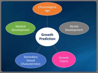
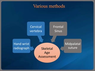
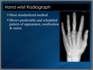
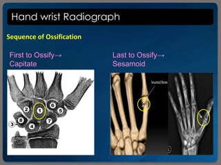

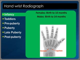


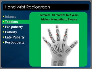

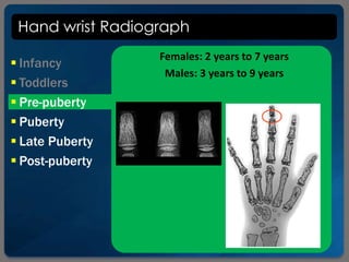
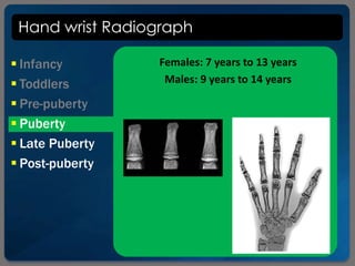
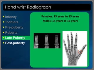


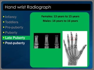

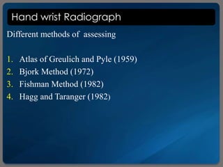

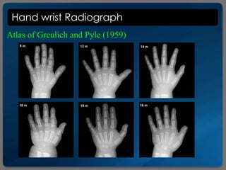
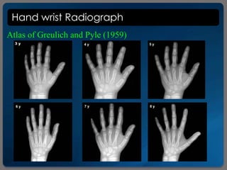
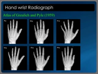
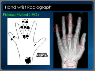
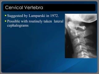
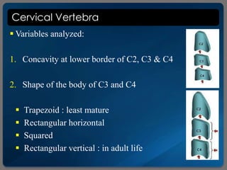
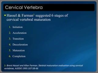
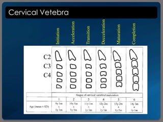
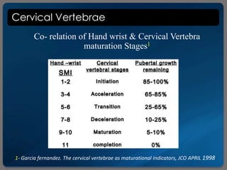
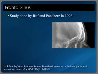
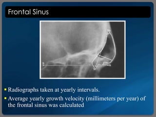
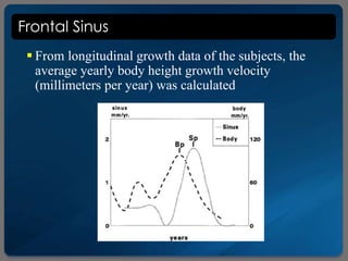
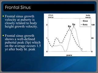
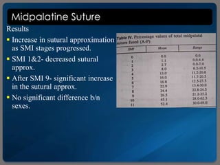
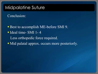
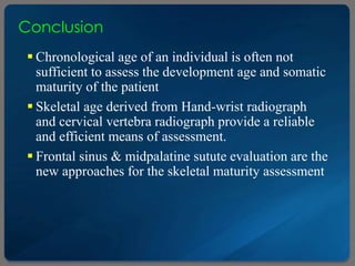


This document discusses methods for assessing skeletal age through radiographic examination of bones and structures. It describes using hand-wrist radiographs according to Greulich and Pyle's atlas to determine skeletal age based on ossification patterns. Cervical vertebrae morphology is also assessed in six stages of maturation. Additional methods examined are frontal sinus size relative to growth velocity and midpalatal suture approximation correlated to hand-wrist development. Skeletal age assessment provides a more reliable indicator of maturation than chronological age alone for orthodontic treatment planning.


