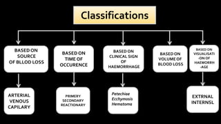
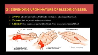
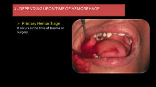
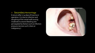
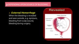
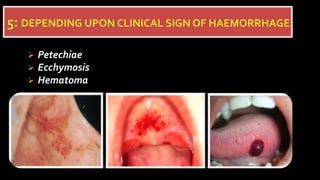
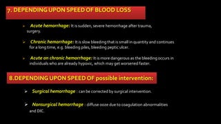
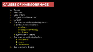
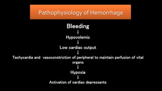
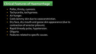
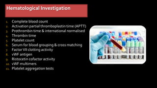
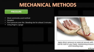
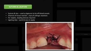
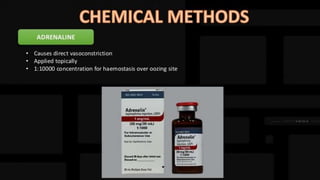
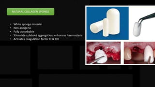
Haemorrhage
Hemorrhage
 Hemorrhage ( hemorrhage ) denotes the escape of
blood from a blood vessel
Note
the word hemorrhage is synonymous bleeding
Classifications
BASED ON
SOURCE
OF BLLOD LOSS
BASED ON
TIME OF
OCCURENCE
BASED ON
CLINICAL SIGN
OF
HAEMORRHAGE
BASED ON
VISUALISATI
-ON OF
HAEMORRH
-AGE
ARTERIAL
VENOUS
CAPILARY
PRIMERY
SECONDARY
REACTIONARY
Petechiae
Ecchymosis
Hematoma
EXTRNAL
INTERNSL
BASED ON
VOLUMEOF
BLOOD LOSS
 Arterial is bright red in colour,The blood is emitted as a jet with each heartbeat.
 Venous is dark red, steady and continuous flow
 Capillary: Here bleeding is rapid and bright red.There is generalized ooze of blood
2. DEPENDINGUPONTIMEOF HEMORRHAGE
 Primary Hemorrhage
It occurs at the time of trauma or
surgery.
 Reactionary Hemorrhage
It occurs within 24 hours of trauma or operation .of blood clot or slippage of ligature.
The precipitating factors are:
‱ Rise in blood pressure during recovery from shock.
‱ Rise in venous pressure due to coughing, vomiting, etc.
 Secondary Hemorrhage
It occurs after 7-14 days of trauma or
operation. It is due to infection and
sloughing of the vessel wall causing
moderate to severe bleeding and
pericepated by factors such as infection
pressure necrosis such ( drain or
malignancy )
 External Hemorrhage
When the bleeding is revealed
and seen outside, e.g. epistaxis,
bleeding from scalp wound,
bleeding during surgery.
3:DEPENDING UPON SOURCEOF BLEEDING
 Internal Hemorrhage
When the bleeding is
concealed and not seen
outside, e.g. intracranial
hematoma
4: DEPENDINGUPON SOURCEOF BLEEDING
 Petechiae
 Ecchymosis
 Hematoma
6.DEPENDING UPON VOLUME OF BLOOD LOSS
 Mild Hemorrhage ( class I )
 Moderate Hemorrhage ( class II & III )
 Severe Hemorrhage ( class IV )
7. DEPENDING UPON SPEED OF BLOOD LOSS
 Acute hemorrhage: It is sudden, severe hemorrhage after trauma,
surgery.
 Chronic hemorrhage: It is slow bleeding that is small in quantity and continues
for a long time, e.g. bleeding piles, bleeding peptic ulcer.
 Acute on chronic hemorrhage: It is more dangerous as the bleeding occurs in
individuals who are already hypoxic, which may get worsened faster.
8.DEPENDING UPON SPEED OF possible intervention:
 Surgical hemorrhage : can be corrected by surgical intervention.
 Nonsurgical hemorrhage : diffuse ooze due to coagulation abnormalities
and DIC.
CAUSES OF HAEMORRHAGE
1. Trauma
2. Infections
3. Local irritant
4. Congenital malformations
5. Surgical
6. Due to abnormalities in clotting factors
a clotting factor deficiencies
- hereditary
- anticoagulation therapy
- liver disease
b dysfunction of clotting
7. Due to abnormalities in platelets
a deficiencies
b excess
c dysfunction
8. Due to systemic disease
Pathophysiology of Hemorrhage
Bleeding
↓
Hypovolemic
↓
Low cardiac output
↓
Tachycardia and vasoconstriction of peripheral to maintain perfusion of vital
organs
↓
Hypoxia
↓
Activation of cardiac depressants
↓
Anaerobic metabolism
↓
Hyponatraemic, hyperkalaemic, hypocalcaemic metabolic acidosis
↓
Lysosomes of cell get lysed releasing powerful enzymes
which is lethal to cell itself
↓
SICK CELL SYNDROME
Platelets and coagulants are activated leading to formation of
small clots DIC and further bleeding.
↓
Progressive haemodilution leading to total circulatory Failure.
Clinical Features of Haemorrhage
 Pallor, thirsty, cyanosis.
 Tachycardia, tachypnoea.
 Air hunger.
 Cold clammy skin due to vasoconstriction.
 Dry face, dry mouth and goose skin appearance (due to
contraction of arrector pilorum).
 Rapid thready pulse, hypotension.
 Oliguria.
 Features related to specific causes.
Hematological Investigation
1. Complete blood count
2. Activation partial thromboplastin time (APTT)
3. Prothrombin time & international normalised
4. Thrombin time
5. Platelet count
6. Serum for blood grouping & cross matching
7. FactorVII clotting activity
8. vWF antigen
9. Ristocetin cofactor activity
10. vWF multimers
11. Platelet aggregation tests
CLINICAL FEATURESOF ACUTE BLOOD LOSS
1. Increasing pallor
2. Increasing pulse rate
3. Restlessness
4. Air hunger
5. Cold clammy skin
6. Thirst
7. Tinnitus
8. Blindness
9. Blood pressure – normal / slightly raised
10. Urinary output – reduced
11. Haemoglobin level – may fall after some hours
ThankYou

hemorrhage 22 .pptx

  • 1.
  • 2.
    Hemorrhage  Hemorrhage (hemorrhage ) denotes the escape of blood from a blood vessel Note the word hemorrhage is synonymous bleeding
  • 4.
    Classifications BASED ON SOURCE OF BLLODLOSS BASED ON TIME OF OCCURENCE BASED ON CLINICAL SIGN OF HAEMORRHAGE BASED ON VISUALISATI -ON OF HAEMORRH -AGE ARTERIAL VENOUS CAPILARY PRIMERY SECONDARY REACTIONARY Petechiae Ecchymosis Hematoma EXTRNAL INTERNSL BASED ON VOLUMEOF BLOOD LOSS
  • 5.
     Arterial isbright red in colour,The blood is emitted as a jet with each heartbeat.  Venous is dark red, steady and continuous flow  Capillary: Here bleeding is rapid and bright red.There is generalized ooze of blood
  • 6.
    2. DEPENDINGUPONTIMEOF HEMORRHAGE Primary Hemorrhage It occurs at the time of trauma or surgery.
  • 7.
     Reactionary Hemorrhage Itoccurs within 24 hours of trauma or operation .of blood clot or slippage of ligature. The precipitating factors are: ‱ Rise in blood pressure during recovery from shock. ‱ Rise in venous pressure due to coughing, vomiting, etc.
  • 8.
     Secondary Hemorrhage Itoccurs after 7-14 days of trauma or operation. It is due to infection and sloughing of the vessel wall causing moderate to severe bleeding and pericepated by factors such as infection pressure necrosis such ( drain or malignancy )
  • 9.
     External Hemorrhage Whenthe bleeding is revealed and seen outside, e.g. epistaxis, bleeding from scalp wound, bleeding during surgery. 3:DEPENDING UPON SOURCEOF BLEEDING
  • 10.
     Internal Hemorrhage Whenthe bleeding is concealed and not seen outside, e.g. intracranial hematoma 4: DEPENDINGUPON SOURCEOF BLEEDING
  • 11.
  • 12.
    6.DEPENDING UPON VOLUMEOF BLOOD LOSS  Mild Hemorrhage ( class I )  Moderate Hemorrhage ( class II & III )  Severe Hemorrhage ( class IV )
  • 13.
    7. DEPENDING UPONSPEED OF BLOOD LOSS  Acute hemorrhage: It is sudden, severe hemorrhage after trauma, surgery.  Chronic hemorrhage: It is slow bleeding that is small in quantity and continues for a long time, e.g. bleeding piles, bleeding peptic ulcer.  Acute on chronic hemorrhage: It is more dangerous as the bleeding occurs in individuals who are already hypoxic, which may get worsened faster. 8.DEPENDING UPON SPEED OF possible intervention:  Surgical hemorrhage : can be corrected by surgical intervention.  Nonsurgical hemorrhage : diffuse ooze due to coagulation abnormalities and DIC.
  • 14.
    CAUSES OF HAEMORRHAGE 1.Trauma 2. Infections 3. Local irritant 4. Congenital malformations 5. Surgical 6. Due to abnormalities in clotting factors a clotting factor deficiencies - hereditary - anticoagulation therapy - liver disease b dysfunction of clotting 7. Due to abnormalities in platelets a deficiencies b excess c dysfunction 8. Due to systemic disease
  • 15.
    Pathophysiology of Hemorrhage Bleeding ↓ Hypovolemic ↓ Lowcardiac output ↓ Tachycardia and vasoconstriction of peripheral to maintain perfusion of vital organs ↓ Hypoxia ↓ Activation of cardiac depressants
  • 16.
    ↓ Anaerobic metabolism ↓ Hyponatraemic, hyperkalaemic,hypocalcaemic metabolic acidosis ↓ Lysosomes of cell get lysed releasing powerful enzymes which is lethal to cell itself ↓ SICK CELL SYNDROME Platelets and coagulants are activated leading to formation of small clots DIC and further bleeding. ↓ Progressive haemodilution leading to total circulatory Failure.
  • 17.
    Clinical Features ofHaemorrhage  Pallor, thirsty, cyanosis.  Tachycardia, tachypnoea.  Air hunger.  Cold clammy skin due to vasoconstriction.  Dry face, dry mouth and goose skin appearance (due to contraction of arrector pilorum).  Rapid thready pulse, hypotension.  Oliguria.  Features related to specific causes.
  • 18.
    Hematological Investigation 1. Completeblood count 2. Activation partial thromboplastin time (APTT) 3. Prothrombin time & international normalised 4. Thrombin time 5. Platelet count 6. Serum for blood grouping & cross matching 7. FactorVII clotting activity 8. vWF antigen 9. Ristocetin cofactor activity 10. vWF multimers 11. Platelet aggregation tests
  • 19.
    CLINICAL FEATURESOF ACUTEBLOOD LOSS 1. Increasing pallor 2. Increasing pulse rate 3. Restlessness 4. Air hunger 5. Cold clammy skin 6. Thirst 7. Tinnitus 8. Blindness 9. Blood pressure – normal / slightly raised 10. Urinary output – reduced 11. Haemoglobin level – may fall after some hours
  • 35.