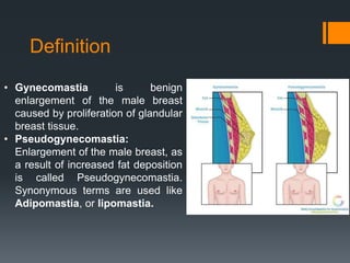
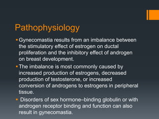
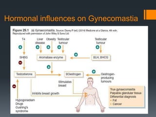
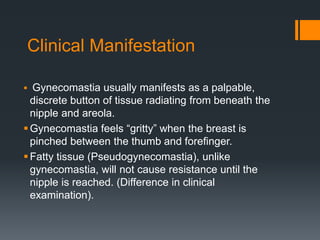
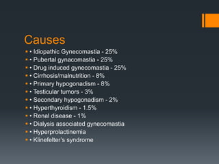
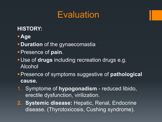
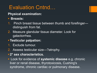
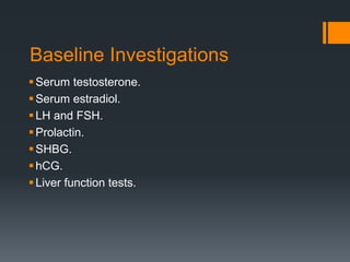
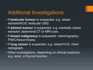
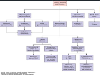
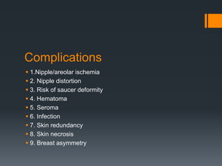
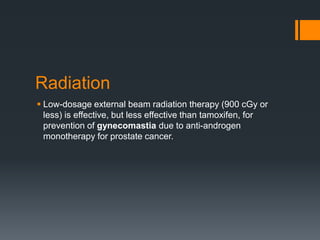
GYNECOMASTIA
DR. SANDHYA GUPTA
Definition
• Gynecomastia is benign
enlargement of the male breast
caused by proliferation of glandular
breast tissue.
• Pseudogynecomastia:
Enlargement of the male breast, as
a result of increased fat deposition
is called Pseudogynecomastia.
Synonymous terms are used like
Adipomastia, or lipomastia.
Pathophysiology
Gynecomastia results from an imbalance between
the stimulatory effect of estrogen on ductal
proliferation and the inhibitory effect of androgen
on breast development.
The imbalance is most commonly caused by
increased production of estrogens, decreased
production of testosterone, or increased
conversion of androgens to estrogens in peripheral
tissue.
 Disorders of sex hormone–binding globulin or with
androgen receptor binding and function can also
result in gynecomastia.
Hormonal influences on Gynecomastia
Clinical Manifestation
 Gynecomastia usually manifests as a palpable,
discrete button of tissue radiating from beneath the
nipple and areola.
Gynecomastia feels “gritty” when the breast is
pinched between the thumb and forefinger.
Fatty tissue (Pseudogynecomastia), unlike
gynecomastia, will not cause resistance until the
nipple is reached. (Difference in clinical
examination).
Examination
 The examination is performed by
having the patient lie on his back
with his hands behind his head.
The examiner then places his or
her thumb and forefinger on each
side of the breast and slowly
brings them together
 Gynecomastia is appreciated as
a concentric, rubbery to firm disk
of tissue, often mobile, located
directly beneath the areolar area.
 Pseudogynecomastia presents
no discrete mass,
 Other masses due to disorders
such as cancertend to be
eccentrically positioned.
Prevalence
 Asymptomatic palpable breast tissue is common in normal
males, particularly in the
neonate(60%–90%)
at puberty (60%–70%), and
with increasing age (20%–65%, >50 years)
 Because of this high prevalence, gynecomastia is
considered a relatively normal finding during these periods of
life and is often called physiological at these ages.
Physiological gynecomastia in
different age group
Neonatal gynecomastia results from placental transfer of
estrogens.
During early puberty, production of estrogens begins sooner
than testosterone production, thus causing an imbalance in the
ratio of estrogens to androgens.
With aging, testosterone production decreases, and peripheral
androgen to estrogen conversion often increases because of
an age-related increase in adipose tissue. Drugs and medical
conditions may also contribute.
Causes
 • Idiopathic Gynecomastia - 25%
 • Pubertal gynacomastia - 25%
 • Drug induced gynecomastia - 25%
 • Cirrhosis/malnutrition - 8%
 • Primary hypogonadism - 8%
 • Testicular tumors - 3%
 • Secondary hypogonadism - 2%
 • Hyperthyroidism - 1.5%
 • Renal disease - 1%
 • Dialysis associated gynecomastia
 • Hyperprolactinemia
 • Klinefelter’s syndrome
Classification
Simon Classified gynaecomastia into the following groups:
 Stage-1: A slight volume increase with no excess skin.
 Stage-2a: A moderate volume increase with no excess skin.
 Stage-2b: A moderate volume increase with excess skin.
 Stage-3: A marked volume increase with excess skin
Histopathological Classification
Three histologic patterns of gynecomastia have been
recognized.
1. Florid pattern: consists of an increase in the number of
budding ducts, proliferation of the ductal epithelium.
2. Fibrous type: has dilated ducts, minimal duct epithelial
proliferation, no periductal edema, and a virtually acellular
fibrous stroma.
3. An intermediate pattern contains features of both types.
The duration of gynecomastia is the most important factor in
determining the pathologic picture.
Florid pattern response well to medical treatment, while
fibrous type needs surgical management.
Questions to be answered
Gynecomastia / Pseudogynecomastia/Breast
Cancer?
Characteristics: Unilateral / Bilateral, Tenderness,
Size, progression
Physiological / Pathological
Possible cause of gynecomastia, eg: Systemic
cause, Drugs
Any underlying hormonal derangement, eg:
Hypogonadism
Associated malignancy, eg: hCG-secreting testicular
tumor.
Management
Evaluation
HISTORY:
Age
Duration of the gynaecomastia
Presence of pain.
Use of drugs including recreation drugs e.g.
Alcohol
Presence of symptoms suggestive of pathological
cause.
1. Symptome of hypogonadism - reduced libido,
erectile dysfunction, virilization.
2. Systemic disease: Hepatic, Renal, Endocrine
disease. (Thyrotoxicosis, Cushing syndrome).
Evaluation Cntnd…
Physical examination:
 Breasts:
1. Pinch breast tissue between thumb and forefinger—
distinguish from fat.
2. Measure glandular tissue diameter. Look for
galactorrhea.
Testicular palpation:
1. Exclude tumour.
2. Assess testicular size—?atrophy.
2° sex characteristics.
 Look for evidence of systemic disease e.g. chronic
liver or renal disease, thyrotoxicosis, Cushing's
syndrome, chronic cardiac or pulmonary disease.
Baseline Investigations
Serum testosterone.
Serum estradiol.
LH and FSH.
Prolactin.
SHBG.
hCG.
Liver function tests.
Additional Investigations
If testicular tumour is suspected, e.g. raised
estradiol/hCG: testicular USG.
If adrenal tumour is suspected, e.g. markedly raised
estradiol: abdominal CT or MRI scan.
If breast malignancy is suspected: mammography;
FNAC/tissue biopsy.
If lung cancer is suspected, e.g. raised hCG: chest
radiograph.
Other investigations, depending on clinical suspicion,
e.g. renal or thyroid function .
Management
 Medical
 Surgical
Medical
 The underlying disease should be corrected if possible, and
offending drugs should be discontinued.
 ER antagonists (tamoxifen, 10 to 20 mg daily, or
raloxifene, 60 mg daily) are effective in treating pubertal and
adult gynecomastia and preventing gynecomastia induced by
androgen deprivation therapy.
 Other drugs can be used, such as Clomifene (50–
100mg/day), Danazol (300– 600mg/day), Testolactone
(450mg/day), Anastrozole (1mg/day).
 Medical therapy is ineffective for chronic, fibrous
gynecomastia.
Surgical
 With long standing gynecomastia (more than 1 year):
surgical reduction mammoplasty (i.e. removal of breast
tissue with or without periareolar adipose tissue) is necessary
if breast enlargement is severe, painful, socially
embarrassing or disfiguring.
Surgeries
 Open subcutaneous mastectomy by a circum areolar incision
extending from 3 to 9’O clock position
 2. Endoscopic – assisted subcutaneous mastectomy (very small
distant incision)
 3. Liposuction – assisted mastectomy (for pseudogynecomastia)
 4. Ultrasound – assisted liposuction.
Complications
 1.Nipple/areolar ischemia
 2. Nipple distortion
 3. Risk of saucer deformity
 4. Hematoma
 5. Seroma
 6. Infection
 7. Skin redundancy
 8. Skin necrosis
 9. Breast asymmetry
Radiation
 Low-dosage external beam radiation therapy (900 cGy or
less) is effective, but less effective than tamoxifen, for
prevention of gynecomastia due to anti-androgen
monotherapy for prostate cancer.
THANK YOU

Gynecomastia final

  • 1.
  • 2.
    Definition • Gynecomastia isbenign enlargement of the male breast caused by proliferation of glandular breast tissue. • Pseudogynecomastia: Enlargement of the male breast, as a result of increased fat deposition is called Pseudogynecomastia. Synonymous terms are used like Adipomastia, or lipomastia.
  • 3.
    Pathophysiology Gynecomastia results froman imbalance between the stimulatory effect of estrogen on ductal proliferation and the inhibitory effect of androgen on breast development. The imbalance is most commonly caused by increased production of estrogens, decreased production of testosterone, or increased conversion of androgens to estrogens in peripheral tissue.  Disorders of sex hormone–binding globulin or with androgen receptor binding and function can also result in gynecomastia.
  • 4.
  • 5.
    Clinical Manifestation  Gynecomastiausually manifests as a palpable, discrete button of tissue radiating from beneath the nipple and areola. Gynecomastia feels “gritty” when the breast is pinched between the thumb and forefinger. Fatty tissue (Pseudogynecomastia), unlike gynecomastia, will not cause resistance until the nipple is reached. (Difference in clinical examination).
  • 6.
    Examination  The examinationis performed by having the patient lie on his back with his hands behind his head. The examiner then places his or her thumb and forefinger on each side of the breast and slowly brings them together  Gynecomastia is appreciated as a concentric, rubbery to firm disk of tissue, often mobile, located directly beneath the areolar area.  Pseudogynecomastia presents no discrete mass,  Other masses due to disorders such as cancertend to be eccentrically positioned.
  • 7.
    Prevalence  Asymptomatic palpablebreast tissue is common in normal males, particularly in the neonate(60%–90%) at puberty (60%–70%), and with increasing age (20%–65%, >50 years)  Because of this high prevalence, gynecomastia is considered a relatively normal finding during these periods of life and is often called physiological at these ages.
  • 8.
    Physiological gynecomastia in differentage group Neonatal gynecomastia results from placental transfer of estrogens. During early puberty, production of estrogens begins sooner than testosterone production, thus causing an imbalance in the ratio of estrogens to androgens. With aging, testosterone production decreases, and peripheral androgen to estrogen conversion often increases because of an age-related increase in adipose tissue. Drugs and medical conditions may also contribute.
  • 9.
    Causes  • IdiopathicGynecomastia - 25%  • Pubertal gynacomastia - 25%  • Drug induced gynecomastia - 25%  • Cirrhosis/malnutrition - 8%  • Primary hypogonadism - 8%  • Testicular tumors - 3%  • Secondary hypogonadism - 2%  • Hyperthyroidism - 1.5%  • Renal disease - 1%  • Dialysis associated gynecomastia  • Hyperprolactinemia  • Klinefelter’s syndrome
  • 10.
    Classification Simon Classified gynaecomastiainto the following groups:  Stage-1: A slight volume increase with no excess skin.  Stage-2a: A moderate volume increase with no excess skin.  Stage-2b: A moderate volume increase with excess skin.  Stage-3: A marked volume increase with excess skin
  • 11.
    Histopathological Classification Three histologicpatterns of gynecomastia have been recognized. 1. Florid pattern: consists of an increase in the number of budding ducts, proliferation of the ductal epithelium. 2. Fibrous type: has dilated ducts, minimal duct epithelial proliferation, no periductal edema, and a virtually acellular fibrous stroma. 3. An intermediate pattern contains features of both types. The duration of gynecomastia is the most important factor in determining the pathologic picture. Florid pattern response well to medical treatment, while fibrous type needs surgical management.
  • 12.
    Questions to beanswered Gynecomastia / Pseudogynecomastia/Breast Cancer? Characteristics: Unilateral / Bilateral, Tenderness, Size, progression Physiological / Pathological Possible cause of gynecomastia, eg: Systemic cause, Drugs Any underlying hormonal derangement, eg: Hypogonadism Associated malignancy, eg: hCG-secreting testicular tumor. Management
  • 13.
    Evaluation HISTORY: Age Duration of thegynaecomastia Presence of pain. Use of drugs including recreation drugs e.g. Alcohol Presence of symptoms suggestive of pathological cause. 1. Symptome of hypogonadism - reduced libido, erectile dysfunction, virilization. 2. Systemic disease: Hepatic, Renal, Endocrine disease. (Thyrotoxicosis, Cushing syndrome).
  • 14.
    Evaluation Cntnd… Physical examination: Breasts: 1. Pinch breast tissue between thumb and forefinger— distinguish from fat. 2. Measure glandular tissue diameter. Look for galactorrhea. Testicular palpation: 1. Exclude tumour. 2. Assess testicular size—?atrophy. 2° sex characteristics.  Look for evidence of systemic disease e.g. chronic liver or renal disease, thyrotoxicosis, Cushing's syndrome, chronic cardiac or pulmonary disease.
  • 15.
    Baseline Investigations Serum testosterone. Serumestradiol. LH and FSH. Prolactin. SHBG. hCG. Liver function tests.
  • 16.
    Additional Investigations If testiculartumour is suspected, e.g. raised estradiol/hCG: testicular USG. If adrenal tumour is suspected, e.g. markedly raised estradiol: abdominal CT or MRI scan. If breast malignancy is suspected: mammography; FNAC/tissue biopsy. If lung cancer is suspected, e.g. raised hCG: chest radiograph. Other investigations, depending on clinical suspicion, e.g. renal or thyroid function .
  • 18.
  • 19.
    Medical  The underlyingdisease should be corrected if possible, and offending drugs should be discontinued.  ER antagonists (tamoxifen, 10 to 20 mg daily, or raloxifene, 60 mg daily) are effective in treating pubertal and adult gynecomastia and preventing gynecomastia induced by androgen deprivation therapy.  Other drugs can be used, such as Clomifene (50– 100mg/day), Danazol (300– 600mg/day), Testolactone (450mg/day), Anastrozole (1mg/day).  Medical therapy is ineffective for chronic, fibrous gynecomastia.
  • 20.
    Surgical  With longstanding gynecomastia (more than 1 year): surgical reduction mammoplasty (i.e. removal of breast tissue with or without periareolar adipose tissue) is necessary if breast enlargement is severe, painful, socially embarrassing or disfiguring.
  • 21.
    Surgeries  Open subcutaneousmastectomy by a circum areolar incision extending from 3 to 9’O clock position  2. Endoscopic – assisted subcutaneous mastectomy (very small distant incision)  3. Liposuction – assisted mastectomy (for pseudogynecomastia)  4. Ultrasound – assisted liposuction.
  • 22.
    Complications  1.Nipple/areolar ischemia 2. Nipple distortion  3. Risk of saucer deformity  4. Hematoma  5. Seroma  6. Infection  7. Skin redundancy  8. Skin necrosis  9. Breast asymmetry
  • 23.
    Radiation  Low-dosage externalbeam radiation therapy (900 cGy or less) is effective, but less effective than tamoxifen, for prevention of gynecomastia due to anti-androgen monotherapy for prostate cancer.
  • 24.