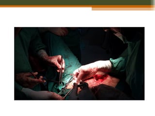
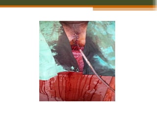
20. Clinical Anatomy
• 12-15 cm from anal verge.
• Diameter
▫ 4 cm (upper part)
▫ Dilated (lower part)
• Posterior part of the
lesser pelvis and in front
of lower three pieces of
sacrum and the coccyx
• Begins at the
rectosigmoid junction, at
level of third sacral
vertebra
• Ends at the anorectal
junction, 2-3 cm in front
of and a little below the
coccyx
21. • Divided into 3 parts
• Upper third
• Middle third
• Lower third
• 3 distinct intraluminal
curves ( Valves of
Houston)
22. • Superior 1/3rd of the rectum
▫ Covered by peritoneum on the
anterior and lateral surfaces
• Middle 1/3rd of the rectum
▫ Covered by peritoneum on the
anterior surface
• Inferior 1/3rd of the rectum
▫ Devoid of peritoneum
▫ Close proximity to adjacent
structure including boney pelvis.
Peritoneal Relations
24. Lymphatic drainage
• Upper and middle rectum
▫ Pararectal lymph nodes,
located directly on the muscle
layer of the rectum
▫ Inferior mesenteric lymph
nodes, via the nodes along the
superior rectal vessels
• Lower rectum
▫ Sacral group of lymph nodes or
Internal iliac lymph nodes
• Below the dentate line
▫ Inguinal nodes and external iliac
chain
25. Epidemiology
• Colorectal caner is the third most frequently diagnosed cancer
in the US men and women.
• 108,070 new cases of colon cancer and 40,740 new cases of
rectal cancer in the US in 2008. Combined mortality for
colorectal cancer 49,960 in 2008.
• Worldwide approx. 1 million new cases p.a. are diagnosed,
with 529,000 deaths.
• Incidence rate in India is quite low about 2 to 8 per 100,000
• Incidence in Wah Cantt, Taxilla and surrounding Sub-urban
areas is 55---60 cases per year.
• Median age- 7th
decade but can occur any time in adulthood.
29. • Signs
▫ Pallor
▫ Abdominal mass
▫ PR mass
▫ Jaundice
▫ Nodular liver
▫ Ascites
30. Pathological features
WHO Classification
• Adenocarcinoma in situ
• Adenocarcinoma
• Mucinous (colloid) adenocarcinoma (>50% mucinous)
• Signet ring cell carcinoma (>50% signet ring cells)
• Squamous cell (epidermoid) carcinoma
• Adenosquamous carcinoma
• Small-cell (oat cell) carcinoma
• Medullary carcinoma
• Undifferentiated Carcinoma
31. Dukes classification-
Dukes A: Invasion into but not through the bowel wall.
Dukes B: Invasion through the bowel wall but not involving lymph
nodes.
Dukes C: Involvement of lymph nodes
Dukes D: Widespread metastases
Modified astler coller classification-
Stage A : Limited to mucosa.
Stage B1 : Extending into muscularis propria but not penetrating
through it; nodes not involved.
Stage B2 : Penetrating through muscularis propria; nodes not
involved
Stage C1 : Extending into muscularis propria but not penetrating
through it. Nodes involved
Stage C2 : Penetrating through muscularis propria. Nodes
involved
Stage D: Distant metastatic spread
32. Tis TTis T11 TT22 TT33 TT44
MucosaMucosa
Muscularis mucosaeMuscularis mucosae
SubmucosaSubmucosa
Muscularis propriaMuscularis propria
SubserosaSubserosa
SerosaSerosa
TNM Classification
TX Primary tumor cannot be assessed
T0 No evidence of primary tumor
Tis Carcinoma in situ: intraepithelial or invasion of lamina propria
T1 Tumor invades submucosa
T2 Tumor invades muscularis propria
T3 Tumor invades through the muscularis propria into pericolorectal tissues
T4a Tumor penetrates to the surface of the visceral peritoneum
T4b Tumor directly invades or is adherent to other organs or structures
35. Prognostic factors
Good prognostic
factors
Old age
Gender(F>M)
Asymptomatic pts
Polypoidal lesions
Diploid
Poor prognostic
factors
Obstruction
Perforation
Ulcerative lesion
Adjacent structures
involvement
Positive margins
LVSI
Signet cell carcinoma
High CEA
Tethered and fixed
cancer
36. Stage and Prognosis
Stage 5-year Survival (%)
0,1 Tis,T1;No;Mo > 90
I T2;No;Mo 80-85
II T3-4;No;Mo 70-75
III T2;N1-3;Mo 70-75
III T3;N1-3;Mo 50-65
III T4;N1-2;Mo 25-45
IV M1 <3
37. Diagnostic Workup
• History—including family history of colorectal cancer
or polyps
• Physical examinations including DRE and complete
pelvic examination in women: size, location, ulceration,
mobile vs. tethered vs. fixed, distance from anal verge
and sphincter functions.
• Proctoscopy—including assessment of mobility,
minimum diameter of the lumen, and distance from the
anal verge
• Biopsy of the primary tumor
38. Colonoscopy or barium enema
Figure: Carcinoma of the rectum. Double-
contrast barium enema shows a long
segment of concentric luminal narrowing
(arrows) along the rectum with minimal
irregularity of the mucosal surface.
To evaluate remainder of large bowel to rule out synchronous
tumor or presence of polyp syndrome.
39. Transrectal ultrasound –EUS
• use for clinical staging.
• 80-95% accurate in tumor staging
• 70-75% accurate in mesorectal lymph
node staging
• Very good at demonstrating layers of
rectal wall
• Use is limited to lesion < 14 cm from
anus, not applicable for upper rectum,
for stenosing tumor
• Very useful in determining extension of
disease into anal canal (clinical
important for planning sphincter
preserving surgery)
Figure. Endorectal
ultrasound of a T3 tumor of
the rectum, extension
through the muscularis
propria, and into perirectal
fat.
40. CT scan
• Part of routine workup of patients
• Useful in identifying enlarged pelvic lymph-nodes and
metastasis outside the pelvis than the extent or stage of
primary tumor
• Limited utility in small primary cancer
• Sensitivity 50-80%
• Specificity 30-80%
• Ability to detect pelvic and para-aortic lymph nodes is
higher than peri-rectal lymph nodes.
41. Magnetic Resonance Imaging (MRI)
• Greater accuracy in defining extent of rectal cancer
extension and also location & stage of tumor
• Also helpful in lateral extension of disease, critical in
predicting circumferential margin for surgical
excision.
• Different approaches (body coils, endorectal MRI &
phased array technique)
42. Figure: Mucinous adenocarcinoma of
the rectum. T2-weighted MRI shows high
signal intensity (arrowheads) of the
cancer lesion in right anterolateral side
of the rectal wall.
Figure: Normal rectal and perirectal
anatomy on high-resolution T2-weighted
MRI. Rectal mucosa (M), submucosa
(SM), and muscularis propria (PM) are
well discriminated. Mesorectal fascia
appears as a thin, low-signal-intensity
structure (arrowheads) and fuses with the
remnant of urogenital septum making
Denonvilliers fascia (arrows).
43. PET
• Shows promise as the most sensitive study
for the detection of metastatic disease in
the liver and elsewhere.
• Sensitivity of 97% and specificity of 76% in
evaluating for recurrent colorectal cancer.
cancer
rectum
prostate pubic bone
bladder
Small bowel
44. • CEA: High CEA levels associated with poorer
survival
• Routine investigation
▫ Complete blood count, RFT, LFT
▫ Chest X-ray
45.
46. Surgery
• Surgery is the mainstay of treatment of RC
• Local recurrence after conventional surgery:
▫ 20%-50% (average of 35%)**
• Radiotherapy significantly reduces the number
of local recurrences
47. Types of Surgery
• Local excision- reserved for superficially
invasive (T1) tumors with low likelihood of LN
metastases
• Should be considered a total biopsy, with further
treatment based on pathology
• With unfavorable pathology patient should undergo
total mesorectal excision with or without sphincter-
preservation:
▫ positive margin (or <2 mm), lymphovascular invasion,
▫ poorly differentiated tumors, T2 lesion
48. • Low Anterior Resection - for tumors in upper/mid
rectum; allows preservation of anal sphincter
• Abdominoperineal resection
▫ for tumors of distal rectum with distal edge up to 6 cm from anal
verge
▫ associated with permanent colostomy and high incidence of
sexual and genitourinary dysfunction
51. Purpose of Radio(chemo)therapy in Rectal
Cancer
• To lower local failure rates and improve survival in
resectable cancers
• to allow surgery in primarly inoperable cancers
• to facilitate a sphincter-preserving procedure
• to cure patients without surgery: very small cancer
or very high surgical risk
53. Radiotherapy
• Preoperative radiotherapy
▫ Short course: 25 Gy in 5 daily fractions of 5 Gy given in 1 week.
▫ Long course
Phase 1
45 Gy in 25 daily fractions of 1.8 Gy given in 5 weeks.
Phase 2 (optional)
5.4–9 Gy in 3–5 daily fractions of 1.8 Gy
• Postoperative radiotherapy
Phase 1
45 Gy in 25 daily fractions of 1.8 Gy given in 5 weeks.
Phase 2 (optional)
5.4–9 Gy in 3–5 daily fractions of 1.8 Gy.
• Palliative radiotherapy
55. Pre-op RT vs. surgery alone
Swedish Rectal Cancer Trial(NEJM 1997;336:980 ): 1168 patients
randomised to 25 Gy (5x5) PRT or no RT.
Surgery alone Preop. RT
Rate of local recurrence 27% 11% p<0.001
5-year overall survival 48% 58% p=0.004
Dutch Colorectal Cancer Group (Kapiteijn E. NEJM 2001;345:638):
1861 patients randomised TME vs PRT+TME
TME PRT+TME
Recurrence rate 2.4% 8.2%
OS ns ns
56. Pre-op vs. post-op Chemo RT
Randomized trial of the German Rectal Cancer study
Group (Sauer R et al. N Engl J Med 2004;351:1731-40):
▫ cT3 or cT4 or node-positive rectal cancer
▫ 50,4 Gy (1.8 Gy per day)
▫ 5-FU: 1000 mg/m2
per day (d1-5) during 1. and 5. week
Preop CRT Postop CRT
Patients N=415 N=384
5 y. OS 76% 74% p=0.8
5 y. local relapse 6% 13% p=0.006
G3,4 toxic effects 27% 40% p=0.001
• Increase in sphincter-preserving surgery with preop Th.
There are interesting things to be found when researching information on the internet to include in a presentation. The Colossal Colon is a replica of the human colon that is four feet wide. It was modeled from colonoscopy footage. It has traveled across the U.S. to inform the public about colon health. People can crawl through the colon or view through windows on the outside. It shows healthy colon tissue as well as diseased tissue including polyps and colon cancer. This picture was taken at a mall near the Creighton University Medical Center. It’s a fun way to spread information about colon health.