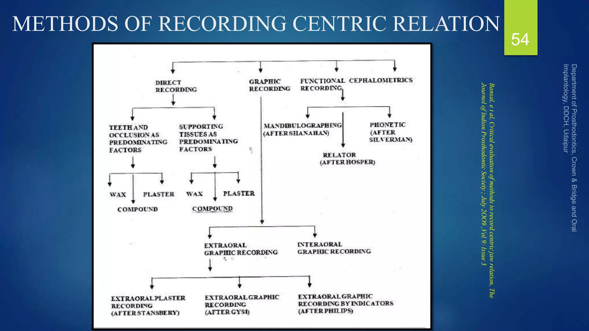
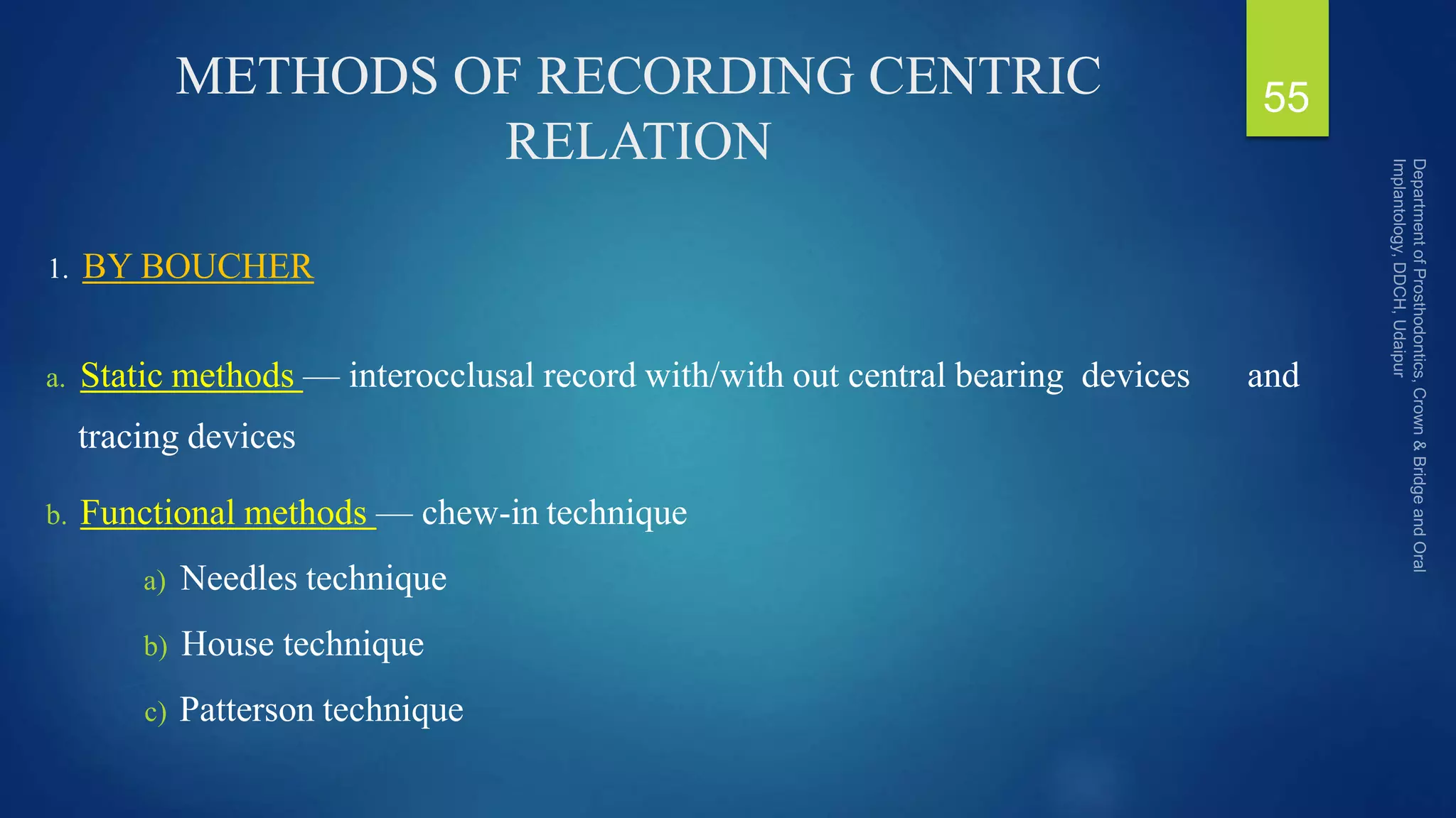
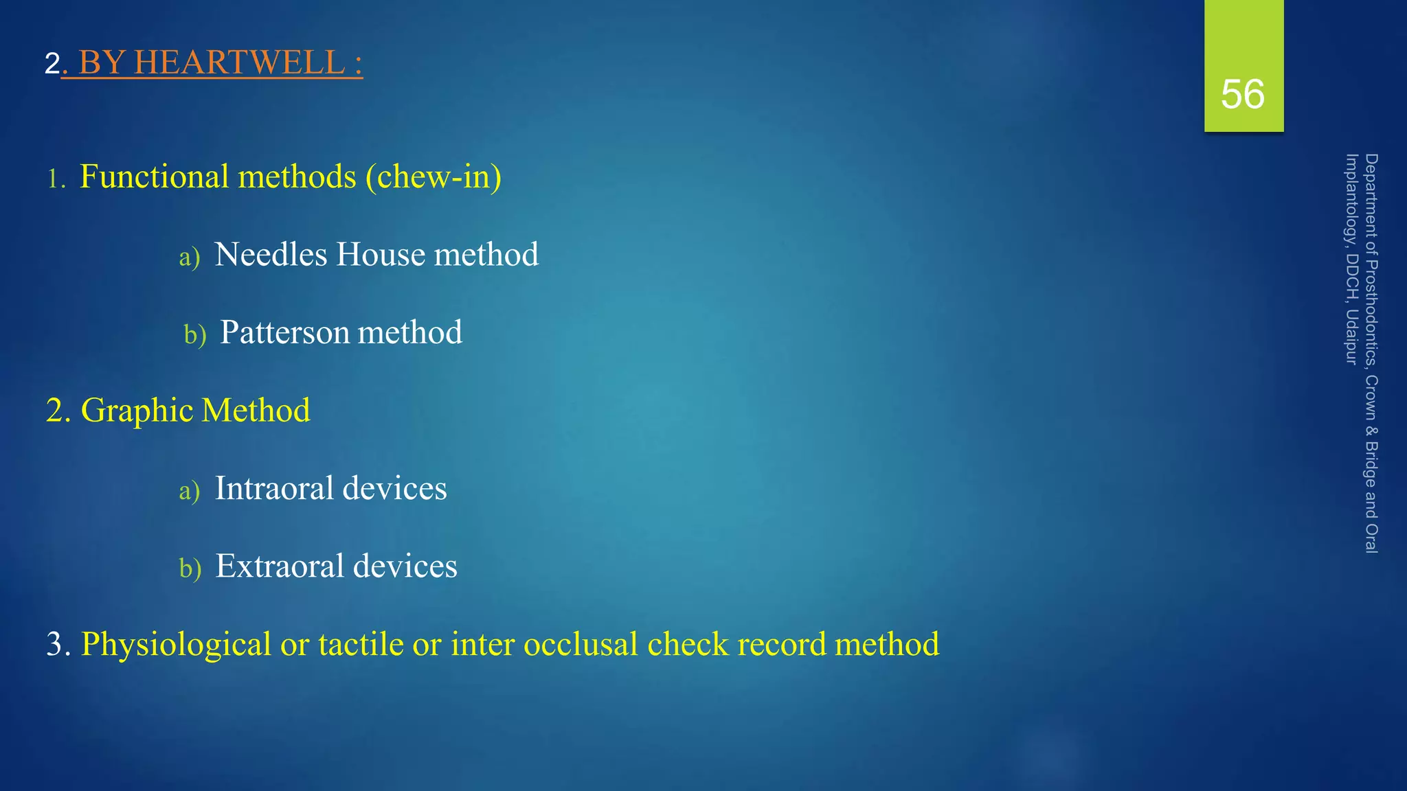
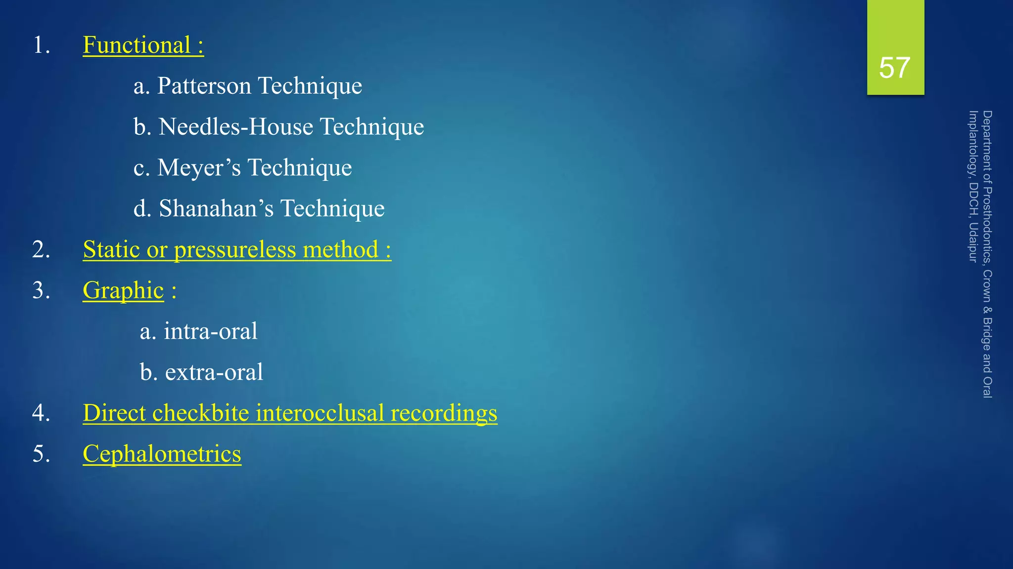
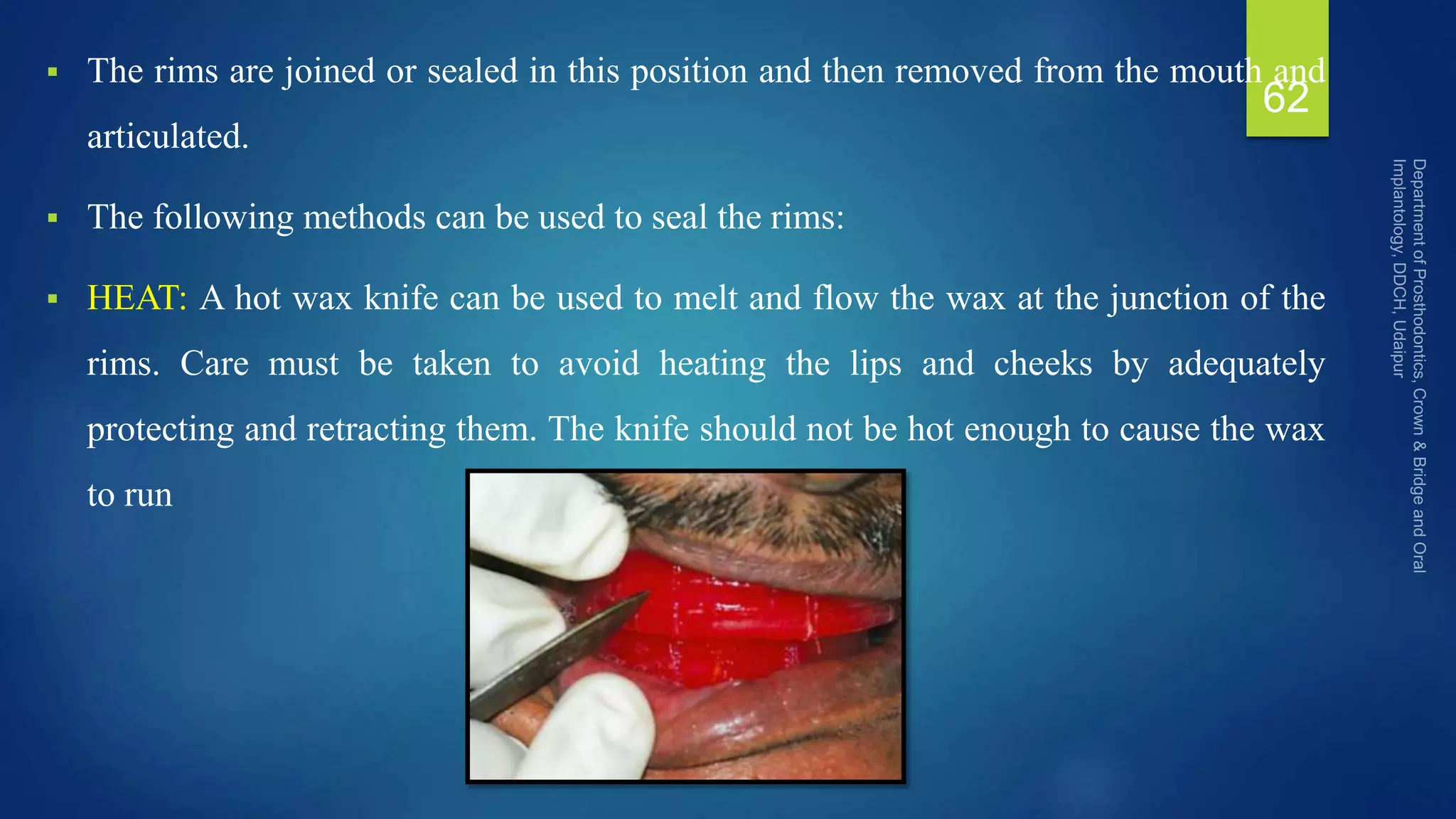
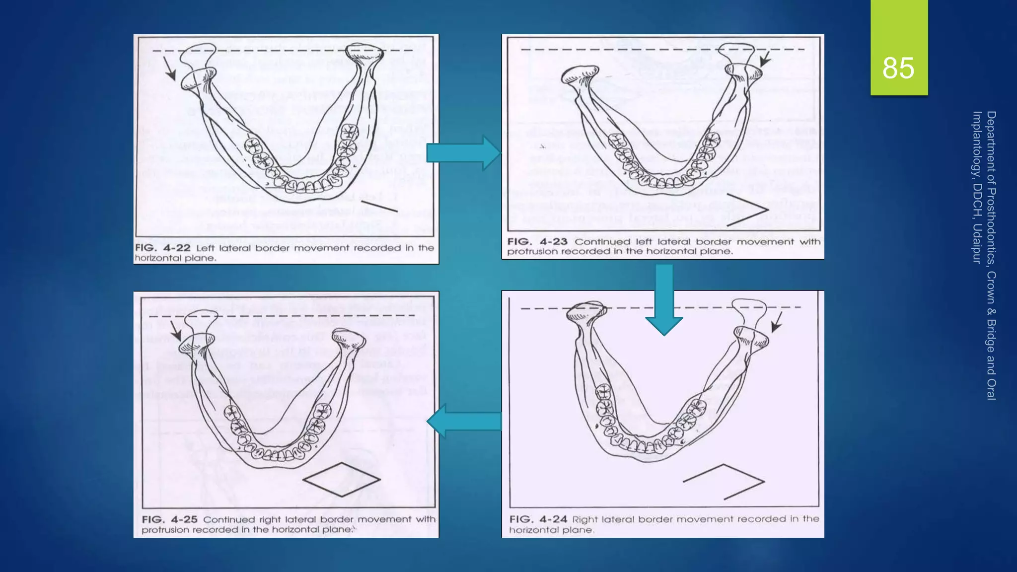
This document discusses the concept of centric relation, which refers to the position of the mandible relative to the maxilla. It provides an overview of the many definitions that have been proposed for centric relation over time from 1929 to present. There has been significant debate and controversy around defining this term. More recently, centric relation is understood to refer to the most anterior-superior position of the condyles in the glenoid fossa, rather than the most retruded position as was previously believed. The document also discusses methods for recording centric relation in patients.
![CENTRIC RELATION FROM 1929 TO 1970’S
Hanau [1929] defined CR as ‘the position of the mandible in which the condylar
heads are resting upon the menisci in the sockets of the glenoid fossa, regardless
of the opening of the jaws’.
Goodfriend [1933] considered the ‘centricity of the condyles in centric relation to
be an abnormal position’.
Niswonger [1934] described CR as a position where the patient can ‘clench the
back teeth’
Schuyler [1935] defined CR as ‘upper lingual cusps are resting in the central
fossae of the opposing lower bicuspids and molars’
9](https://image.slidesharecdn.com/centricjawrelation1autosaved-220205182724/75/Centric-jaw-relation-9-2048.jpg)
![CENTRIC RELATION FROM 1929 TO 1970’S
Thompson [1946] stated that ‘some believed that, in CR, the condyles are in the
most retruded position in the fossae, while others maintained they are not’.
Robinson [1951] stated that the mandible ‘can be retruded beyond what we should
consider centric into a strained retruded position’.
McCollum and Stuart [1955] proposed a definition for CR in which the condyles
are in a ‘rearmost, uppermost and midmost (RUM) position in the glenoid fossae’
Moyers [1956] defined CR as ‘the position of the mandible as determined by the
neuromuscular reflex first learned for controlling the mandibular position when
the primary teeth were in occlusion’
10](https://image.slidesharecdn.com/centricjawrelation1autosaved-220205182724/75/Centric-jaw-relation-10-2048.jpg)
![CENTRIC RELATION FROM 1929 TO 1970’S
Stallard [1959] defined CR of the mandible as ‘the rearmost, midmost,
untranslated hinged position. It is a strained relation as are all border relations. It is
the only maxillamandibular relation that can be statically repeated’
Avant [1960] declared the ‘seven definitions of CR’ that appeared in GPT-2
[1960], as ‘regrettable’ and stated that CR is a bone-to-bone (mandible to maxilla)
relation, whereas centric occlusion is a tooth-to-tooth (mandibular teeth to
maxillary teeth) relation.
McCollum [1960] defined CR position as ‘the most retruded position of the idle
condyles in the glenoid fossa’
11](https://image.slidesharecdn.com/centricjawrelation1autosaved-220205182724/75/Centric-jaw-relation-11-2048.jpg)
![CENTRIC RELATION FROM 1929 TO 1970’S
Boucher [1964] stated ‘CR is the most posterior relation of the mandible to
maxillae at the established vertical relation’ .
Graber [1966] thought that CR was an ‘unstrained, neutral position of the
mandible and is deviating neither to the right nor to the left and is neither
protruded nor retruded’ .
Glickman [1966] stated that CR was ‘the most retruded position to which the
mandible can be carried by the patient’s musculature’
12](https://image.slidesharecdn.com/centricjawrelation1autosaved-220205182724/75/Centric-jaw-relation-12-2048.jpg)
![CENTRIC RELATION FROM 1929 TO 1970’S
Goldman and Cohen [1968] defined CR as ‘the most posterior relation of the
mandible to maxilla from which lateral movements can be made’
Debate on the definition of CR escalated.
Posterior border closure, relaxed closure, bracing position, hinge position,
ligamentous position, retruded contact position, terminal hinge position added
confusion to term
CR. Schweitzer [1969] gave ‘almost 40 definitions of CR
13](https://image.slidesharecdn.com/centricjawrelation1autosaved-220205182724/75/Centric-jaw-relation-13-2048.jpg)
![CENTRIC RELATION FROM 1970 TO 1980’S
Dawson [1973] defined CR as ‘the most superior position the condyle can assume
in the glenoid fossa and it is not unstrained’ .
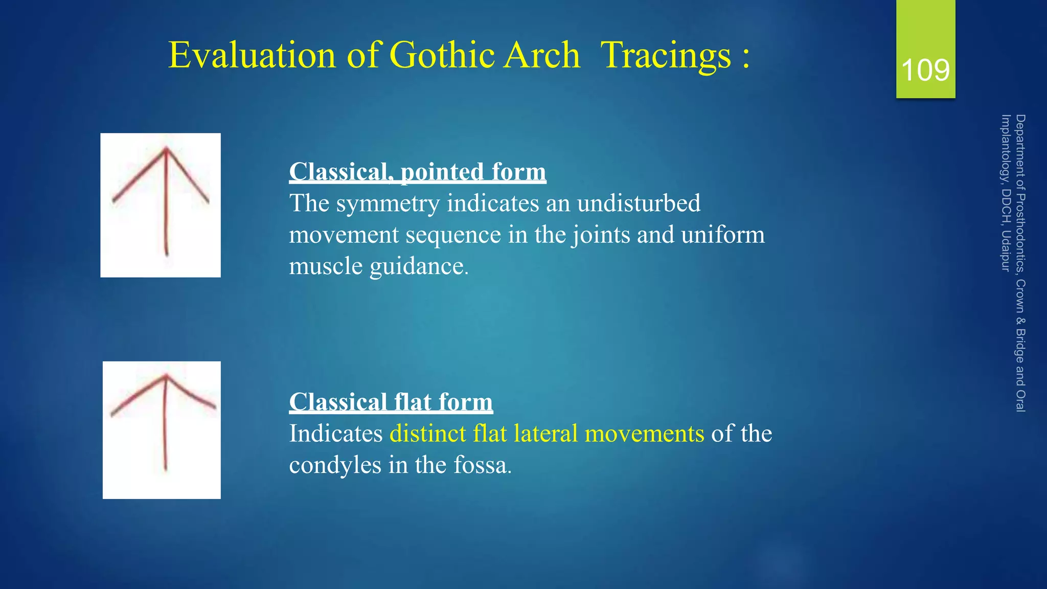
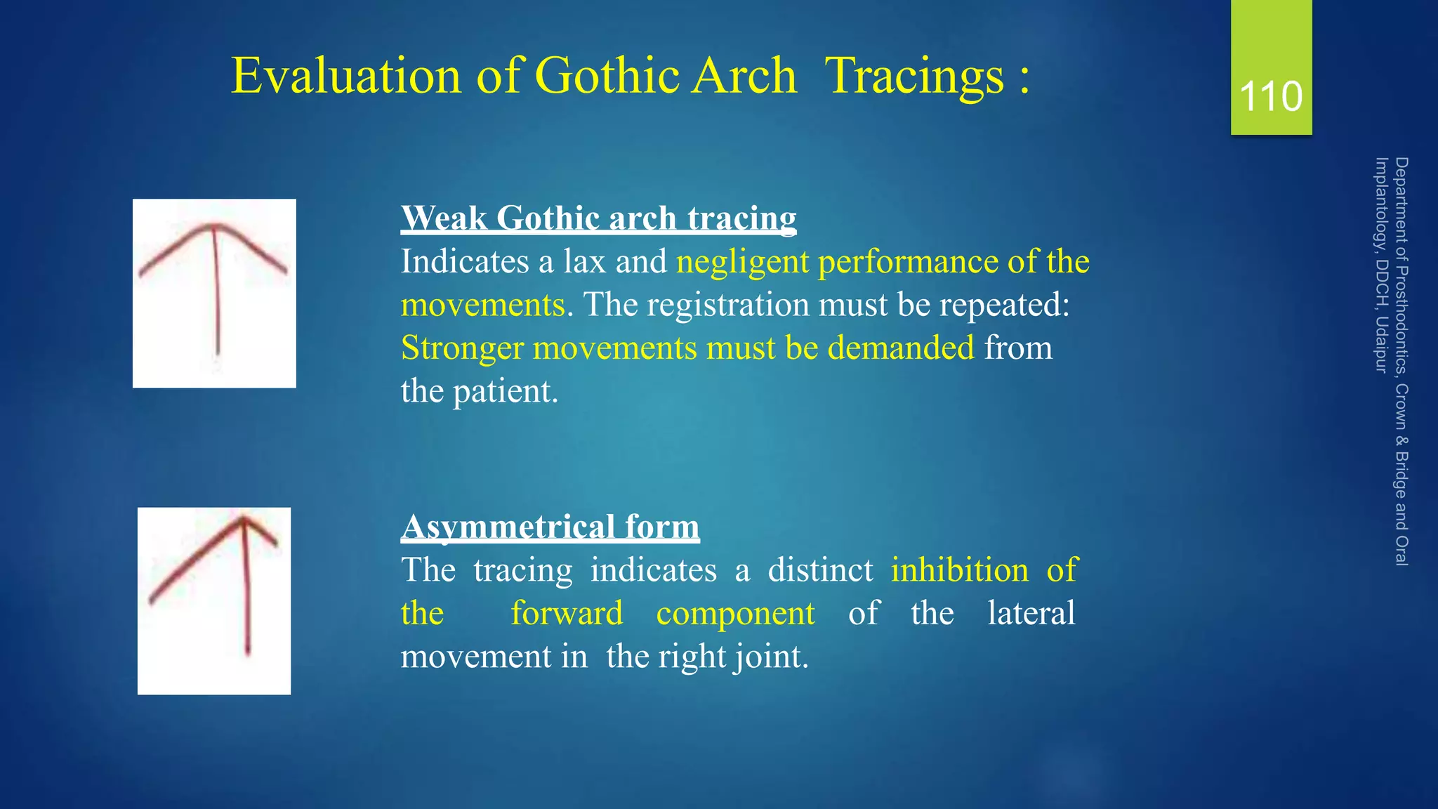
Smith [1975] considered CR to be ‘the most retruded position of the mandible’
and concluded that the gothic arch tracing provides the most retruded and most
repeatable position and thus was the most precise method.
Williamson et al. [1977] stated that the hinge axis and CR are the same; adding
that this axis occurs when the mandible is in CR and a pure rotational movement
of the mandible is produced in the sagittal plane
14](https://image.slidesharecdn.com/centricjawrelation1autosaved-220205182724/75/Centric-jaw-relation-14-2048.jpg)
![CENTRIC RELATION FROM 1970 TO 1980’S
Lucia [1979] stated that ‘the mandible is in CR when the centers of vertical and
lateral motion are in the terminal hinge position’ .
Myers et al. [1980] defined CR as ‘the most posterior unstrained relation of the
mandible to the maxilla at a given degree of jaw separation’. They stated that the
more posterior the condyles, the more acceptable the position
15](https://image.slidesharecdn.com/centricjawrelation1autosaved-220205182724/75/Centric-jaw-relation-15-2048.jpg)
![CENTRIC RELATION FROM 1980 TO 2010
Gilbe [1983] defined CR as ‘the most superior position of the mandibular
condyles with the central bearing area of the disc in contact with the articular
surface of the condyle and the articular eminence. This position may not always be
possible to attain due to anterior dislocation of the disc .
Dawson in 1985 stated that ‘CR is achieved when the properly aligned condyle-
disk assemblies are in the most superior position against the eminentia irrespective
of tooth position or vertical dimension’
16](https://image.slidesharecdn.com/centricjawrelation1autosaved-220205182724/75/Centric-jaw-relation-16-2048.jpg)
![CENTRIC RELATION FROM 1980 TO 2010
American College of Prosthodontist [1994] defined CR as ‘the spatial relationship
between the maxilla and mandible where the condyles relate to the articular
eminence in a ventro-cranial position with the pars intermedia of the disc’
17](https://image.slidesharecdn.com/centricjawrelation1autosaved-220205182724/75/Centric-jaw-relation-17-2048.jpg)
![GPT DEFINITIONS
GPT-1 [1956] defined CR as ‘the most retruded relation of the mandible to the
maxilla when the condyles are in the most posterior unstrained position in the
glenoid fossa from which lateral movements can be made, at any given degrees of
jaw separation’
18](https://image.slidesharecdn.com/centricjawrelation1autosaved-220205182724/75/Centric-jaw-relation-18-2048.jpg)
![GPT DEFINITIONS
GPT-2 [1960] defined the CR as ‘the most posterior relation of the mandible to the
maxilla at the established vertical relation’,
GPT-3 [1968] defined CR as ‘the most retruded physiologic relation of the
mandible to the maxilla and from which the individual can make lateral
movements’. It is a condition that can exist at various degrees of jaw separation. It
occurs around the terminal hinge axis .
19](https://image.slidesharecdn.com/centricjawrelation1autosaved-220205182724/75/Centric-jaw-relation-19-2048.jpg)
![GPT DEFINITIONS
GPT-4 [1977] defined CR as ‘the jaw relation when the condyles are in the most
posterior, unstrained position in the glenoid fossa at any given degree of jaw
separation from which lateral movements can be made’
20](https://image.slidesharecdn.com/centricjawrelation1autosaved-220205182724/75/Centric-jaw-relation-20-2048.jpg)
![GPT DEFINITIONS
GPT-5 and 6 [1987, 1994] defined the CR as ‘the relation of the mandible to the
maxilla when the condyles are in their most posterior position in the glenoid fossa
from which unstrained lateral movements can be made at occluding vertical
dimension normal for the individual’
21](https://image.slidesharecdn.com/centricjawrelation1autosaved-220205182724/75/Centric-jaw-relation-21-2048.jpg)
![GPT DEFINITIONS
GPT-7 [1999] defined centric relation as ‘a maxillomandibular relationship in
which the condyles articulate with the thinnest avascular portion of their
respective disks with the complex in the anterosuperior position against the shapes
of the articular eminences. This position is independent of tooth contact. This
position is clinically discernible when the mandible is directed superiorly and
anteriorly and restricted to a purely rotary movement about a transverse horizontal
axis’
22](https://image.slidesharecdn.com/centricjawrelation1autosaved-220205182724/75/Centric-jaw-relation-22-2048.jpg)